3CQJ
 
 | |
3CQH
 | |
2HXW
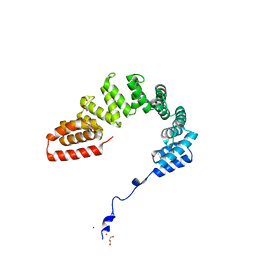
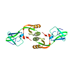
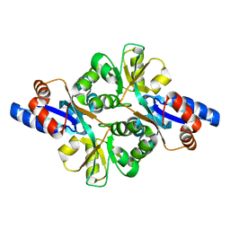
 | | Crystal Structure of Peb3 from Campylobacter jejuni | | 分子名称: | CITRATE ANION, Major antigenic peptide PEB3 | | 著者 | Rangarajan, E.S, Bhatia, S, Watson, D.C, Munger, C, Cygler, M, Matte, A, Young, N.M, Montreal-Kingston Bacterial Structural Genomics Initiative (BSGI) | | 登録日 | 2006-08-04 | | 公開日 | 2007-05-01 | | 最終更新日 | 2022-12-21 | | 実験手法 | X-RAY DIFFRACTION (1.6 Å) | | 主引用文献 | Structural context for protein N-glycosylation in bacteria: The structure of PEB3, an adhesin from Campylobacter jejuni.
Protein Sci., 16, 2007
|
|
3CQI
 | |
3CPT
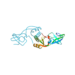
 | | MP1-p14 Scaffolding complex | | 分子名称: | Mitogen-activated protein kinase kinase 1-interacting protein 1, Mitogen-activated protein-binding protein-interacting protein | | 著者 | Schrag, J.D, Cygler, M, Munger, C, Magloire, A. | | 登録日 | 2008-04-01 | | 公開日 | 2008-07-01 | | 最終更新日 | 2024-05-29 | | 実験手法 | X-RAY DIFFRACTION (1.9 Å) | | 主引用文献 | Molecular dynamics-solvated interaction energy studies of protein-protein interactions: the MP1-p14 scaffolding complex.
J.Mol.Biol., 379, 2008
|
|
2I6R
 | | Crystal structure of E. coli HypE, a hydrogenase maturation protein | | 分子名称: | HypE protein | | 著者 | Rangarajan, E.S, Proteau, A, Iannuzzi, P, Matte, A, Cygler, M, Montreal-Kingston Bacterial Structural Genomics Initiative (BSGI) | | 登録日 | 2006-08-29 | | 公開日 | 2007-10-23 | | 最終更新日 | 2024-02-21 | | 実験手法 | X-RAY DIFFRACTION (2.51 Å) | | 主引用文献 | Structure of [NiFe] hydrogenase maturation protein HypE from Escherichia coli and its interaction with HypF.
J.Bacteriol., 190, 2008
|
|
6CDW
 | |
6CGK
 | | Structure of the HAD domain of effector protein Lem4 (lpg1101) from Legionella pneumophila (inactive mutant)with phosphate bound in the active site | | 分子名称: | GLYCEROL, MAGNESIUM ION, PHOSPHATE ION, ... | | 著者 | Beyrakhova, K.A, Xu, C, Cygler, M. | | 登録日 | 2018-02-20 | | 公開日 | 2018-07-18 | | 最終更新日 | 2023-10-04 | | 実験手法 | X-RAY DIFFRACTION (1.668 Å) | | 主引用文献 | Legionella pneumophilaeffector Lem4 is a membrane-associated protein tyrosine phosphatase.
J. Biol. Chem., 293, 2018
|
|
6CGI
 | | Structure of Salmonella Effector SseK3 | | 分子名称: | Type III secretion system effector protein, URIDINE-5'-DIPHOSPHATE | | 著者 | Chung, I.Y.W, Cygler, M. | | 登録日 | 2018-02-20 | | 公開日 | 2018-02-28 | | 最終更新日 | 2024-03-13 | | 実験手法 | X-RAY DIFFRACTION (2.3 Å) | | 主引用文献 | Salmonella Effectors SseK1 and SseK3 Target Death Domain Proteins in the TNF and TRAIL Signaling Pathways.
Mol.Cell Proteomics, 18, 2019
|
|
6CGJ
 | | Structure of the HAD domain of effector protein Lem4 (lpg1101) from Legionella pneumophila | | 分子名称: | ACETATE ION, GLYCEROL, MAGNESIUM ION, ... | | 著者 | Beyrakhova, K.A, Xu, C, Cygler, M. | | 登録日 | 2018-02-20 | | 公開日 | 2018-07-18 | | 最終更新日 | 2024-03-13 | | 実験手法 | X-RAY DIFFRACTION (1.75 Å) | | 主引用文献 | Legionella pneumophilaeffector Lem4 is a membrane-associated protein tyrosine phosphatase.
J. Biol. Chem., 293, 2018
|
|
6D2C
 | |
6DEH
 | | Structure of LpnE Effector Protein from Legionella pneumophila (sp. Philadelphia) | | 分子名称: | DI(HYDROXYETHYL)ETHER, GLYCEROL, NICKEL (II) ION, ... | | 著者 | Voth, K, Chung, I.Y.W, van Straaten, K.E, Cygler, M. | | 登録日 | 2018-05-11 | | 公開日 | 2018-12-19 | | 最終更新日 | 2023-10-11 | | 実験手法 | X-RAY DIFFRACTION (1.8 Å) | | 主引用文献 | The structure of Legionella effector protein LpnE provides insights into its interaction with Oculocerebrorenal syndrome of Lowe (OCRL) protein.
FEBS J., 286, 2019
|
|
6DUS
 | | Structure of Salmonella Effector SseK3 E258Q mutant | | 分子名称: | (4S)-2-METHYL-2,4-PENTANEDIOL, 2-AMINO-2-HYDROXYMETHYL-PROPANE-1,3-DIOL, 2-acetamido-2-deoxy-beta-D-glucopyranose, ... | | 著者 | Chung, I.Y.W, Cygler, M. | | 登録日 | 2018-06-21 | | 公開日 | 2018-07-18 | | 最終更新日 | 2024-03-13 | | 実験手法 | X-RAY DIFFRACTION (2.6 Å) | | 主引用文献 | Salmonella Effectors SseK1 and SseK3 Target Death Domain Proteins in the TNF and TRAIL Signaling Pathways.
Mol.Cell Proteomics, 18, 2019
|
|
6D3U
 | | Complex structure of Ulvan lyase from Nonlaben Ulvanivorans- NLR48 | | 分子名称: | 1,2-ETHANEDIOL, 4-deoxy-alpha-L-threo-hex-4-enopyranuronic acid-(1-4)-3-O-sulfo-alpha-L-rhamnopyranose-(1-4)-beta-D-glucopyranuronic acid-(1-4)-3-O-sulfo-alpha-L-rhamnopyranose, CALCIUM ION, ... | | 著者 | Ulaganathan, T, Cygler, M. | | 登録日 | 2018-04-16 | | 公開日 | 2018-06-06 | | 最終更新日 | 2020-07-29 | | 実験手法 | X-RAY DIFFRACTION (2.21 Å) | | 主引用文献 | Structural and functional characterization of PL28 family ulvan lyase NLR48 fromNonlabens ulvanivorans.
J. Biol. Chem., 293, 2018
|
|
3FJ7
 | |
3LA0
 | | Crystal Structure of UreE from Helicobacter pylori (metal of unknown identity bound) | | 分子名称: | UNKNOWN ATOM OR ION, Urease accessory protein ureE | | 著者 | Shi, R, Munger, C, Assinas, A, Matte, A, Cygler, M, Montreal-Kingston Bacterial Structural Genomics Initiative (BSGI) | | 登録日 | 2010-01-06 | | 公開日 | 2010-08-25 | | 最終更新日 | 2023-09-06 | | 実験手法 | X-RAY DIFFRACTION (2.86 Å) | | 主引用文献 | Crystal Structures of Apo and Metal-Bound Forms of the UreE Protein from Helicobacter pylori: Role of Multiple Metal Binding Sites
Biochemistry, 49, 2010
|
|
3L9Z
 | | Crystal Structure of UreE from Helicobacter pylori (apo form) | | 分子名称: | Urease accessory protein ureE | | 著者 | Shi, R, Munger, C, Assinas, A, Matte, A, Cygler, M, Montreal-Kingston Bacterial Structural Genomics Initiative (BSGI) | | 登録日 | 2010-01-06 | | 公開日 | 2010-08-25 | | 最終更新日 | 2024-02-21 | | 実験手法 | X-RAY DIFFRACTION (2.08 Å) | | 主引用文献 | Crystal Structures of Apo and Metal-Bound Forms of the UreE Protein from Helicobacter pylori: Role of Multiple Metal Binding Sites
Biochemistry, 49, 2010
|
|
3FJG
 | | Crystal structure of 3PG bound PEB3 | | 分子名称: | 3-PHOSPHOGLYCERIC ACID, Major antigenic peptide PEB3 | | 著者 | Min, T, Matte, A, Cygler, M. | | 登録日 | 2008-12-14 | | 公開日 | 2009-03-10 | | 最終更新日 | 2023-09-06 | | 実験手法 | X-RAY DIFFRACTION (2.2 Å) | | 主引用文献 | Specificity of Campylobacter jejuni adhesin PEB3 for phosphates and structural differences among its ligand complexes.
Biochemistry, 48, 2009
|
|
3FIR
 | | Crystal structure of Glycosylated K135E PEB3 | | 分子名称: | 2-acetamido-2-deoxy-alpha-L-glucopyranose-(1-3)-2,4-bisacetamido-2,4,6-trideoxy-beta-D-glucopyranose, CITRATE ANION, Major antigenic peptide PEB3 | | 著者 | Min, T, Matte, A, Cygler, M. | | 登録日 | 2008-12-12 | | 公開日 | 2009-03-10 | | 最終更新日 | 2023-09-06 | | 実験手法 | X-RAY DIFFRACTION (2 Å) | | 主引用文献 | Specificity of Campylobacter jejuni adhesin PEB3 for phosphates and structural differences among its ligand complexes.
Biochemistry, 48, 2009
|
|
3FJM
 | | crystal structure of phosphate bound PEB3 | | 分子名称: | Major antigenic peptide PEB3, PHOSPHATE ION | | 著者 | Min, T, Matte, A, Cygler, M. | | 登録日 | 2008-12-14 | | 公開日 | 2009-03-10 | | 最終更新日 | 2023-09-06 | | 実験手法 | X-RAY DIFFRACTION (1.6 Å) | | 主引用文献 | Specificity of Campylobacter jejuni adhesin PEB3 for phosphates and structural differences among its ligand complexes.
Biochemistry, 48, 2009
|
|
3G05
 | | Crystal structure of N-terminal domain (2-550) of E.coli MnmG | | 分子名称: | SULFATE ION, tRNA uridine 5-carboxymethylaminomethyl modification enzyme mnmG | | 著者 | Shi, R, Matte, A, Cygler, M, Montreal-Kingston Bacterial Structural Genomics Initiative (BSGI) | | 登録日 | 2009-01-27 | | 公開日 | 2009-10-20 | | 最終更新日 | 2023-09-06 | | 実験手法 | X-RAY DIFFRACTION (3.49 Å) | | 主引用文献 | Structure-function analysis of Escherichia coli MnmG (GidA), a highly conserved tRNA-modifying enzyme.
J.Bacteriol., 191, 2009
|
|
2LIP
 | |
1MFD
 | |
1DBG
 | | CRYSTAL STRUCTURE OF CHONDROITINASE B | | 分子名称: | 4-deoxy-alpha-D-glucopyranose-(1-3)-[beta-D-glucopyranose-(1-4)]2-O-methyl-beta-L-fucopyranose-(1-4)-beta-D-xylopyranose-(1-4)-alpha-D-glucopyranuronic acid-(1-2)-[alpha-L-rhamnopyranose-(1-4)]alpha-D-mannopyranose, CHONDROITINASE B | | 著者 | Huang, W, Matte, A, Li, Y, Kim, Y.S, Linhardt, R.J, Su, H, Cygler, M. | | 登録日 | 1999-11-02 | | 公開日 | 2000-01-12 | | 最終更新日 | 2020-07-29 | | 実験手法 | X-RAY DIFFRACTION (1.7 Å) | | 主引用文献 | Crystal structure of chondroitinase B from Flavobacterium heparinum and its complex with a disaccharide product at 1.7 A resolution.
J.Mol.Biol., 294, 1999
|
|
1EOJ
 | | Design of P1' and P3' residues of trivalent thrombin inhibitors and their crystal structures | | 分子名称: | ALPHA THROMBIN, THROMBIN INHIBITOR P798 | | 著者 | Slon-Usakiewicz, J.J, Sivaraman, J, Li, Y, Cygler, M, Konishi, Y. | | 登録日 | 2000-03-23 | | 公開日 | 2000-05-03 | | 最終更新日 | 2023-11-15 | | 実験手法 | X-RAY DIFFRACTION (2.1 Å) | | 主引用文献 | Design of P1' and P3' residues of trivalent thrombin inhibitors and their crystal structures.
Biochemistry, 39, 2000
|
|


