Loading
PDBj
MenuPDBj@FacebookPDBj@TwitterPDBj@YouTubewwPDB FoundationwwPDB
RCSB PDBPDBeBMRBAdv. SearchSearch help
PDB: 2 results

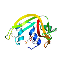
1H1H
DownloadVisualize
BU of 1h1h by Molmil
Crystal Structure of Eosinophil Cationic Protein in Complex with 2',5'-ADP at 2.0 A resolution Reveals the Details of the Ribonucleolytic Active site
Descriptor: ADENOSINE-2'-5'-DIPHOSPHATE, EOSINOPHIL CATIONIC PROTEIN
Authors:Mohan, C.G, Boix, E, Evans, H.R, Nikolovski, Z, Nogues, M.V, Cuchillo, C.M, Acharya, K.R.
Deposit date:2002-07-15
Release date:2002-10-03
Last modified:2023-12-13
Method:X-RAY DIFFRACTION (2 Å)
Cite:The Crystal Structure of Eosinophil Cationic Protein in Complex with 2'5'-Adp at 2.0 A Resolution Reveals the Details of the Ribonucleolytic Active Site
Biochemistry, 41, 2002
1RCN
DownloadVisualize
BU of 1rcn by Molmil
CRYSTAL STRUCTURE OF THE RIBONUCLEASE A D(APTPAPAPG) COMPLEX : DIRECT EVIDENCE FOR EXTENDED SUBSTRATE RECOGNITION
Descriptor: DNA (5'-D(*AP*TP*AP*A)-3'), PROTEIN (RIBONUCLEASE A (E.C.3.1.27.5))
Authors:Fontecilla-Camps, J.C, De Llorens, R, Le Du, M.H, Cuchillo, C.M.
Deposit date:1994-05-27
Release date:1994-09-30
Last modified:2011-07-13
Method:X-RAY DIFFRACTION (2.32 Å)
Cite:Crystal structure of ribonuclease A.d(ApTpApApG) complex. Direct evidence for extended substrate recognition.
J.Biol.Chem., 269, 1994

222624

数据于2024-07-17公开中

PDB statisticsPDBj update infoContact PDBjnumon