Loading
PDBj
MenuPDBj@FacebookPDBj@TwitterPDBj@YouTubewwPDB FoundationwwPDB
RCSB PDBPDBeBMRBAdv. SearchSearch help
PDB: 153 results

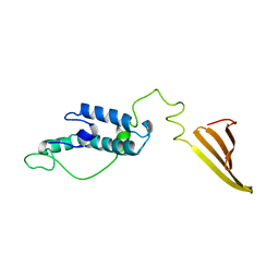
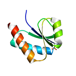
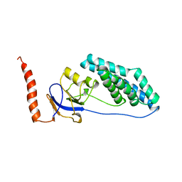
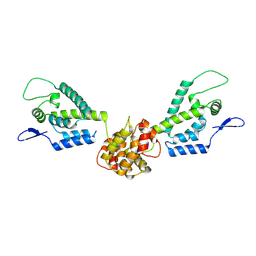
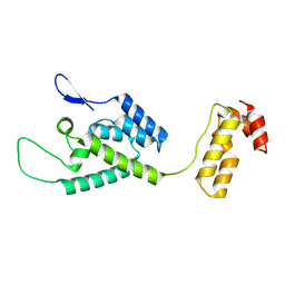
6U3R
DownloadVisualize
BU of 6u3r by Molmil
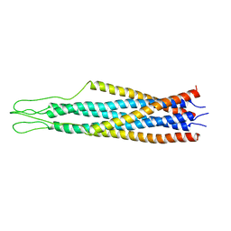
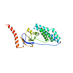
Solution NMR structure of the DNAJB6b deltaST variant (Aligned on the J domain)
Descriptor: DnaJ homolog subfamily B member 6,DnaJ homolog subfamily B member 6
Authors:Karamanos, T.K, Clore, G.M.
Deposit date:2019-08-22
Release date:2019-10-09
Last modified:2024-05-15
Method:SOLUTION NMR
Cite:Unraveling the structure and dynamics of the human DNAJB6b chaperone by NMR reveals insights into Hsp40-mediated proteostasis.
Proc.Natl.Acad.Sci.USA, 116, 2019
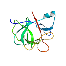
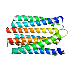
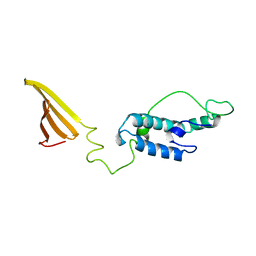
1VMC
DownloadVisualize
BU of 1vmc by Molmil
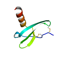
STROMA CELL-DERIVED FACTOR-1ALPHA (SDF-1ALPHA)
Descriptor: Stromal cell-derived factor 1
Authors:Gozansky, E.K, Clore, G.M.
Deposit date:2004-09-20
Release date:2005-03-01
Last modified:2023-12-27
Method:SOLUTION NMR
Cite:Mapping the binding of the N-terminal extracellular tail of the CXCR4 receptor to stromal cell-derived factor-1alpha.
J.Mol.Biol., 345, 2005
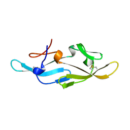
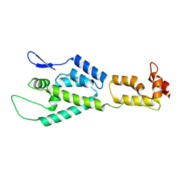
6U3S
DownloadVisualize
BU of 6u3s by Molmil
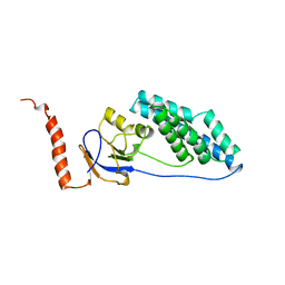
Solution NMR structure of the DNAJB6b deltaST variant (Aligned on the CTD domain)
Descriptor: DnaJ homolog subfamily B member 6
Authors:Karamanos, T.K, Clore, G.M.
Deposit date:2019-08-22
Release date:2019-10-09
Last modified:2024-05-15
Method:SOLUTION NMR
Cite:Unraveling the structure and dynamics of the human DNAJB6b chaperone by NMR reveals insights into Hsp40-mediated proteostasis.
Proc.Natl.Acad.Sci.USA, 116, 2019
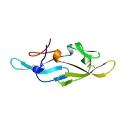
1IOB
DownloadVisualize
BU of 1iob by Molmil
INTERLEUKIN-1 BETA FROM JOINT X-RAY AND NMR REFINEMENT
Descriptor: INTERLEUKIN-1 BETA
Authors:Shaanan, B, Clore, G.M.
Deposit date:1996-03-14
Release date:1996-08-17
Last modified:2024-05-01
Method:SOLUTION NMR (2 Å), X-RAY DIFFRACTION
Cite:Combining experimental information from crystal and solution studies: joint X-ray and NMR refinement.
Science, 257, 1992
2EZM
DownloadVisualize
BU of 2ezm by Molmil
SOLUTION NMR STRUCTURE OF CYANOVIRIN-N, RESTRAINED REGULARIZED MEAN COORDINATES
Descriptor: CYANOVIRIN-N
Authors:Bewley, C.A, Gronenborn, A.M, Clore, G.M.
Deposit date:1998-05-06
Release date:1999-05-11
Last modified:2022-03-09
Method:SOLUTION NMR
Cite:Solution structure of cyanovirin-N, a potent HIV-inactivating protein.
Nat.Struct.Biol., 5, 1998
2EZN
DownloadVisualize
BU of 2ezn by Molmil
SOLUTION NMR STRUCTURE OF CYANOVIRIN-N ENSEMBLE OF 40 SIMULATED ANNEALING STRUCTURES
Descriptor: CYANOVIRIN-N
Authors:Bewley, C.A, Gronenborn, A.M, Clore, G.M.
Deposit date:1998-05-06
Release date:1999-05-11
Last modified:2022-03-09
Method:SOLUTION NMR
Cite:Solution structure of cyanovirin-N, a potent HIV-inactivating protein.
Nat.Struct.Biol., 5, 1998
1VRV
DownloadVisualize
BU of 1vrv by Molmil
Structure of phosphorylated IIB (C384(SEP)) domain of the mannitol-specific permease enzyme II
Descriptor: mannitol-specific PTS system enzyme IIABC components
Authors:Suh, J.Y, Tang, C, Cai, M, Clore, G.M.
Deposit date:2005-06-17
Release date:2005-11-22
Last modified:2023-12-27
Method:SOLUTION NMR
Cite:Visualization of the Phosphorylated Active Site Loop of the Cytoplasmic B Domain of the Mannitol Transporter II(Mannitol) of the Escherichia coli Phosphotransferase System by NMR Spectroscopy and Residual Dipolar Couplings.
J.Mol.Biol., 353, 2005
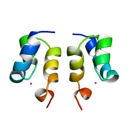
1WJE
DownloadVisualize
BU of 1wje by Molmil
SOLUTION STRUCTURE OF H12C MUTANT OF THE N-TERMINAL ZN BINDING DOMAIN OF HIV-1 INTEGRASE COMPLEXED TO CADMIUM, NMR, MINIMIZED AVERAGE STRUCTURE
Descriptor: CADMIUM ION, HIV-1 INTEGRASE
Authors:Cai, M, Gronenborn, A.M, Clore, G.M.
Deposit date:1998-06-11
Release date:1998-12-16
Last modified:2024-05-01
Method:SOLUTION NMR
Cite:Solution structure of the His12 --> Cys mutant of the N-terminal zinc binding domain of HIV-1 integrase complexed to cadmium.
Protein Sci., 7, 1998
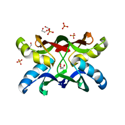
1ZHQ
DownloadVisualize
BU of 1zhq by Molmil
Crystal structure of apo MVL
Descriptor: 1,2-ETHANEDIOL, PHOSPHATE ION, mannan-binding lectin
Authors:Williams, D.C, Lee, J.Y, Cai, M, Bewley, C.A, Clore, G.M.
Deposit date:2005-04-26
Release date:2005-06-07
Last modified:2024-02-14
Method:X-RAY DIFFRACTION (1.9 Å)
Cite:Crystal Structures of the HIV-1 Inhibitory Cyanobacterial Protein MVL Free and Bound to Man3GlcNAc2: STRUCTURAL BASIS FOR SPECIFICITY AND HIGH-AFFINITY BINDING TO THE CORE PENTASACCHARIDE FROM N-LINKED OLIGOMANNOSIDE.
J.Biol.Chem., 280, 2005
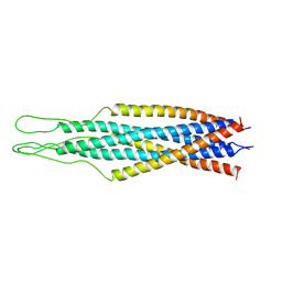
2EZQ
DownloadVisualize
BU of 2ezq by Molmil
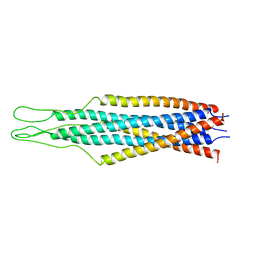
SOLUTION NMR STRUCTURE OF ECTODOMAIN OF SIV GP41, MODELS 11-20 OF AN ENSEMBLE OF 40 SIMULATED ANNEALING STRUCTURES
Descriptor: GP41
Authors:Caffrey, M, Gronenborn, A.M, Clore, G.M.
Deposit date:1998-05-20
Release date:1998-10-14
Last modified:2024-05-01
Method:SOLUTION NMR
Cite:Three-dimensional solution structure of the 44 kDa ectodomain of SIV gp41.
EMBO J., 17, 1998
2EZP
DownloadVisualize
BU of 2ezp by Molmil
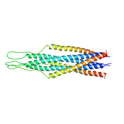
SOLUTION NMR STRUCTURE OF ECTODOMAIN OF SIV GP41, MODELS 1-10 OF AN ENSEMBLE OF 40 SIMULATED ANNEALING STRUCTURES
Descriptor: GP41
Authors:Caffrey, M, Gronenborn, A.M, Clore, G.M.
Deposit date:1998-05-20
Release date:1998-10-14
Last modified:2024-05-01
Method:SOLUTION NMR
Cite:Three-dimensional solution structure of the 44 kDa ectodomain of SIV gp41.
EMBO J., 17, 1998
2EZS
DownloadVisualize
BU of 2ezs by Molmil
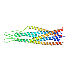
SOLUTION NMR STRUCTURE OF ECTODOMAIN OF SIV GP41, MODELS 31-40 OF AN ENSEMBLE OF 40 SIMULATED ANNEALING STRUCTURES
Descriptor: GP41
Authors:Caffrey, M, Gronenborn, A.M, Clore, G.M.
Deposit date:1998-05-20
Release date:1998-10-14
Last modified:2024-05-01
Method:SOLUTION NMR
Cite:Three-dimensional solution structure of the 44 kDa ectodomain of SIV gp41.
EMBO J., 17, 1998
2EZO
DownloadVisualize
BU of 2ezo by Molmil
SOLUTION NMR STRUCTURE OF ECTODOMAIN OF SIV GP41, RESTRAINED REGULARIZED MEAN STRUCTURE
Descriptor: GP41
Authors:Caffrey, M, Gronenborn, A.M, Clore, G.M.
Deposit date:1998-05-20
Release date:1998-10-14
Last modified:2024-05-29
Method:SOLUTION NMR
Cite:Three-dimensional solution structure of the 44 kDa ectodomain of SIV gp41.
EMBO J., 17, 1998
2EZR
DownloadVisualize
BU of 2ezr by Molmil
SOLUTION NMR STRUCTURE OF ECTODOMAIN OF SIV GP41, MODELS 21-30 OF AN ENSEMBLE OF 40 SIMULATED ANNEALING STRUCTURES
Descriptor: GP41
Authors:Caffrey, M, Gronenborn, A.M, Clore, G.M.
Deposit date:1998-05-20
Release date:1998-10-14
Last modified:2024-05-01
Method:SOLUTION NMR
Cite:Three-dimensional solution structure of the 44 kDa ectodomain of SIV gp41.
EMBO J., 17, 1998
1ZHS
DownloadVisualize
BU of 1zhs by Molmil
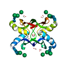
Crystal structure of MVL bound to Man3GlcNAc2
Descriptor: 1,2-ETHANEDIOL, PHOSPHATE ION, alpha-D-mannopyranose-(1-3)-[alpha-D-mannopyranose-(1-6)]beta-D-mannopyranose-(1-4)-2-acetamido-2-deoxy-beta-D-glucopyranose-(1-4)-2-acetamido-2-deoxy-beta-D-glucopyranose, ...
Authors:Williams, D.C, Lee, J.Y, Cai, M, Bewley, C.A, Clore, G.M.
Deposit date:2005-04-26
Release date:2005-06-07
Last modified:2024-02-14
Method:X-RAY DIFFRACTION (1.8 Å)
Cite:Crystal Structures of the HIV-1 Inhibitory Cyanobacterial Protein MVL Free and Bound to Man3GlcNAc2: STRUCTURAL BASIS FOR SPECIFICITY AND HIGH-AFFINITY BINDING TO THE CORE PENTASACCHARIDE FROM N-LINKED OLIGOMANNOSIDE.
J.Biol.Chem., 280, 2005
1EZB
DownloadVisualize
BU of 1ezb by Molmil
AMINO TERMINAL DOMAIN OF ENZYME I FROM ESCHERICHIA COLI, NMR, 17 STRUCTURES
Descriptor: ENZYME I
Authors:Garrett, D.S, Gronenborn, A.M, Clore, G.M.
Deposit date:1997-01-01
Release date:1998-01-07
Last modified:2024-05-01
Method:SOLUTION NMR
Cite:Solution structure of the 30 kDa N-terminal domain of enzyme I of the Escherichia coli phosphoenolpyruvate:sugar phosphotransferase system by multidimensional NMR.
Biochemistry, 36, 1997
1EZA
DownloadVisualize
BU of 1eza by Molmil
AMINO TERMINAL DOMAIN OF ENZYME I FROM ESCHERICHIA COLI NMR, RESTRAINED REGULARIZED MEAN STRUCTURE
Descriptor: ENZYME I
Authors:Garrett, D.S, Gronenborn, A.M, Clore, G.M.
Deposit date:1997-01-01
Release date:1998-01-07
Last modified:2024-05-01
Method:SOLUTION NMR
Cite:Solution structure of the 30 kDa N-terminal domain of enzyme I of the Escherichia coli phosphoenolpyruvate:sugar phosphotransferase system by multidimensional NMR.
Biochemistry, 36, 1997
1EZD
DownloadVisualize
BU of 1ezd by Molmil
AMINO TERMINAL DOMAIN OF ENZYME I FROM ESCHERICHIA COLI NMR, 16 STRUCTURES
Descriptor: ENZYME I
Authors:Garrett, D.S, Gronenborn, A.M, Clore, G.M.
Deposit date:1997-01-01
Release date:1998-01-07
Last modified:2024-05-01
Method:SOLUTION NMR
Cite:Solution structure of the 30 kDa N-terminal domain of enzyme I of the Escherichia coli phosphoenolpyruvate:sugar phosphotransferase system by multidimensional NMR.
Biochemistry, 36, 1997
1EZC
DownloadVisualize
BU of 1ezc by Molmil
AMINO TERMINAL DOMAIN OF ENZYME I FROM ESCHERICHIA COLI, NMR, 17 STRUCTURES
Descriptor: ENZYME I
Authors:Garrett, D.S, Gronenborn, A.M, Clore, G.M.
Deposit date:1997-01-01
Release date:1998-01-07
Last modified:2024-05-01
Method:SOLUTION NMR
Cite:Solution structure of the 30 kDa N-terminal domain of enzyme I of the Escherichia coli phosphoenolpyruvate:sugar phosphotransferase system by multidimensional NMR.
Biochemistry, 36, 1997
1WCR
DownloadVisualize
BU of 1wcr by Molmil
Trimeric Structure of the Enzyme IIA from Escherichia coli Phosphotransferase System Specific for N,N'-Diacetylchitobiose
Descriptor: PTS SYSTEM, N, N'-DIACETYLCHITOBIOSE-SPECIFIC IIA COMPONENT
Authors:Tang, C, Clore, G.M.
Deposit date:2004-11-19
Release date:2005-01-19
Last modified:2024-05-15
Method:SOLUTION NMR
Cite:Solution Structure of Enzyme Iiachitobiose from the N,N'-Diacetylchitobiose Branch of the Escherichia Coli Phosphotransferase System
J.Biol.Chem., 280, 2005
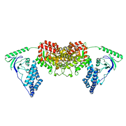
2L5H
DownloadVisualize
BU of 2l5h by Molmil
Solution Structure of the H189Q mutant of the Enzyme I dimer Using Residual Dipolar Couplings and Small Angle X-Ray Scattering
Descriptor: Phosphoenolpyruvate-protein phosphotransferase
Authors:Takayama, Y.D, Schwieters, C.D, Grishaev, A, Guirlando, R, Clore, G.
Deposit date:2010-11-01
Release date:2011-01-12
Last modified:2024-05-01
Method:SOLUTION NMR, SOLUTION SCATTERING
Cite:Combined Use of Residual Dipolar Couplings and Solution X-ray Scattering To Rapidly Probe Rigid-Body Conformational Transitions in a Non-phosphorylatable Active-Site Mutant of the 128 kDa Enzyme I Dimer.
J.Am.Chem.Soc., 133, 2011
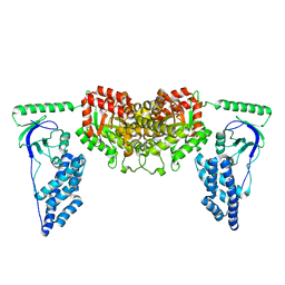
2KX9
DownloadVisualize
BU of 2kx9 by Molmil
Solution Structure of the Enzyme I dimer Using Residual Dipolar Couplings and Small Angle X-Ray Scattering
Descriptor: Phosphoenolpyruvate-protein phosphotransferase
Authors:Schwieters, C.D, Suh, J, Grishaev, A, Takayama, Y, Guirlando, R, Clore, G.
Deposit date:2010-04-29
Release date:2010-09-15
Last modified:2024-05-01
Method:SOLUTION NMR, SOLUTION SCATTERING
Cite:Solution structure of the 128 kDa enzyme I dimer from Escherichia coli and its 146 kDa complex with HPr using residual dipolar couplings and small- and wide-angle X-ray scattering.
J.Am.Chem.Soc., 132, 2010
2M8L
DownloadVisualize
BU of 2m8l by Molmil
HIV capsid dimer structure
Descriptor: Capsid protein p24
Authors:Deshmukh, L, Schwieters, C.D, Grishaev, A, Clore, G, Ghirlando, R.
Deposit date:2013-05-23
Release date:2013-11-20
Last modified:2023-06-14
Method:SOLUTION NMR, SOLUTION SCATTERING
Cite:Structure and Dynamics of Full-Length HIV-1 Capsid Protein in Solution.
J.Am.Chem.Soc., 135, 2013
2M8P
DownloadVisualize
BU of 2m8p by Molmil
The structure of the W184AM185A mutant of the HIV-1 capsid protein
Descriptor: Capsid protein p24
Authors:Deshmukh, L, Schwieters, C.D, Grishaev, A, Clore, G, Ghirlando, R.
Deposit date:2013-05-24
Release date:2013-11-20
Last modified:2023-06-14
Method:SOLUTION NMR, SOLUTION SCATTERING
Cite:Structure and Dynamics of Full-Length HIV-1 Capsid Protein in Solution.
J.Am.Chem.Soc., 135, 2013
2M8N
DownloadVisualize
BU of 2m8n by Molmil
HIV-1 capsid monomer structure
Descriptor: Capsid protein p24
Authors:Deshmukh, L, Schwieters, C.D, Grishaev, A, Clore, G, Ghirlando, R.
Deposit date:2013-05-24
Release date:2013-11-20
Last modified:2023-06-14
Method:SOLUTION NMR, SOLUTION SCATTERING
Cite:Structure and Dynamics of Full-Length HIV-1 Capsid Protein in Solution.
J.Am.Chem.Soc., 135, 2013

222926

數據於2024-07-24公開中

PDB statisticsPDBj update infoContact PDBjnumon