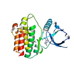
8X2Q
 
 | | The Crystal Structure of APC from Biortus. | | 分子名称: | 1,2-ETHANEDIOL, Adenomatous polyposis coli protein | | 著者 | Wang, F, Cheng, W, Lv, Z, Ju, C, Bao, C. | | 登録日 | 2023-11-10 | | 公開日 | 2023-11-22 | | 実験手法 | X-RAY DIFFRACTION (2 Å) | | 主引用文献 | The Crystal Structure of APC from Biortus.
To Be Published
|
|
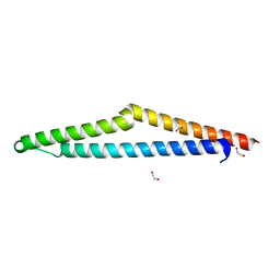
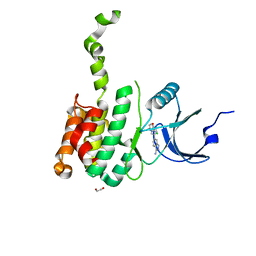
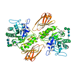
8X5Z
 | |
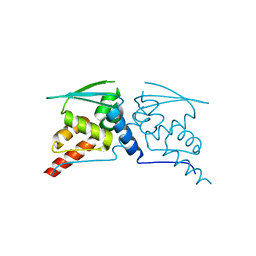
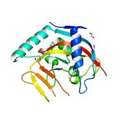
8WF7
 | | The Crystal Structure of integrase from Biortus | | 分子名称: | ACETATE ION, Integrase, SULFATE ION | | 著者 | Wang, F, Cheng, W, Yuan, Z, Qi, J, Li, J. | | 登録日 | 2023-09-19 | | 公開日 | 2023-10-04 | | 実験手法 | X-RAY DIFFRACTION (1.55 Å) | | 主引用文献 | The Crystal Structure of integrase from Biortus
To Be Published
|
|
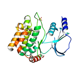
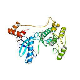
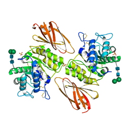
8X87
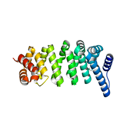 | |
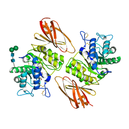
8X88
 | |
8XPX
 | | The Crystal Structure of PARP12 from Biortus. | | 分子名称: | 1,2-ETHANEDIOL, ACETATE ION, DI(HYDROXYETHYL)ETHER, ... | | 著者 | Wang, F, Cheng, W, Lv, Z, Qi, J, Shen, Z. | | 登録日 | 2024-01-04 | | 公開日 | 2024-03-06 | | 実験手法 | X-RAY DIFFRACTION (1.75 Å) | | 主引用文献 | The Crystal Structure of PARP12 from Biortus.
To Be Published
|
|
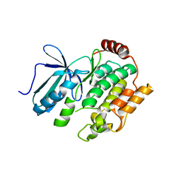
8WFG
 | |
8X5M
 | | The Crystal Structure of JNK1 from Biortus. | | 分子名称: | 1,2-ETHANEDIOL, 3-[4-(dimethylamino)butanoylamino]-~{N}-[3-methyl-4-[(4-pyridin-3-ylpyrimidin-2-yl)amino]phenyl]benzamide, GLYCEROL, ... | | 著者 | Wang, F, Cheng, W, Yuan, Z, Qi, J, Li, J. | | 登録日 | 2023-11-17 | | 公開日 | 2023-12-27 | | 実験手法 | X-RAY DIFFRACTION (2 Å) | | 主引用文献 | The Crystal Structure of JNK1 from Biortus.
To Be Published
|
|
8WGF
 | | The Crystal Structure of JNK3 from Biortus. | | 分子名称: | MAGNESIUM ION, Mitogen-activated protein kinase 10, PHOSPHOAMINOPHOSPHONIC ACID-ADENYLATE ESTER | | 著者 | Wang, F, Cheng, W, Lv, Z, Ju, C, Wang, J. | | 登録日 | 2023-09-21 | | 公開日 | 2023-11-22 | | 実験手法 | X-RAY DIFFRACTION (1.85 Å) | | 主引用文献 | The Crystal Structure of JNK3 from Biortus.
To Be Published
|
|
8X72
 | | The Crystal Structure of PLK1 from Biortus. | | 分子名称: | 1,2-ETHANEDIOL, ACETATE ION, DI(HYDROXYETHYL)ETHER, ... | | 著者 | Wang, F, Cheng, W, Yuan, Z, Lin, D, Wu, B. | | 登録日 | 2023-11-22 | | 公開日 | 2023-12-27 | | 実験手法 | X-RAY DIFFRACTION (2.2 Å) | | 主引用文献 | The Crystal Structure of PLK1 from Biortus.
To Be Published
|
|
8XB9
 | | The Crystal Structure of polo-box domain of PLK1 from Biortus. | | 分子名称: | 1,2-ETHANEDIOL, Serine/threonine-protein kinase PLK1 | | 著者 | Wang, F, Cheng, W, Yuan, Z, Meng, Q, Zhang, B. | | 登録日 | 2023-12-06 | | 公開日 | 2023-12-27 | | 実験手法 | X-RAY DIFFRACTION (1.95 Å) | | 主引用文献 | The Crystal Structure of polo-box domain of PLK1 from Biortus
To Be Published
|
|
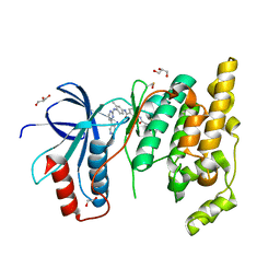
8ZWV
 | | The Crystal Structure of carbonic anhydrase II from Biortus. | | 分子名称: | 1,2-ETHANEDIOL, Carbonic anhydrase 2, ZINC ION, ... | | 著者 | Wang, F, Cheng, W, Lv, Z, Ju, C, Bao, C. | | 登録日 | 2024-06-13 | | 公開日 | 2024-07-03 | | 実験手法 | X-RAY DIFFRACTION (1.5 Å) | | 主引用文献 | The Crystal Structure of carbonic anhydrase II from Biortus.
To Be Published
|
|
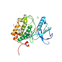
8X5K
 | | The Crystal Structure of SYK from Biortus. | | 分子名称: | 1,2-ETHANEDIOL, 2-{[(1R,2S)-2-aminocyclohexyl]amino}-4-{[3-(2H-1,2,3-triazol-2-yl)phenyl]amino}pyrimidine-5-carboxamide, Tyrosine-protein kinase SYK | | 著者 | Wang, F, Cheng, W, Yuan, Z, Qi, J, Shen, Z. | | 登録日 | 2023-11-17 | | 公開日 | 2023-12-27 | | 実験手法 | X-RAY DIFFRACTION (1.8 Å) | | 主引用文献 | The Crystal Structure of SYK from Biortus.
To Be Published
|
|
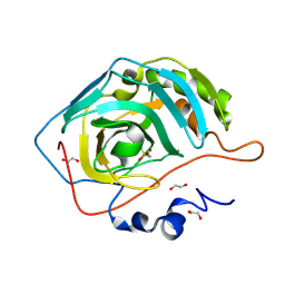
8XFM
 | | The Crystal Structure of MNK2 from Biortus. | | 分子名称: | 1,2-ETHANEDIOL, 5-(3-azanyl-1~{H}-indazol-6-yl)-1-[(3-chlorophenyl)methyl]pyridin-2-one, MAP kinase-interacting serine/threonine-protein kinase 2, ... | | 著者 | Wang, F, Cheng, W, Yuan, Z, Qi, J, Li, J. | | 登録日 | 2023-12-14 | | 公開日 | 2023-12-27 | | 実験手法 | X-RAY DIFFRACTION (2.6 Å) | | 主引用文献 | The Crystal Structure of MNK2 from Biortus.
To Be Published
|
|
8X5J
 | | The Crystal Structure of PARP5A from Biortus. | | 分子名称: | 1,2-ETHANEDIOL, L(+)-TARTARIC ACID, Poly [ADP-ribose] polymerase, ... | | 著者 | Wang, F, Cheng, W, Yuan, Z, Qi, J, Wu, B. | | 登録日 | 2023-11-17 | | 公開日 | 2023-12-27 | | 実験手法 | X-RAY DIFFRACTION (1.7 Å) | | 主引用文献 | The Crystal Structure of PARP5A from Biortus.
To Be Published
|
|
8XFL
 | |
8J2I
 | | Crystal structure of a bright green fluorescent protein (StayGold) with single mutation (Q140S) in jellyfish Cytaeis uchidae from Biortus | | 分子名称: | 1,2-ETHANEDIOL, StayGold(Q140S) | | 著者 | Wu, J, Wang, F, Gui, W, Cheng, W, Yang, Y. | | 登録日 | 2023-04-14 | | 公開日 | 2023-12-13 | | 実験手法 | X-RAY DIFFRACTION (1.75 Å) | | 主引用文献 | Crystal structure of a bright green fluorescent protein (StayGold) in jellyfish Cytaeis uchidae from Biortus
To Be Published
|
|
8J3J
 | | Crystal structure of a bright green fluorescent protein (StayGold) with double mutations (Q140S, Y187F) in jellyfish Cytaeis uchidae from Biortus | | 分子名称: | 1,2-ETHANEDIOL, StayGold(Q140S, Y187F) | | 著者 | Wu, J, Wang, F, Gui, W, Cheng, W, Yang, Y. | | 登録日 | 2023-04-17 | | 公開日 | 2023-12-13 | | 実験手法 | X-RAY DIFFRACTION (1.7 Å) | | 主引用文献 | Crystal structure of a bright green fluorescent protein (StayGold) in jellyfish Cytaeis uchidae from Biortus
To Be Published
|
|
8J2L
 | | Crystal structure of a bright green fluorescent protein (StayGold) with double mutations (N137A, Y187F) in jellyfish Cytaeis uchidae from Biortus | | 分子名称: | 4-(2-HYDROXYETHYL)-1-PIPERAZINE ETHANESULFONIC ACID, GLYCEROL, SODIUM ION, ... | | 著者 | Wu, J, Wang, F, Gui, W, Cheng, W, Yang, Y. | | 登録日 | 2023-04-14 | | 公開日 | 2023-12-13 | | 実験手法 | X-RAY DIFFRACTION (1.7 Å) | | 主引用文献 | Crystal structure of a bright green fluorescent protein (StayGold) in jellyfish Cytaeis uchidae from Biortus
To Be Published
|
|
8J2H
 | | Crystal structure of a bright green fluorescent protein (StayGold) with single mutation (N137A) in jellyfish Cytaeis uchidae from Biortus | | 分子名称: | GLYCEROL, SODIUM ION, StayGold(N137A) | | 著者 | Wu, J, Wang, F, Gui, W, Cheng, W, Yang, Y. | | 登録日 | 2023-04-14 | | 公開日 | 2023-12-13 | | 実験手法 | X-RAY DIFFRACTION (1.7 Å) | | 主引用文献 | Crystal structure of a bright green fluorescent protein (StayGold) in jellyfish Cytaeis uchidae from Biortus
To Be Published
|
|
8J2K
 | | Crystal structure of a bright green fluorescent protein (StayGold) with double mutation (N137A, Q140S) in jellyfish Cytaeis uchidae from Biortus | | 分子名称: | 1,2-ETHANEDIOL, StayGold(N137A, Q140S) | | 著者 | Wu, J, Wang, F, Gui, W, Cheng, W, Yang, Y. | | 登録日 | 2023-04-14 | | 公開日 | 2023-12-13 | | 実験手法 | X-RAY DIFFRACTION (1.7 Å) | | 主引用文献 | Crystal structure of a bright green fluorescent protein (StayGold) in jellyfish Cytaeis uchidae from Biortus
To Be Published
|
|
8J2J
 | | Crystal structure of a bright green fluorescent protein (StayGold) with single mutation (Y187F) in jellyfish Cytaeis uchidae from Biortus | | 分子名称: | 1,2-ETHANEDIOL, SULFATE ION, StayGold(Y187F) | | 著者 | Wu, J, Wang, F, Gui, W, Cheng, W, Yang, Y. | | 登録日 | 2023-04-14 | | 公開日 | 2023-12-13 | | 実験手法 | X-RAY DIFFRACTION (1.9 Å) | | 主引用文献 | Crystal structure of a bright green fluorescent protein (StayGold) in jellyfish Cytaeis uchidae from Biortus
To Be Published
|
|
8K7I
 | | Crystal structure of human lysosomal alpha-galactosidase A in complex with (2R,3S,4R,5R)-2-(hydroxymethyl)-5-((methylamino)methyl)pyrrolidine-3,4-diol | | 分子名称: | (2~{R},3~{S},4~{R},5~{R})-2-(hydroxymethyl)-5-(methylaminomethyl)pyrrolidine-3,4-diol, 2-acetamido-2-deoxy-beta-D-glucopyranose, 2-acetamido-2-deoxy-beta-D-glucopyranose-(1-4)-2-acetamido-2-deoxy-beta-D-glucopyranose, ... | | 著者 | Li, H.Y, Huang, K.F, Ko, T.P, Cheng, W.C. | | 登録日 | 2023-07-26 | | 公開日 | 2024-04-10 | | 実験手法 | X-RAY DIFFRACTION (2.12 Å) | | 主引用文献 | Mechanistic Insights into Dibasic Iminosugars as pH-Selective Pharmacological Chaperones to Stabilize Human alpha-Galactosidase.
Jacs Au, 4, 2024
|
|
8K7J
 | | Crystal structure of human lysosomal alpha-galactosidase A in complex with (2R,3R,4S,5R)-2-((dimethylamino)methyl)-5-(hydroxymethyl)pyrrolidine-3,4-diol | | 分子名称: | (2~{R},3~{R},4~{S},5~{R})-2-[(dimethylamino)methyl]-5-(hydroxymethyl)pyrrolidine-3,4-diol, 2-acetamido-2-deoxy-beta-D-glucopyranose, 2-acetamido-2-deoxy-beta-D-glucopyranose-(1-4)-2-acetamido-2-deoxy-beta-D-glucopyranose, ... | | 著者 | Li, H.Y, Huang, K.F, Ko, T.P, Cheng, W.C. | | 登録日 | 2023-07-26 | | 公開日 | 2024-04-10 | | 実験手法 | X-RAY DIFFRACTION (2.01 Å) | | 主引用文献 | Mechanistic Insights into Dibasic Iminosugars as pH-Selective Pharmacological Chaperones to Stabilize Human alpha-Galactosidase.
Jacs Au, 4, 2024
|
|
8K7H
 | | Crystal structure of human lysosomal alpha-galactosidase A in complex with (2R,3S,4R)-2-(hydroxymethyl)-1-methylpyrrolidine-3,4-diol | | 分子名称: | (2~{R},3~{S},4~{R})-2-(hydroxymethyl)-1-methyl-pyrrolidine-3,4-diol, 2-acetamido-2-deoxy-beta-D-glucopyranose, 2-acetamido-2-deoxy-beta-D-glucopyranose-(1-4)-2-acetamido-2-deoxy-beta-D-glucopyranose, ... | | 著者 | Li, H.Y, Huang, K.F, Ko, T.P, Cheng, W.C. | | 登録日 | 2023-07-26 | | 公開日 | 2024-04-10 | | 実験手法 | X-RAY DIFFRACTION (2.28 Å) | | 主引用文献 | Mechanistic Insights into Dibasic Iminosugars as pH-Selective Pharmacological Chaperones to Stabilize Human alpha-Galactosidase.
Jacs Au, 4, 2024
|
|


