2A9M
 
 | |
2A9N
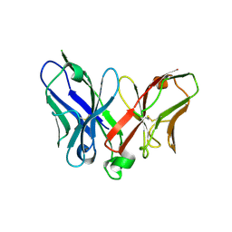
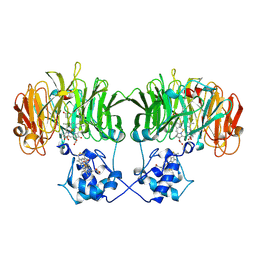
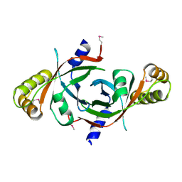
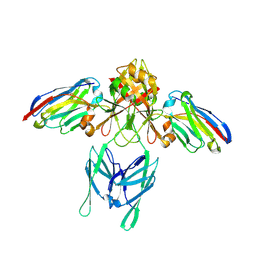
 | | A Mutation Designed to Alter Crystal Packing Permits Structural Analysis of a Tight-binding Fluorescein-scFv complex | | 分子名称: | 4-(2,7-DIFLUORO-6-HYDROXY-3-OXO-3H-XANTHEN-9-YL)ISOPHTHALIC ACID, fluorescein-scfv | | 著者 | Cambillau, C, Spinelli, S, Honegger, A, Pluckthun, A. | | 登録日 | 2005-07-12 | | 公開日 | 2005-10-25 | | 最終更新日 | 2024-04-03 | | 実験手法 | X-RAY DIFFRACTION (3 Å) | | 主引用文献 | A mutation designed to alter crystal packing permits structural analysis of a tight-binding fluorescein-scFv complex.
Protein Sci., 14, 2005
|
|
1N8O
 | |
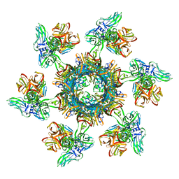
4V5I
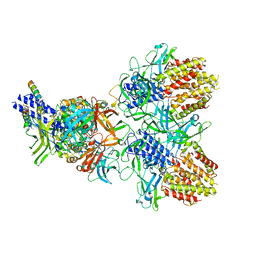
 | | Structure of the Phage P2 Baseplate in its Activated Conformation with Ca | | 分子名称: | CALCIUM ION, ORF15, ORF16, ... | | 著者 | Sciara, G, Bebeacua, C, Bron, P, Tremblay, D, Ortiz-Lombardia, M, Lichiere, J, van Heel, M, Campanacci, V, Moineau, S, Cambillau, C. | | 登録日 | 2010-02-05 | | 公開日 | 2014-07-09 | | 最終更新日 | 2024-01-10 | | 実験手法 | X-RAY DIFFRACTION (5.464 Å) | | 主引用文献 | Structure of Lactococcal Phage P2 Baseplate and its Mechanism of Activation.
Proc.Natl.Acad.Sci.USA, 107, 2010
|
|
1N15
 | |
1N50
 | |
4EQQ
 | | Structure of Ltp, a superinfection exclusion protein from the Streptococcus thermophilus temperate phage TP-J34 | | 分子名称: | PHOSPHATE ION, Putative host cell surface-exposed lipoprotein, SULFATE ION | | 著者 | Bebeacua, C, Lorenzo, C, Blangy, S, Spinelli, S, Heller, K, Cambillau, C. | | 登録日 | 2012-04-19 | | 公開日 | 2013-06-05 | | 最終更新日 | 2024-02-28 | | 実験手法 | X-RAY DIFFRACTION (2.05 Å) | | 主引用文献 | X-ray structure of a superinfection exclusion lipoprotein from phage TP-J34 and identification of the tape measure protein as its target.
Mol.Microbiol., 89, 2013
|
|
3U6X
 | | Phage TP901-1 baseplate tripod | | 分子名称: | BPP, BROMIDE ION, ORF48 | | 著者 | Veesler, D, Spinelli, S, Mahony, J, Lichiere, J, Blangy, S, Bricogne, G, Legrand, P, Ortiz-Lombardia, M, Campanacci, V.I, van Sinderen, D, Cambillau, C. | | 登録日 | 2011-10-13 | | 公開日 | 2012-07-04 | | 最終更新日 | 2023-09-13 | | 実験手法 | X-RAY DIFFRACTION (2.6 Å) | | 主引用文献 | Structure of the phage TP901-1 1.8 MDa baseplate suggests an alternative host adhesion mechanism.
Proc.Natl.Acad.Sci.USA, 109, 2012
|
|
3CAB
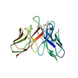
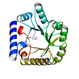
 | | Crystal structure of a pheromone binding protein from Apis mellifera soaked at pH 7.0 | | 分子名称: | GLYCEROL, Pheromone-binding protein ASP1 | | 著者 | Pesenti, M.E, Spinelli, S, Bezirard, V, Briand, L, Pernollet, J.C, Tegoni, M, Cambillau, C. | | 登録日 | 2008-02-19 | | 公開日 | 2008-06-10 | | 最終更新日 | 2023-11-01 | | 実験手法 | X-RAY DIFFRACTION (1.95 Å) | | 主引用文献 | Structural basis of the honey bee PBP pheromone and pH-induced conformational change
J.Mol.Biol., 380, 2008
|
|
3CDN
 | | Crystal structure of a pheromone binding protein from Apis mellifera soaked at pH 4.0 | | 分子名称: | CHLORIDE ION, GLYCEROL, Pheromone-binding protein ASP1 | | 著者 | Pesenti, M.E, Spinelli, S, Bezirard, V, Briand, L, Pernollet, J.C, Tegoni, M, Cambillau, C. | | 登録日 | 2008-02-27 | | 公開日 | 2008-06-10 | | 最終更新日 | 2023-11-01 | | 実験手法 | X-RAY DIFFRACTION (2 Å) | | 主引用文献 | Structural basis of the honey bee PBP pheromone and pH-induced conformational change
J.Mol.Biol., 380, 2008
|
|
2FSD
 | |
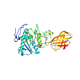
1W8G
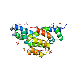
 | | CRYSTAL STRUCTURE OF E. COLI K-12 YGGS | | 分子名称: | HYPOTHETICAL UPF0001 PROTEIN YGGS, ISOCITRIC ACID, PYRIDOXAL-5'-PHOSPHATE | | 著者 | Sulzenbacher, G, Gruez, A, Spinelli, S, Roig-Zamboni, V, Pagot, F, Bignon, C, Vincentelli, R, Cambillau, C. | | 登録日 | 2004-09-21 | | 公開日 | 2006-07-05 | | 最終更新日 | 2023-12-13 | | 実験手法 | X-RAY DIFFRACTION (2 Å) | | 主引用文献 | Crystal Structure of E. Coli K-12 Yggs
To be Published
|
|
1W9A
 | | Crystal structure of Rv1155 from Mycobacterium tuberculosis | | 分子名称: | PUTATIVE PYRIDOXINE/PYRIDOXAMINE 5'-PHOSPHATE OXIDASE | | 著者 | Cannan, S, Sulzenbacher, G, Roig-Zamboni, V, Scappuccini, L, Frassinetti, F, Maurien, D, Cambillau, C, Bourne, Y. | | 登録日 | 2004-10-07 | | 公開日 | 2005-01-06 | | 最終更新日 | 2013-03-06 | | 実験手法 | X-RAY DIFFRACTION (1.8 Å) | | 主引用文献 | Crystal Structure of the Conserved Hypothetical Protein Rv1155 from Mycobacterium Tuberculosis
FEBS Lett., 579, 2005
|
|
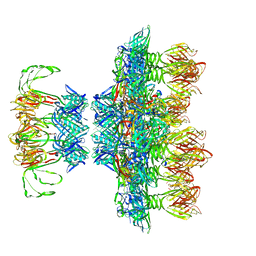
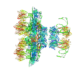
4V96
 | | The structure of a 1.8 MDa viral genome injection device suggests alternative infection mechanisms | | 分子名称: | BPP, ORF46, ORF48 | | 著者 | Veesler, D, Spinelli, S, Mahony, J, Lichiere, J, Blangy, S, Bricogne, G, Legrand, P, Ortiz-Lombardia, M, Campanacci, V, van Sinderen, D, Cambillau, C. | | 登録日 | 2012-02-01 | | 公開日 | 2014-07-09 | | 最終更新日 | 2024-02-28 | | 実験手法 | X-RAY DIFFRACTION (3.8 Å) | | 主引用文献 | Structure of the phage TP901-1 1.8 MDa baseplate suggests an alternative host adhesion mechanism.
Proc.Natl.Acad.Sci.USA, 109, 2012
|
|
6N38
 | | Structure of the type VI secretion system TssK-TssF-TssG baseplate subcomplex revealed by cryo-electron microscopy - full map sharpened | | 分子名称: | Putative type VI secretion protein, Unassigned protein | | 著者 | Park, Y.J, Lacourse, K.D, Cambillau, C, Seattle Structural Genomics Center for Infectious Disease (SSGCID), DiMaio, F, Mougous, J.D, Veesler, D. | | 登録日 | 2018-11-14 | | 公開日 | 2018-12-26 | | 最終更新日 | 2024-03-20 | | 実験手法 | ELECTRON MICROSCOPY (3.7 Å) | | 主引用文献 | Structure of the type VI secretion system TssK-TssF-TssG baseplate subcomplex revealed by cryo-electron microscopy.
Nat Commun, 9, 2018
|
|
5EFV
 | | The host-recognition device of Staphylococcus aureus phage Phi11 | | 分子名称: | FE (III) ION, Phi ETA orf 56-like protein | | 著者 | Koc, C, Kuhner, P, Xia, G, Spinelli, S, Roussel, A, Cambillau, C, Stehle, T. | | 登録日 | 2015-10-26 | | 公開日 | 2016-05-25 | | 最終更新日 | 2024-05-08 | | 実験手法 | X-RAY DIFFRACTION (2.2 Å) | | 主引用文献 | Structure of the host-recognition device of Staphylococcus aureus phage 11.
Sci Rep, 6, 2016
|
|
6ZIG
 | |
6ZJJ
 | |
6ZIH
 | |
2PVS
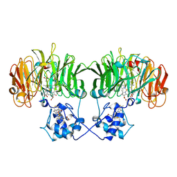
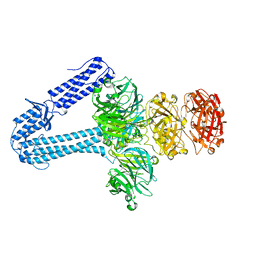
 | | Structure of human pancreatic lipase related protein 2 mutant N336Q | | 分子名称: | CALCIUM ION, Pancreatic lipase-related protein 2, SULFATE ION | | 著者 | Spinelli, S, Eydoux, C, Carriere, F, Cambillau, C. | | 登録日 | 2007-05-10 | | 公開日 | 2007-12-18 | | 最終更新日 | 2023-08-30 | | 実験手法 | X-RAY DIFFRACTION (3 Å) | | 主引用文献 | Structure of human pancreatic lipase-related protein 2 with the lid in an open conformation.
Biochemistry, 47, 2008
|
|
6EY5
 | |
6EY6
 | |
6TOP
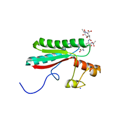 | | Structure of the PorE C-terminal domain, a protein of T9SS from Porphyromonas gingivalis | | 分子名称: | GLCNAC(BETA1-4)-MURNAC(1,6-ANHYDRO)-L-ALA-GAMMA-D-GLU-MESO-A2PM-D-ALA, OmpA family protein, SODIUM ION | | 著者 | Trinh, T.N, Cambillau, C, Roussel, A, Leone, P. | | 登録日 | 2019-12-11 | | 公開日 | 2020-06-17 | | 最終更新日 | 2024-05-15 | | 実験手法 | X-RAY DIFFRACTION (1.55 Å) | | 主引用文献 | Crystal structure of Type IX secretion system PorE C-terminal domain from Porphyromonas gingivalis in complex with a peptidoglycan fragment.
Sci Rep, 10, 2020
|
|
5LMJ
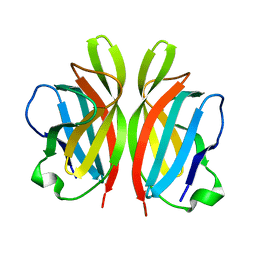
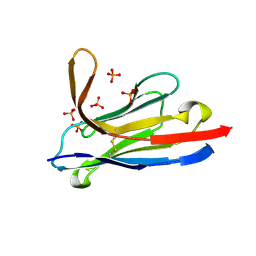 | | Llama nanobody PorM_19 | | 分子名称: | PHOSPHATE ION, nanobody | | 著者 | Leone, P, Cambillau, C, Roussel, A. | | 登録日 | 2016-08-01 | | 公開日 | 2017-05-03 | | 最終更新日 | 2024-01-10 | | 実験手法 | X-RAY DIFFRACTION (2.1 Å) | | 主引用文献 | Camelid nanobodies used as crystallization chaperones for different constructs of PorM, a component of the type IX secretion system from Porphyromonas gingivalis.
Acta Crystallogr F Struct Biol Commun, 73, 2017
|
|
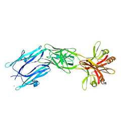
6EY0
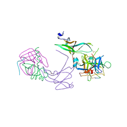
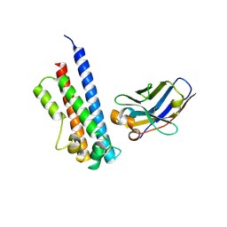 | | N-terminal part (residues 30-212) of PorM with the llama nanobody nb01 | | 分子名称: | T9SS component cytoplasmic membrane protein PorM, llama nanobody nb01 | | 著者 | Leone, P, Roche, J, Cambillau, C, Roussel, A. | | 登録日 | 2017-11-10 | | 公開日 | 2018-02-07 | | 最終更新日 | 2024-01-17 | | 実験手法 | X-RAY DIFFRACTION (2.4 Å) | | 主引用文献 | Type IX secretion system PorM and gliding machinery GldM form arches spanning the periplasmic space.
Nat Commun, 9, 2018
|
|


