5JJD
 
 | |
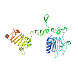
3PXJ
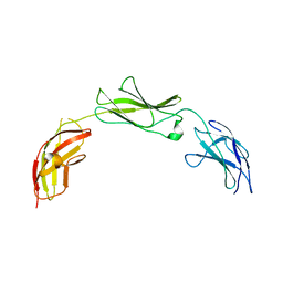
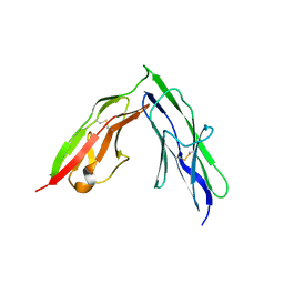 | | Tandem Ig repeats of Dlar | | 分子名称: | Tyrosine-protein phosphatase Lar | | 著者 | Biersmith, B.H, Bouyain, S. | | 登録日 | 2010-12-10 | | 公開日 | 2011-03-23 | | 最終更新日 | 2023-09-13 | | 実験手法 | X-RAY DIFFRACTION (2.3003 Å) | | 主引用文献 | The Immunoglobulin-like Domains 1 and 2 of the Protein Tyrosine Phosphatase LAR Adopt an Unusual Horseshoe-like Conformation.
J.Mol.Biol., 408, 2011
|
|
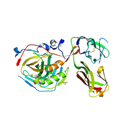
3PXH
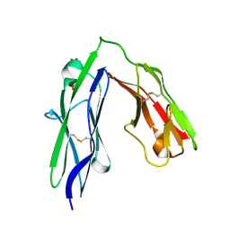 | | Tandem Ig domains of tyrosine phosphatase LAR | | 分子名称: | Receptor-type tyrosine-protein phosphatase F, SULFATE ION | | 著者 | Biersmith, B.H, Bouyain, S. | | 登録日 | 2010-12-09 | | 公開日 | 2011-03-23 | | 最終更新日 | 2023-09-13 | | 実験手法 | X-RAY DIFFRACTION (2.0009 Å) | | 主引用文献 | The Immunoglobulin-like Domains 1 and 2 of the Protein Tyrosine Phosphatase LAR Adopt an Unusual Horseshoe-like Conformation.
J.Mol.Biol., 408, 2011
|
|
5I99
 | |
3U2P
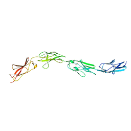
 | | Crystal structure of N-terminal three extracellular domains of ErbB4/Her4 | | 分子名称: | 2-acetamido-2-deoxy-beta-D-glucopyranose, Receptor tyrosine-protein kinase erbB-4 | | 著者 | Liu, P, Bouyain, S, Elgenbrot, C, Leahy, D.J. | | 登録日 | 2011-10-04 | | 公開日 | 2011-11-09 | | 最終更新日 | 2023-09-13 | | 実験手法 | X-RAY DIFFRACTION (2.57 Å) | | 主引用文献 | The ErbB4 extracellular region retains a tethered-like conformation in the absence of the tether.
Protein Sci., 21, 2012
|
|
5E53
 | |
5E5R
 | |
3U7U
 | | Crystal structure of extracellular region of human epidermal growth factor receptor 4 in complex with neuregulin-1 beta | | 分子名称: | 2-acetamido-2-deoxy-beta-D-glucopyranose, Neuregulin 1, Receptor tyrosine-protein kinase erbB-4 | | 著者 | Liu, P, Cleveland IV, T.E, Bouyain, S, Longo, P.A, Leahy, D.J. | | 登録日 | 2011-10-14 | | 公開日 | 2012-08-29 | | 最終更新日 | 2023-09-13 | | 実験手法 | X-RAY DIFFRACTION (3.03 Å) | | 主引用文献 | A single ligand is sufficient to activate EGFR dimers.
Proc.Natl.Acad.Sci.USA, 109, 2012
|
|
4IT4
 | |
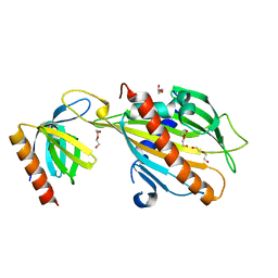
3I86
 | | Crystal structure of the P60 Domain from M. avium subspecies paratuberculosis antigen MAP1204 | | 分子名称: | ISOPROPYL ALCOHOL, Putative uncharacterized protein | | 著者 | Ramyar, K.X, Lingle, C.K, McWhorter, W.J, Bouyain, S, Bannantine, J.P, Geisbrecht, B.V. | | 登録日 | 2009-07-09 | | 公開日 | 2010-07-21 | | 最終更新日 | 2023-09-06 | | 実験手法 | X-RAY DIFFRACTION (2.4 Å) | | 主引用文献 | Atypical structural features of two P60 family members from Mycobacterium avium subspecies paratuberculosis
To be Published
|
|
5E4Q
 | |
5E55
 | |
5E4S
 | |
5E52
 | |
5E7L
 | |
5E4I
 | |
5E5U
 | |


