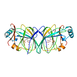
2ISZ
 
 | | Crystal structure of a two-domain IdeR-DNA complex crystal form I | | 分子名称: | Iron-dependent repressor ideR, NICKEL (II) ION, SODIUM ION, ... | | 著者 | Wisedchaisri, G, Chou, C.J, Wu, M, Roach, C, Rice, A.E, Holmes, R.K, Beeson, C, Hol, W.G. | | 登録日 | 2006-10-18 | | 公開日 | 2007-02-13 | | 最終更新日 | 2023-08-30 | | 実験手法 | X-RAY DIFFRACTION (2.403 Å) | | 主引用文献 | Crystal structures, metal activation, and DNA-binding properties of two-domain IdeR from Mycobacterium tuberculosis
Biochemistry, 46, 2007
|
|
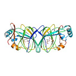
2ISY
 | | Crystal structure of the nickel-activated two-domain iron-dependent regulator (IdeR) | | 分子名称: | Iron-dependent repressor ideR, NICKEL (II) ION, PHOSPHATE ION | | 著者 | Wisedchaisri, G, Chou, C.J, Wu, M, Roach, C, Rice, A.E, Holmes, R.K, Beeson, C, Hol, W.G. | | 登録日 | 2006-10-18 | | 公開日 | 2007-02-13 | | 最終更新日 | 2023-11-15 | | 実験手法 | X-RAY DIFFRACTION (1.955 Å) | | 主引用文献 | Crystal structures, metal activation, and DNA-binding properties of two-domain IdeR from Mycobacterium tuberculosis
Biochemistry, 46, 2007
|
|
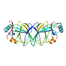
2IT0
 | | Crystal structure of a two-domain IdeR-DNA complex crystal form II | | 分子名称: | ACETATE ION, Iron-dependent repressor ideR, NICKEL (II) ION, ... | | 著者 | Wisedchaisri, G, Chou, C.J, Wu, M, Roach, C, Rice, A.E, Holmes, R.K, Beeson, C, Hol, W.G. | | 登録日 | 2006-10-18 | | 公開日 | 2007-02-13 | | 最終更新日 | 2023-08-30 | | 実験手法 | X-RAY DIFFRACTION (2.6 Å) | | 主引用文献 | Crystal structures, metal activation, and DNA-binding properties of two-domain IdeR from Mycobacterium tuberculosis
Biochemistry, 46, 2007
|
|
1QXR
 | | Crystal structure of phosphoglucose isomerase from Pyrococcus furiosus in complex with 5-phosphoarabinonate | | 分子名称: | 5-PHOSPHOARABINONIC ACID, Glucose-6-phosphate isomerase, NICKEL (II) ION | | 著者 | Swan, M.K, Solomons, J.T.G, Beeson, C.C, Hansen, P, Schonheit, P, Davies, C. | | 登録日 | 2003-09-08 | | 公開日 | 2003-12-09 | | 最終更新日 | 2023-08-23 | | 実験手法 | X-RAY DIFFRACTION (1.7 Å) | | 主引用文献 | Structural evidence for a hydride transfer mechanism of catalysis in phosphoglucose isomerase from Pyrococcus furiosus
J.Biol.Chem., 278, 2003
|
|
1QY4
 | | Crystal structure of phosphoglucose isomerase from Pyrococcus furiosus in complex with gluconate 6-phosphate | | 分子名称: | 6-PHOSPHOGLUCONIC ACID, Glucose-6-phosphate isomerase, NICKEL (II) ION | | 著者 | Swan, M.K, Solomons, J.T.G, Beeson, C.C, Hansen, T, Schonheit, P, Davies, C. | | 登録日 | 2003-09-09 | | 公開日 | 2003-12-09 | | 最終更新日 | 2023-08-23 | | 実験手法 | X-RAY DIFFRACTION (1.8 Å) | | 主引用文献 | Structural evidence for a hydride transfer mechanism of catalysis in phosphoglucose isomerase from Pyrococcus furiosus
J.Biol.Chem., 278, 2003
|
|
1QXJ
 | | Crystal structure of native phosphoglucose isomerase from Pyrococcus furiosus | | 分子名称: | Glucose-6-phosphate isomerase, NICKEL (II) ION | | 著者 | Swan, M.K, Solomons, J.T.G, Beeson, C.C, Hansen, T, Schonheit, P, Davies, C. | | 登録日 | 2003-09-07 | | 公開日 | 2003-12-09 | | 最終更新日 | 2023-08-23 | | 実験手法 | X-RAY DIFFRACTION (1.8 Å) | | 主引用文献 | Structural evidence for a hydride transfer mechanism of catalysis in phosphoglucose isomerase from Pyrococcus furiosus
J.Biol.Chem., 278, 2003
|
|


