Loading
PDBj
MenuPDBj@FacebookPDBj@TwitterPDBj@YouTubewwPDB FoundationwwPDB
RCSB PDBPDBeBMRBAdv. SearchSearch help
PDB: 66 results

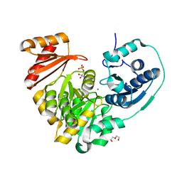
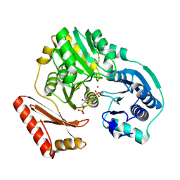
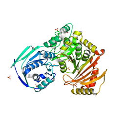
6NOL
DownloadVisualize
BU of 6nol by Molmil
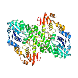
Xanthomonas citri Dephospho-PGM in complex with mannose-1-phosphate
Descriptor: 1-O-phosphono-alpha-D-mannopyranose, DI(HYDROXYETHYL)ETHER, MAGNESIUM ION, ...
Authors:Stiers, K.M, Beamer, L.J.
Deposit date:2019-01-16
Release date:2019-04-10
Last modified:2023-10-11
Method:X-RAY DIFFRACTION (1.73 Å)
Cite:Structural and dynamical description of the enzymatic reaction of a phosphohexomutase.
Struct Dyn., 6, 2019
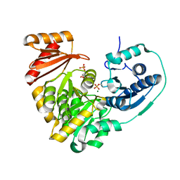
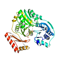
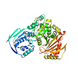
6NNO
DownloadVisualize
BU of 6nno by Molmil
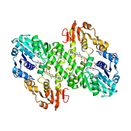
Xanthomonas citri Phospho-PGM in complex with glucose-1-phosphate
Descriptor: 1-O-phosphono-alpha-D-glucopyranose, CALCIUM ION, DI(HYDROXYETHYL)ETHER, ...
Authors:Stiers, K.M, Beamer, L.J.
Deposit date:2019-01-15
Release date:2019-04-10
Last modified:2023-10-11
Method:X-RAY DIFFRACTION (1.57 Å)
Cite:Structural and dynamical description of the enzymatic reaction of a phosphohexomutase.
Struct Dyn., 6, 2019
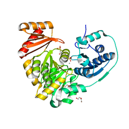
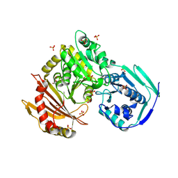
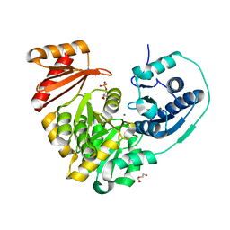
6NNN
DownloadVisualize
BU of 6nnn by Molmil
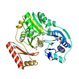
Xanthomonas citri Dephospho-PGM in complex with glucose-1-phosphate
Descriptor: 1-O-phosphono-alpha-D-glucopyranose, DI(HYDROXYETHYL)ETHER, MAGNESIUM ION, ...
Authors:Stiers, K.M, Beamer, L.J.
Deposit date:2019-01-15
Release date:2019-04-10
Last modified:2023-10-11
Method:X-RAY DIFFRACTION (1.35 Å)
Cite:Structural and dynamical description of the enzymatic reaction of a phosphohexomutase.
Struct Dyn., 6, 2019
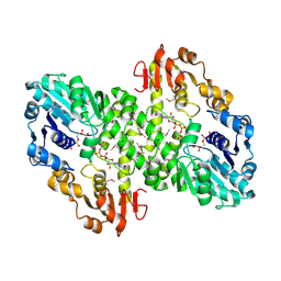
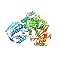
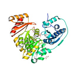
6NOQ
DownloadVisualize
BU of 6noq by Molmil
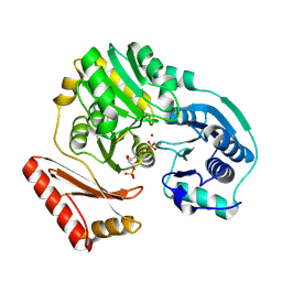
Xanthomonas citri Phospho-PGM in complex with mannose-1-phosphate
Descriptor: 1-O-phosphono-alpha-D-mannopyranose, MAGNESIUM ION, Phosphoglucomutase
Authors:Stiers, K.M, Beamer, L.J.
Deposit date:2019-01-16
Release date:2019-04-10
Last modified:2020-07-29
Method:X-RAY DIFFRACTION (1.61 Å)
Cite:Structural and dynamical description of the enzymatic reaction of a phosphohexomutase.
Struct Dyn., 6, 2019
6NP8
DownloadVisualize
BU of 6np8 by Molmil
Xanthomonas citri Phospho-PGM in complex with mannose-6-phosphate
Descriptor: 6-O-phosphono-alpha-D-mannopyranose, CALCIUM ION, DI(HYDROXYETHYL)ETHER, ...
Authors:Stiers, K.M, Beamer, L.J.
Deposit date:2019-01-17
Release date:2019-04-10
Last modified:2024-11-06
Method:X-RAY DIFFRACTION (1.41 Å)
Cite:Structural and dynamical description of the enzymatic reaction of a phosphohexomutase.
Struct Dyn., 6, 2019
1MUU
DownloadVisualize
BU of 1muu by Molmil
2.0 A crystal structure of GDP-mannose dehydrogenase
Descriptor: GDP-mannose 6-dehydrogenase, GUANOSINE 5'-(TRIHYDROGEN DIPHOSPHATE), P'-D-MANNOPYRANOSYL ESTER, ...
Authors:Snook, C.F, Tipton, P.A, Beamer, L.J.
Deposit date:2002-09-24
Release date:2003-05-06
Last modified:2024-10-30
Method:X-RAY DIFFRACTION (2.02 Å)
Cite:Crystal structure of GDP-mannose dehydrogenase: A key enzyme of alginate biosynthesis in P. aeruginosa
Biochemistry, 42, 2003
1MFZ
DownloadVisualize
BU of 1mfz by Molmil
Partially refined 2.8 A Crystal structure of GDP-mannose dehydrogenase from P. aeruginosa
Descriptor: GDP-mannose 6-dehydrogenase, GUANOSINE 5'-(TRIHYDROGEN DIPHOSPHATE), P'-D-MANNOPYRANOSYL ESTER
Authors:Snook, C.F, Tipton, P.A, Beamer, L.J.
Deposit date:2002-08-14
Release date:2003-05-06
Last modified:2024-10-16
Method:X-RAY DIFFRACTION (2.8 Å)
Cite:The crystal structure of GDP-mannose dehydrogenase: A key enzyme in alginate biosynthesis of P. aeruginosa
Biochemistry, 42, 2003
1MV8
DownloadVisualize
BU of 1mv8 by Molmil
1.55 A crystal structure of a ternary complex of GDP-mannose dehydrogenase from Psuedomonas aeruginosa
Descriptor: (4S)-2-METHYL-2,4-PENTANEDIOL, ACETIC ACID, GDP-mannose 6-dehydrogenase, ...
Authors:Snook, C.F, Tipton, P.A, Beamer, L.J.
Deposit date:2002-09-24
Release date:2003-05-06
Last modified:2024-02-14
Method:X-RAY DIFFRACTION (1.55 Å)
Cite:The crystal structure of GDP-mannose dehydrogenase: A key enzyme in alginate biosynthesis of P. aeruginosa
Biochemistry, 42, 2003
1P5G
DownloadVisualize
BU of 1p5g by Molmil
Enzyme-ligand complex of P. aeruginosa PMM/PGM
Descriptor: 6-O-phosphono-alpha-D-glucopyranose, Phosphomannomutase, ZINC ION
Authors:Regni, C, Tipton, P.A, Beamer, L.J.
Deposit date:2003-04-26
Release date:2004-01-20
Last modified:2023-08-16
Method:X-RAY DIFFRACTION (1.61 Å)
Cite:Structural basis of diverse substrate recognition by the enzyme PMM/PGM from P. aeruginosa.
Structure, 12, 2004
1PCJ
DownloadVisualize
BU of 1pcj by Molmil
Enzyme-ligand complex of P. aeruginosa PMM/PGM
Descriptor: 1-O-phosphono-alpha-D-mannopyranose, Phosphomannomutase, ZINC ION
Authors:Regni, C, Tipton, P.A, Beamer, L.J.
Deposit date:2003-05-16
Release date:2004-01-20
Last modified:2024-11-06
Method:X-RAY DIFFRACTION (2 Å)
Cite:Structural basis of diverse substrate recognition by the enzyme PMM/PGM from P. aeruginosa.
Structure, 12, 2004
1PCM
DownloadVisualize
BU of 1pcm by Molmil
Enzyme-ligand complex of P. aeruginosa PMM/PGM
Descriptor: 6-O-phosphono-alpha-D-mannopyranose, Phosphomannomutase, ZINC ION
Authors:Regni, C, Tipton, P.A, Beamer, L.J.
Deposit date:2003-05-16
Release date:2004-01-20
Last modified:2024-10-30
Method:X-RAY DIFFRACTION (1.9 Å)
Cite:Structural basis of diverse substrate recognition by the enzyme PMM/PGM from P. aeruginosa.
Structure, 12, 2004
1P5D
DownloadVisualize
BU of 1p5d by Molmil
Enzyme-ligand complex of P. aeruginosa PMM/PGM
Descriptor: 1-O-phosphono-alpha-D-glucopyranose, Phosphomannomutase, ZINC ION
Authors:Regni, C, Tipton, P.A, Beamer, L.J.
Deposit date:2003-04-25
Release date:2004-01-20
Last modified:2024-10-30
Method:X-RAY DIFFRACTION (1.6 Å)
Cite:Structural basis of diverse substrate recognition by the enzyme PMM/PGM from P. aeruginosa.
Structure, 12, 2004
5VG7
DownloadVisualize
BU of 5vg7 by Molmil
Crystal Structure of the R503Q missense variant of human PGM1
Descriptor: GLYCEROL, MAGNESIUM ION, Phosphoglucomutase-1, ...
Authors:Stiers, K.M, Beamer, L.J.
Deposit date:2017-04-10
Release date:2018-06-20
Last modified:2024-10-30
Method:X-RAY DIFFRACTION (1.95000327 Å)
Cite:A Hotspot for Disease-Associated Variants of Human PGM1 Is Associated with Impaired Ligand Binding and Loop Dynamics.
Structure, 26, 2018
5VEC
DownloadVisualize
BU of 5vec by Molmil
Crystal Structure of the R515L missense variant of human PGM1
Descriptor: GLYCEROL, MAGNESIUM ION, Phosphoglucomutase-1, ...
Authors:Stiers, K.M, Beamer, L.J.
Deposit date:2017-04-04
Release date:2018-06-27
Last modified:2023-10-04
Method:X-RAY DIFFRACTION (2.20001626 Å)
Cite:A Hotspot for Disease-Associated Variants of Human PGM1 Is Associated with Impaired Ligand Binding and Loop Dynamics.
Structure, 26, 2018
5VIN
DownloadVisualize
BU of 5vin by Molmil
Crystal Structure of the R515Q missense variant of human PGM1
Descriptor: COBALT (II) ION, GLYCEROL, Phosphoglucomutase-1, ...
Authors:Stiers, K.M, Beamer, L.J.
Deposit date:2017-04-17
Release date:2018-06-27
Last modified:2023-10-04
Method:X-RAY DIFFRACTION (2.60004282 Å)
Cite:A Hotspot for Disease-Associated Variants of Human PGM1 Is Associated with Impaired Ligand Binding and Loop Dynamics.
Structure, 26, 2018
5VBI
DownloadVisualize
BU of 5vbi by Molmil
Crystal Structure of the R515W missense variant of human PGM1
Descriptor: GLYCEROL, MAGNESIUM ION, Phosphoglucomutase-1, ...
Authors:Stiers, K.M, Beamer, L.J.
Deposit date:2017-03-29
Release date:2018-06-20
Last modified:2024-10-09
Method:X-RAY DIFFRACTION (1.75000083 Å)
Cite:A Hotspot for Disease-Associated Variants of Human PGM1 Is Associated with Impaired Ligand Binding and Loop Dynamics.
Structure, 26, 2018
<123


227111

건을2024-11-06부터공개중

PDB statisticsPDBj update infoContact PDBjnumon