3V7E
 
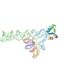 | | Crystal structure of YbxF bound to the SAM-I riboswitch aptamer | | 分子名称: | COBALT HEXAMMINE(III), MAGNESIUM ION, Ribosome-associated protein L7Ae-like, ... | | 著者 | Baird, N.J, Zhang, J, Hamma, T, Ferre-D'Amare, A.R. | | 登録日 | 2011-12-21 | | 公開日 | 2012-03-07 | | 最終更新日 | 2023-09-13 | | 実験手法 | X-RAY DIFFRACTION (2.8 Å) | | 主引用文献 | YbxF and YlxQ are bacterial homologs of L7Ae and bind K-turns but not K-loops.
Rna, 18, 2012
|
|
3V7Q
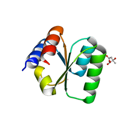 | | Crystal structure of B. subtilis YlxQ at 1.55 A resolution | | 分子名称: | CITRIC ACID, POTASSIUM ION, Probable ribosomal protein ylxQ | | 著者 | Baird, N.J, Zhang, J, Hamma, T, Ferre-D'Amare, A.R. | | 登録日 | 2011-12-21 | | 公開日 | 2012-03-07 | | 最終更新日 | 2023-09-13 | | 実験手法 | X-RAY DIFFRACTION (1.55 Å) | | 主引用文献 | YbxF and YlxQ are bacterial homologs of L7Ae and bind K-turns but not K-loops.
Rna, 18, 2012
|
|
3IWN
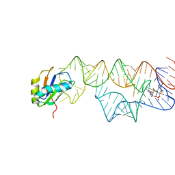 | | Co-crystal structure of a bacterial c-di-GMP riboswitch | | 分子名称: | 9,9'-[(2R,3R,3aS,5S,7aR,9R,10R,10aS,12S,14aR)-3,5,10,12-tetrahydroxy-5,12-dioxidooctahydro-2H,7H-difuro[3,2-d:3',2'-j][1,3,7,9,2,8]tetraoxadiphosphacyclododecine-2,9-diyl]bis(2-amino-1,9-dihydro-6H-purin-6-one), C-di-GMP riboswitch, U1 small nuclear ribonucleoprotein A | | 著者 | Kulshina, N, Baird, N.J, Ferre-D'Amare, A.R. | | 登録日 | 2009-09-02 | | 公開日 | 2009-11-10 | | 最終更新日 | 2024-02-21 | | 実験手法 | X-RAY DIFFRACTION (3.2 Å) | | 主引用文献 | Recognition of the bacterial second messenger cyclic diguanylate by its cognate riboswitch.
Nat.Struct.Mol.Biol., 16, 2009
|
|


