4FFY
 
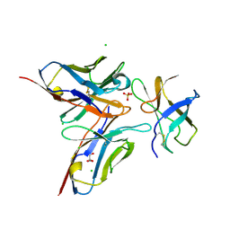 | | Crystal structure of DENV1-E111 single chain variable fragment bound to DENV-1 DIII, strain 16007. | | 分子名称: | CHLORIDE ION, DENV1-E111 single chain variable fragment (heavy chain), DENV1-E111 single chain variable fragment (light chain), ... | | 著者 | Austin, S.K, Nelson, C.A, Fremont, D.H, Center for Structural Genomics of Infectious Diseases (CSGID) | | 登録日 | 2012-06-01 | | 公開日 | 2012-06-20 | | 最終更新日 | 2012-10-31 | | 実験手法 | X-RAY DIFFRACTION (2.5 Å) | | 主引用文献 | Structural Basis of Differential Neutralization of DENV-1 Genotypes by an Antibody that Recognizes a Cryptic Epitope.
Plos Pathog., 8, 2012
|
|
4FFZ
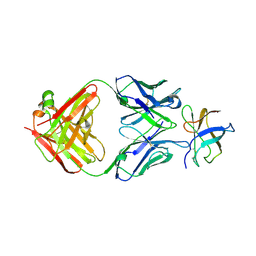 | | Crystal Structure of DENV1-E111 fab fragment bound to DENV-1 DIII (Western Pacific-74 strain). | | 分子名称: | DENV1-E111 fab fragment (heavy chain), DENV1-E111 fab fragment (light chain), Envelope protein E | | 著者 | Austin, S.K, Nelson, C.A, Fremont, D.H, Center for Structural Genomics of Infectious Diseases (CSGID) | | 登録日 | 2012-06-01 | | 公開日 | 2012-06-27 | | 最終更新日 | 2023-09-13 | | 実験手法 | X-RAY DIFFRACTION (3.8 Å) | | 主引用文献 | Structural Basis of Differential Neutralization of DENV-1 Genotypes by an Antibody that Recognizes a Cryptic Epitope.
Plos Pathog., 8, 2012
|
|
3J8D
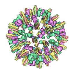 | | Cryoelectron microscopy of dengue-Fab E104 complex at pH 5.5 | | 分子名称: | Envelope protein E, antibody E111 Fab fragment, glycoprotein DIII | | 著者 | Zhang, X.Z, Sheng, J, Austin, S.K, Hoornweg, T, Smit, J.M, Kuhn, R.J, Diamond, M.S, Rossmann, M.G. | | 登録日 | 2014-10-13 | | 公開日 | 2014-11-12 | | 最終更新日 | 2024-02-21 | | 実験手法 | ELECTRON MICROSCOPY (26 Å) | | 主引用文献 | Structure of Acidic pH Dengue Virus Showing the Fusogenic Glycoprotein Trimers.
J.Virol., 89, 2015
|
|
3FMT
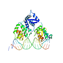 | | Crystal structure of SeqA bound to DNA | | 分子名称: | (4S)-2-METHYL-2,4-PENTANEDIOL, 5'-D(*GP*AP*GP*TP*CP*GP*(6MA)P*TP*CP*GP*GP*CP*GP*GP*GP*(6MA)P*TP*CP*CP*TP*TP*A)-3', 5'-D(*TP*CP*TP*AP*AP*GP*GP*AP*TP*CP*CP*CP*GP*CP*CP*GP*AP*TP*CP*GP*AP*C)-3', ... | | 著者 | Chung, Y.S, Brendler, T, Austin, S, Guarne, A. | | 登録日 | 2008-12-22 | | 公開日 | 2009-04-28 | | 最終更新日 | 2023-09-06 | | 実験手法 | X-RAY DIFFRACTION (2.983 Å) | | 主引用文献 | Structural insights into the cooperative binding of SeqA to a tandem GATC repeat
Nucleic Acids Res., 37, 2009
|
|
1XRX
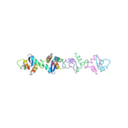 | | Crystal structure of a DNA-binding protein | | 分子名称: | CALCIUM ION, SeqA protein | | 著者 | Guarne, A, Brendler, T, Zhao, Q, Ghirlando, R, Austin, S, Yang, W. | | 登録日 | 2004-10-16 | | 公開日 | 2005-05-10 | | 最終更新日 | 2013-03-06 | | 実験手法 | X-RAY DIFFRACTION (2.15 Å) | | 主引用文献 | Crystal structure of a SeqA-N filament: implications for DNA replication and chromosome organization.
Embo J., 24, 2005
|
|


