Loading
PDBj
MenuPDBj@FacebookPDBj@TwitterPDBj@YouTubewwPDB FoundationwwPDB
RCSB PDBPDBeBMRBAdv. SearchSearch help
PDB: 5 results

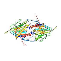
8CCR
DownloadVisualize
BU of 8ccr by Molmil
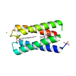
Crystal structure of the T19D mutant of the de novo diheme binding 4D2
Descriptor: (2S)-2-hydroxybutanedioic acid, 4D2 (mutant T19D), CHLORIDE ION, ...
Authors:Barringer, R, Anderson, R.
Deposit date:2023-01-27
Release date:2023-08-09
Last modified:2024-06-19
Method:X-RAY DIFFRACTION (2.1 Å)
Cite:An expandable, modular de novo protein platform for precision redox engineering.
Proc.Natl.Acad.Sci.USA, 120, 2023
1II6
DownloadVisualize
BU of 1ii6 by Molmil
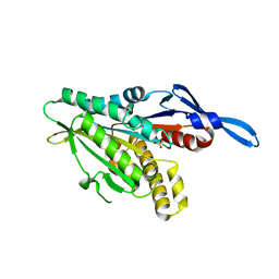
Crystal Structure of the Mitotic Kinesin Eg5 in Complex with Mg-ADP.
Descriptor: ADENOSINE-5'-DIPHOSPHATE, KINESIN-RELATED MOTOR PROTEIN Eg5, MAGNESIUM ION, ...
Authors:Turner, J, Anderson, R, Guo, J, Beraud, C, Sakowicz, R, Fletterick, R.
Deposit date:2001-04-20
Release date:2001-07-18
Last modified:2024-04-03
Method:X-RAY DIFFRACTION (2.1 Å)
Cite:Crystal structure of the mitotic spindle kinesin Eg5 reveals a novel conformation of the neck-linker.
J.Biol.Chem., 276, 2001
1EGY
DownloadVisualize
BU of 1egy by Molmil
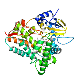
CYTOCHROME P450ERYF WITH 9-AMINOPHENANTHRENE BOUND
Descriptor: 9-AMINOPHENANTHRENE, CYTOCHROME P450ERYF, PROTOPORPHYRIN IX CONTAINING FE
Authors:Cupp-Vickery, J.R, Anderson, R, Hatziris, Z.
Deposit date:2000-02-17
Release date:2000-03-06
Last modified:2024-02-07
Method:X-RAY DIFFRACTION (2.35 Å)
Cite:Crystal structures of ligand complexes of P450eryF exhibiting homotropic cooperativity.
Proc.Natl.Acad.Sci.USA, 97, 2000
1EUP
DownloadVisualize
BU of 1eup by Molmil
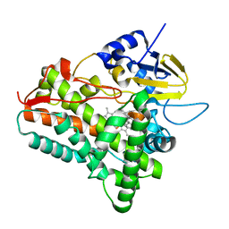
X-RAY CRYSTAL STRUCTURE OF CYTOCHROME P450ERYF WITH ANDROSTENDIONE BOUND
Descriptor: 4-ANDROSTENE-3-17-DIONE, CYTOCHROME P450ERYF, PROTOPORPHYRIN IX CONTAINING FE
Authors:Cupp-Vickery, J.R, Anderson, R, Hatziris, Z.
Deposit date:2000-04-17
Release date:2000-04-28
Last modified:2024-02-07
Method:X-RAY DIFFRACTION (2.1 Å)
Cite:Crystal structures of ligand complexes of P450eryF exhibiting homotropic cooperativity.
Proc.Natl.Acad.Sci.USA, 97, 2000
1RY6
DownloadVisualize
BU of 1ry6 by Molmil
Crystal Structure of Internal Kinesin Motor Domain
Descriptor: INTERNAL KINESIN, SULFATE ION
Authors:Shipley, K, Hekmat-Nejad, M, Turner, J, Moores, C, Anderson, R, Milligan, R, Sakowicz, R, Fletterick, R.
Deposit date:2003-12-19
Release date:2004-04-13
Last modified:2023-09-20
Method:X-RAY DIFFRACTION (1.6 Å)
Cite:Structure of a kinesin microtubule depolymerization machine.
Embo J., 23, 2004

227111

건을2024-11-06부터공개중

PDB statisticsPDBj update infoContact PDBjnumon