Loading
PDBj
MenuPDBj@FacebookPDBj@TwitterPDBj@YouTubewwPDB FoundationwwPDB
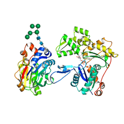
RCSB PDBPDBeBMRBAdv. SearchSearch help
PDB: 263 results

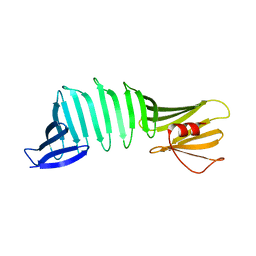
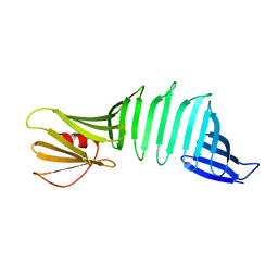
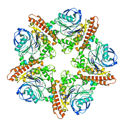
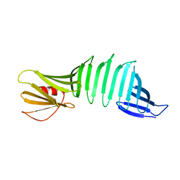
6J5Q
DownloadVisualize
BU of 6j5q by Molmil
OspA mutant, PSAM-VLGDV1-form7, grafted short chameleon sequence from alpha-B crystallin
Descriptor: Outer surface protein A
Authors:Fujiwara, H, Makabe, K.
Deposit date:2019-01-11
Release date:2019-11-27
Last modified:2023-11-22
Method:X-RAY DIFFRACTION (1.798 Å)
Cite:Beta-sheet elasticity of peptide self-assembly mimic, PSAM, with a grafted sequence characterized by comprehensive analyses of isomorphous crystals
J Mol Liq, 290, 2019
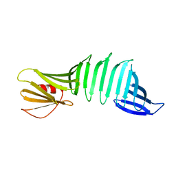
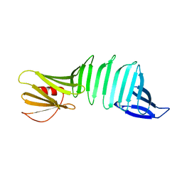
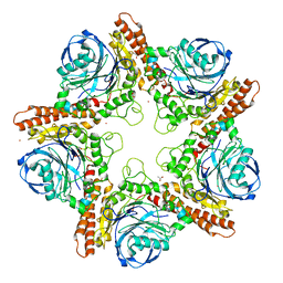
6J6E
DownloadVisualize
BU of 6j6e by Molmil
Borrelia burgdorferi OspA via surface entropy reduction (form5)
Descriptor: Outer surface protein A
Authors:Fujiwara, H, Makabe, K.
Deposit date:2019-01-14
Release date:2019-11-27
Last modified:2023-11-22
Method:X-RAY DIFFRACTION (1.5 Å)
Cite:Beta-sheet elasticity of peptide self-assembly mimic, PSAM, with a grafted sequence characterized by comprehensive analyses of isomorphous crystals
J Mol Liq, 290, 2019
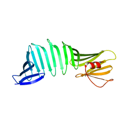
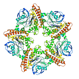
6J5O
DownloadVisualize
BU of 6j5o by Molmil
OspA mutant, PSAM-VLGDV1-form5, grafted short chameleon sequence from alpha-B crystallin
Descriptor: Outer surface protein A
Authors:Fujiwara, H, Makabe, K.
Deposit date:2019-01-11
Release date:2019-11-27
Last modified:2023-11-22
Method:X-RAY DIFFRACTION (1.9 Å)
Cite:Beta-sheet elasticity of peptide self-assembly mimic, PSAM, with a grafted sequence characterized by comprehensive analyses of isomorphous crystals
J Mol Liq, 290, 2019
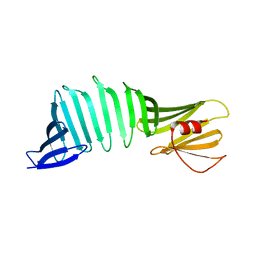
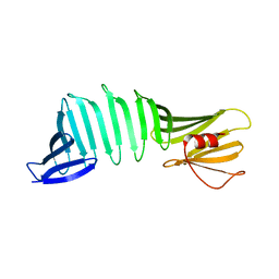
6J6B
DownloadVisualize
BU of 6j6b by Molmil
Borrelia burgdorferi OspA via surface entropy reduction (form2)
Descriptor: Outer surface protein A
Authors:Fujiwara, H, Makabe, K.
Deposit date:2019-01-14
Release date:2019-11-27
Last modified:2023-11-22
Method:X-RAY DIFFRACTION (1.901 Å)
Cite:Beta-sheet elasticity of peptide self-assembly mimic, PSAM, with a grafted sequence characterized by comprehensive analyses of isomorphous crystals
J Mol Liq, 290, 2019
6J5P
DownloadVisualize
BU of 6j5p by Molmil
OspA mutant, PSAM-VLGDV1-form6, grafted short chameleon sequence from alpha-B crystallin
Descriptor: Outer surface protein A
Authors:Fujiwara, H, Makabe, K.
Deposit date:2019-01-11
Release date:2019-11-27
Last modified:2023-11-22
Method:X-RAY DIFFRACTION (1.8 Å)
Cite:Beta-sheet elasticity of peptide self-assembly mimic, PSAM, with a grafted sequence characterized by comprehensive analyses of isomorphous crystals
J Mol Liq, 290, 2019
6J5R
DownloadVisualize
BU of 6j5r by Molmil
OspA mutant, PSAM-VLGDV1-form8, grafted short chameleon sequence from alpha-B crystallin
Descriptor: Outer surface protein A
Authors:Fujiwara, H, Makabe, K.
Deposit date:2019-01-11
Release date:2019-11-27
Last modified:2023-11-22
Method:X-RAY DIFFRACTION (1.85 Å)
Cite:Beta-sheet elasticity of peptide self-assembly mimic, PSAM, with a grafted sequence characterized by comprehensive analyses of isomorphous crystals
J Mol Liq, 290, 2019
6J6D
DownloadVisualize
BU of 6j6d by Molmil
Borrelia burgdorferi OspA via surface entropy reduction (form4)
Descriptor: Outer surface protein A
Authors:Fujiwara, H, Makabe, K.
Deposit date:2019-01-14
Release date:2019-11-27
Last modified:2023-11-22
Method:X-RAY DIFFRACTION (1.9 Å)
Cite:Beta-sheet elasticity of peptide self-assembly mimic, PSAM, with a grafted sequence characterized by comprehensive analyses of isomorphous crystals
J Mol Liq, 290, 2019
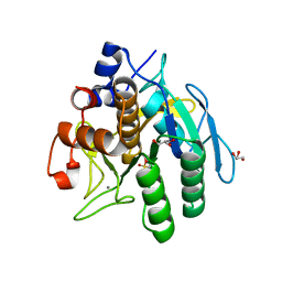
3VYV
DownloadVisualize
BU of 3vyv by Molmil
Crystal structure of subtilisin NAT at 1.36
Descriptor: CALCIUM ION, GLYCEROL, Subtilisin NAT
Authors:Ushijima, H, Fuchita, N, Kajiwara, T, Motoshima, H, Ueno, G, Watanabe, K.
Deposit date:2012-10-03
Release date:2013-10-09
Last modified:2023-11-08
Method:X-RAY DIFFRACTION (1.36 Å)
Cite:Crystal structure of subtilisin NAT at 1.36
TO BE PUBLISHED
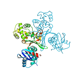
3VPZ
DownloadVisualize
BU of 3vpz by Molmil
Crystal structure of glucokinase from Antarctic psychrotroph at 1.69A
Descriptor: Glucokinase
Authors:Oda, T, Fuchita, N, Motoshima, H, Kawamoto, M, Watanabe, K.
Deposit date:2012-03-16
Release date:2013-03-20
Last modified:2024-10-16
Method:X-RAY DIFFRACTION (1.69 Å)
Cite:Crystal structure of glucokinase from Antarctic psychrotroph at 1.69A
To be Published
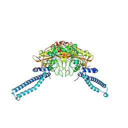
2PI3
DownloadVisualize
BU of 2pi3 by Molmil
The crystal structure of OspA mutant
Descriptor: Outer surface protein A
Authors:Biancalana, M, Makabe, K, Terechko, V, Koide, S.
Deposit date:2007-04-12
Release date:2008-05-27
Last modified:2023-08-30
Method:X-RAY DIFFRACTION (1.4 Å)
Cite:Aromatic cluster mutations produce focal modulations of beta-sheet structure.
Protein Sci., 24, 2015
1WLE
DownloadVisualize
BU of 1wle by Molmil
Crystal Structure of mammalian mitochondrial seryl-tRNA synthetase complexed with seryl-adenylate
Descriptor: SERYL ADENYLATE, Seryl-tRNA synthetase
Authors:Chimnaronk, S, Jeppesen, M.G, Suzuki, T, Nyborg, J, Watanabe, K.
Deposit date:2004-06-25
Release date:2005-09-06
Last modified:2023-10-25
Method:X-RAY DIFFRACTION (1.65 Å)
Cite:Dual-mode recognition of noncanonical tRNAs(Ser) by seryl-tRNA synthetase in mammalian mitochondria
Embo J., 24, 2005
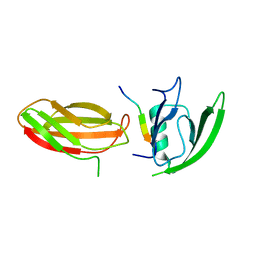
2QBW
DownloadVisualize
BU of 2qbw by Molmil
The crystal structure of PDZ-Fibronectin fusion protein
Descriptor: PDZ-Fibronectin fusion protein, Polypeptide
Authors:Huang, J, Makabe, K, Koide, A, Koide, S.
Deposit date:2007-06-18
Release date:2008-04-22
Last modified:2024-02-21
Method:X-RAY DIFFRACTION (1.8 Å)
Cite:Design of protein function leaps by directed domain interface evolution.
Proc.Natl.Acad.Sci.Usa, 105, 2008
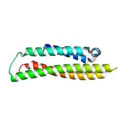
1GS9
DownloadVisualize
BU of 1gs9 by Molmil
Apolipoprotein E4, 22k domain
Descriptor: APOLIPOPROTEIN E
Authors:Verderame, J.R, Kantardjieff, K, Segelke, B, Weisgraber, K, Rupp, B.
Deposit date:2002-01-02
Release date:2003-06-11
Last modified:2023-12-13
Method:X-RAY DIFFRACTION (1.7 Å)
Cite:Crystal Structure of the 22K Domain of Human Apolipoprotein E4
To be Published
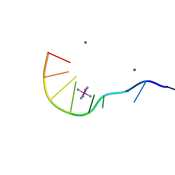
1IXJ
DownloadVisualize
BU of 1ixj by Molmil
Crystal Structure of d(GCGAAAGCT) Containing Parallel-stranded Duplex with Homo Base Pairs and Anti-Parallel Duplex with Watson-Crick Base pairs
Descriptor: 5'-D(*GP*CP*GP*AP*AP*AP*GP*CP*T)-3', COBALT HEXAMMINE(III), MAGNESIUM ION
Authors:Sunami, T, Kondo, J, Kobuna, T, Hirao, I, Watanabe, K, Miura, K, Takenaka, A.
Deposit date:2002-06-22
Release date:2002-12-11
Last modified:2023-12-27
Method:X-RAY DIFFRACTION (2.5 Å)
Cite:Crystal Structure of d(GCGAAAGCT) Containing a Parallel-stranded Duplex with Homo Base Pairs and an Anti-Parallel Duplex with Watson-Crick Base pairs
Nucleic Acids Res., 30, 2002
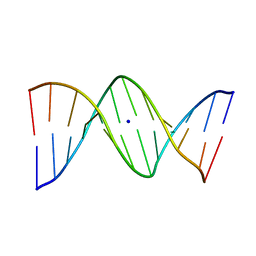
1VE8
DownloadVisualize
BU of 1ve8 by Molmil
X-Ray analyses of oligonucleotides containing 5-formylcytosine, suggesting a structural reason for codon-anticodon recognition of mitochondrial tRNA-Met; Part 1, d(CGCGAATT(f5C)GCG)
Descriptor: 5'-D(*CP*GP*CP*GP*AP*AP*TP*TP*(5FC)P*GP*CP*G)-3', SODIUM ION
Authors:Kimura, K, Ono, A, Watanabe, K, Takenaka, A.
Deposit date:2004-03-29
Release date:2005-06-28
Last modified:2023-12-27
Method:X-RAY DIFFRACTION (1.65 Å)
Cite:X-Ray analyses of oligonucleotides containing 5-formylcytosine, suggest a structural reason for the codon-anticodon recognition of mitochondrial tRNA-Met
To be Published
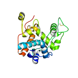
3W3E
DownloadVisualize
BU of 3w3e by Molmil
Structure of Vigna unguiculata chitinase with regulation activity of the plant cell wall
Descriptor: Cotyledoneous yieldin-like protein
Authors:Morohashi, K, Sasaki, K, Sakabe, N, Sakabe, K.
Deposit date:2012-12-20
Release date:2013-01-16
Last modified:2023-11-08
Method:X-RAY DIFFRACTION (1.5 Å)
Cite:Three-dimensional structure analysis of Vigna unguiculata chitinase with regulation activity of the yield threshold of cell wall
To be Published
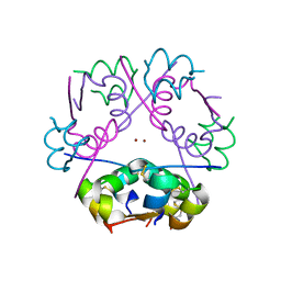
3W80
DownloadVisualize
BU of 3w80 by Molmil
Crystal structure of dodecamer human insulin with double C-axis length of the hexamer 2 Zn insulin cell
Descriptor: Insulin, ZINC ION
Authors:Murayoshi, M, Sasaki, K, Sakabe, N, Sakabe, K.
Deposit date:2013-03-11
Release date:2013-07-03
Last modified:2024-11-06
Method:X-RAY DIFFRACTION (1.4 Å)
Cite:Crystal structure of dodecamer insulin with double C-axis length of the hexamer 2 Zn insulin celll
To be Published
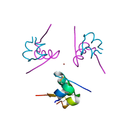
3W7Y
DownloadVisualize
BU of 3w7y by Molmil
0.92A structure of 2Zn human insulin at 100K
Descriptor: Insulin, ZINC ION
Authors:Sakabe, N, Sakabe, K, Sasaki, K, Murayoshi, M.
Deposit date:2013-03-11
Release date:2013-07-03
Last modified:2024-10-30
Method:X-RAY DIFFRACTION (0.92 Å)
Cite:0.92A structure of 2Zn human insulin at 100K
To be Published
3W3D
DownloadVisualize
BU of 3w3d by Molmil
Crystal structure of smooth muscle G actin DNase I complex
Descriptor: ADENOSINE-5'-TRIPHOSPHATE, Actin, gamma-enteric smooth muscle, ...
Authors:Sakabe, N, Sakabe, K, Sasaki, K, Kondo, H, Shimomur, M.
Deposit date:2012-12-20
Release date:2013-01-30
Last modified:2020-07-29
Method:X-RAY DIFFRACTION (1.8 Å)
Cite:Refined structure and solvent network of chicken gizzard G-actin DNase 1 complex at 1.8A resolution
Acta Crystallogr.,Sect.A, 49, 1993
4PDT
DownloadVisualize
BU of 4pdt by Molmil
Japanese Marasmius oreades lectin
Descriptor: Mannose recognizing lectin, SULFATE ION
Authors:Noma, Y, Shimokawa, M, Maeganeku, C, Motoshima, H, Watanabe, K, Minami, Y, Yagi, F.
Deposit date:2014-04-22
Release date:2015-04-29
Last modified:2023-11-08
Method:X-RAY DIFFRACTION (1.4 Å)
Cite:The structure of Japanese Marasmius oreades lectin at 1.40 Angstroms resolution.
To Be Published
5IAU
DownloadVisualize
BU of 5iau by Molmil
A C69-family cysteine dipeptidase from Lactobacillus farciminis
Descriptor: 1,2-ETHANEDIOL, Dipeptidase
Authors:Kono, R, Watanabe, K.
Deposit date:2016-02-21
Release date:2017-03-01
Last modified:2024-03-20
Method:X-RAY DIFFRACTION (1.781 Å)
Cite:A C69-family cysteine dipeptidase from Lactobacillus farciminis; substrate recognition mechanism and autoproteolytic mechanism in enzyme maturation
To Be Published
1RY8
DownloadVisualize
BU of 1ry8 by Molmil
Prostaglandin F synthase complexed with NADPH and rutin
Descriptor: Aldo-keto reductase family 1 member C3, NADPH DIHYDRO-NICOTINAMIDE-ADENINE-DINUCLEOTIDE PHOSPHATE, RUTIN
Authors:Komoto, J, Yamada, T, Watanabe, K, Takusagawa, F.
Deposit date:2003-12-19
Release date:2004-11-02
Last modified:2023-08-23
Method:X-RAY DIFFRACTION (1.69 Å)
Cite:Crystal structure of human prostaglandin F synthase (AKR1C3).
Biochemistry, 43, 2004
5INX
DownloadVisualize
BU of 5inx by Molmil
A C69-family cysteine dipeptidase in complex with Met and Ala from Lactobacillus farciminis
Descriptor: ALANINE, Dipeptidase, METHIONINE
Authors:Kono, R, Watanabe, K.
Deposit date:2016-03-08
Release date:2017-03-15
Last modified:2023-11-08
Method:X-RAY DIFFRACTION (1.681 Å)
Cite:A C69-family cysteine dipeptidase from Lactobacillus farciminis; substrate recognition mechanism and autoproteolytic mechanism in enzyme maturation
To Be Published
5IMH
DownloadVisualize
BU of 5imh by Molmil
D31P mutant of C69-family cysteine dipeptidase from Lactobacillus farciminis
Descriptor: 1,2-ETHANEDIOL, ACETATE ION, COBALT (II) ION, ...
Authors:Kono, R, Watanabe, K.
Deposit date:2016-03-06
Release date:2017-03-15
Last modified:2023-11-08
Method:X-RAY DIFFRACTION (2.471 Å)
Cite:A C69-family cysteine dipeptidase from Lactobacillus farciminis; substrate recognition mechanism and autoproteolytic mechanism in enzyme maturation
To Be Published
5INR
DownloadVisualize
BU of 5inr by Molmil
A C69-family cysteine dipeptidase in complex with Ala-Pro from Lactobacillus farciminis
Descriptor: 1,2-ETHANEDIOL, ACETATE ION, ALANINE, ...
Authors:Kono, R, Watanabe, K.
Deposit date:2016-03-07
Release date:2017-03-15
Last modified:2023-11-08
Method:X-RAY DIFFRACTION (1.652 Å)
Cite:A C69-family cysteine dipeptidase from Lactobacillus farciminis; substrate recognition mechanism and autoproteolytic mechanism in enzyme maturation
To Be Published

227111

数据于2024-11-06公开中

PDB statisticsPDBj update infoContact PDBjnumon