5Y51
 
 | |
5Y57
 | |
5Y5V
 | |
5Y5R
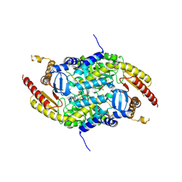
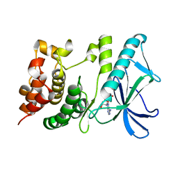
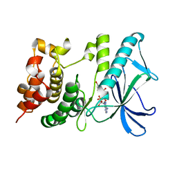
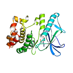
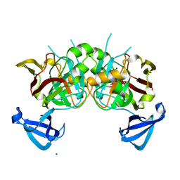
 | | Crystal structure of a novel Pyrethroid Hydrolase PytH with BIF | | 分子名称: | (2-methyl-3-phenyl-phenyl)methyl (1~{S})-3-[(~{E})-2-chloranyl-3,3,3-tris(fluoranyl)prop-1-enyl]-2,2-dimethyl-cyclopropane-1-carboxylate, Pyrethroid hydrolase, SULFATE ION | | 著者 | Xu, D.Q, Ran, T.T, Wang, W.W. | | 登録日 | 2017-08-09 | | 公開日 | 2018-08-15 | | 実験手法 | X-RAY DIFFRACTION (1.899 Å) | | 主引用文献 | Structure and Catalytic Mechanism of a Novel Pyrethroid Hydrolase from Sphingobium faniae JZ-2
To Be Published
|
|
4PPM
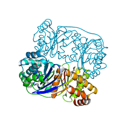
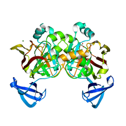
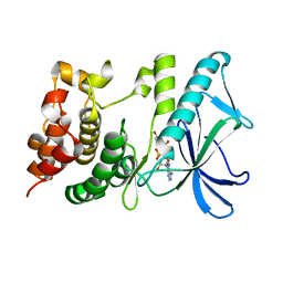
 | | Crystal structure of PigE: a transaminase involved in the biosynthesis of 2-methyl-3-n-amyl-pyrrole (MAP) from Serratia sp. FS14 | | 分子名称: | Aminotransferase, MAGNESIUM ION, PYRIDOXAL-5'-PHOSPHATE | | 著者 | Lou, X.D, Ran, T.T, Xu, D.Q, Wang, W.W. | | 登録日 | 2014-02-27 | | 公開日 | 2015-01-14 | | 実験手法 | X-RAY DIFFRACTION (2.3 Å) | | 主引用文献 | Crystal structure of the catalytic domain of PigE: a transaminase involved in the biosynthesis of 2-methyl-3-n-amyl-pyrrole (MAP) from Serratia sp. FS14
Biochem.Biophys.Res.Commun., 447, 2014
|
|
5HXK
 | |
5ZJK
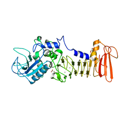
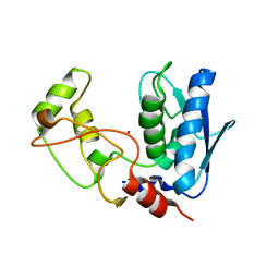 | | Structure of myroilysin | | 分子名称: | Myroilysin, PHOSPHATE ION, ZINC ION | | 著者 | Li, W.D, Ran, T.T, Xu, D.Q, Wang, W.W. | | 登録日 | 2018-03-20 | | 公開日 | 2019-03-20 | | 最終更新日 | 2024-03-27 | | 実験手法 | X-RAY DIFFRACTION (2.6 Å) | | 主引用文献 | Crystal structure of mature myroilysin and implication for its activation mechanism.
Int.J.Biol.Macromol., 2019
|
|
5X1R
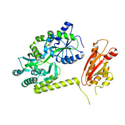
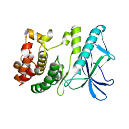 | | PpkA-294 apo form | | 分子名称: | PpkA-294 | | 著者 | Li, P.P, Ran, T.T, Xu, D.Q, Wang, W.W. | | 登録日 | 2017-01-26 | | 公開日 | 2018-01-31 | | 最終更新日 | 2024-03-20 | | 実験手法 | X-RAY DIFFRACTION (1.6 Å) | | 主引用文献 | Crystal structures of the kinase domain of PpkA, a key regulatory component of T6SS, reveal a general inhibitory mechanism.
Biochem.J., 475, 2018
|
|
5X1T
 | | PpkA-294 | | 分子名称: | ADENOSINE-5'-DIPHOSPHATE, PpkA-294 | | 著者 | Li, P.P, Ran, T.T, Xu, D.Q, Wang, W.W. | | 登録日 | 2017-01-26 | | 公開日 | 2018-01-31 | | 最終更新日 | 2024-03-20 | | 実験手法 | X-RAY DIFFRACTION (1.55 Å) | | 主引用文献 | Crystal structures of the kinase domain of PpkA, a key regulatory component of T6SS, reveal a general inhibitory mechanism.
Biochem.J., 475, 2018
|
|
5X1Q
 | | PpkA-294 with ATP and MnCl2 | | 分子名称: | ADENOSINE-5'-TRIPHOSPHATE, GLYCEROL, MANGANESE (II) ION, ... | | 著者 | Li, P.P, Ran, T.T, Xu, D.Q, Wang, W.W. | | 登録日 | 2017-01-26 | | 公開日 | 2018-01-31 | | 最終更新日 | 2024-03-20 | | 実験手法 | X-RAY DIFFRACTION (1.602 Å) | | 主引用文献 | Crystal structures of the kinase domain of PpkA, a key regulatory component of T6SS, reveal a general inhibitory mechanism.
Biochem.J., 475, 2018
|
|
5X1S
 | | PpkA-294 with Amppcp | | 分子名称: | GLYCEROL, PHOSPHOMETHYLPHOSPHONIC ACID ADENYLATE ESTER, PpkA | | 著者 | Li, P.P, Ran, T.T, Xu, D.Q, Wang, W.W. | | 登録日 | 2017-01-26 | | 公開日 | 2018-01-31 | | 最終更新日 | 2024-03-20 | | 実験手法 | X-RAY DIFFRACTION (1.45 Å) | | 主引用文献 | Crystal structures of the kinase domain of PpkA, a key regulatory component of T6SS, reveal a general inhibitory mechanism.
Biochem.J., 475, 2018
|
|
4DBH
 | | Crystal structure of Cg1458 with inhibitor | | 分子名称: | 2-HYDROXYHEPTA-2,4-DIENE-1,7-DIOATE ISOMERASE, MAGNESIUM ION, OXALATE ION | | 著者 | Ran, T.T, Wang, W.W, Xu, D.Q, Gao, Y.Y. | | 登録日 | 2012-01-15 | | 公開日 | 2012-11-28 | | 最終更新日 | 2024-03-20 | | 実験手法 | X-RAY DIFFRACTION (1.94 Å) | | 主引用文献 | Crystal structures of Cg1458 reveal a catalytic lid domain and a common catalytic mechanism for FAH family.
Biochem.J., 449, 2013
|
|
4DBF
 | | Crystal structures of Cg1458 | | 分子名称: | 2-HYDROXYHEPTA-2,4-DIENE-1,7-DIOATE ISOMERASE, MAGNESIUM ION | | 著者 | Ran, T.T, Xu, D.Q, Wang, W.W, Gao, Y.Y, Wang, M.T. | | 登録日 | 2012-01-15 | | 公開日 | 2012-11-28 | | 最終更新日 | 2023-11-08 | | 実験手法 | X-RAY DIFFRACTION (1.9 Å) | | 主引用文献 | Crystal structures of Cg1458 reveal a catalytic lid domain and a common catalytic mechanism for FAH family.
Biochem.J., 449, 2013
|
|
5HNV
 | | Crystal structure of PpkA | | 分子名称: | ADENOSINE-5'-TRIPHOSPHATE, GLYCEROL, MAGNESIUM ION, ... | | 著者 | Li, P.P, Ran, T.T, Wang, W.W. | | 登録日 | 2016-01-19 | | 公開日 | 2017-01-25 | | 最終更新日 | 2024-03-20 | | 実験手法 | X-RAY DIFFRACTION (1.41 Å) | | 主引用文献 | Crystal structures of the kinase domain of PpkA, a key regulatory component of T6SS, reveal a general inhibitory mechanism.
Biochem.J., 475, 2018
|
|
5D7W
 | | Crystal structure of serralysin | | 分子名称: | CALCIUM ION, GLYCEROL, Serralysin, ... | | 著者 | Wu, D, Ran, T, Xu, D.Q, Wang, W. | | 登録日 | 2015-08-14 | | 公開日 | 2016-01-13 | | 最終更新日 | 2023-11-08 | | 実験手法 | X-RAY DIFFRACTION (1.1 Å) | | 主引用文献 | Structure of a thermostable serralysin from Serratia sp. FS14 at 1.1 angstrom resolution.
Acta Crystallogr.,Sect.F, 72, 2016
|
|
6AEO
 | | TssL periplasmic domain | | 分子名称: | GLYCEROL, Maltose/maltodextrin-binding periplasmic protein,TssL | | 著者 | Ran, T.T, Wang, W.W, Wang, X.B, Xu, D.Q. | | 登録日 | 2018-08-06 | | 公開日 | 2019-06-12 | | 最終更新日 | 2024-03-27 | | 実験手法 | X-RAY DIFFRACTION (2.3 Å) | | 主引用文献 | Crystal structure of the periplasmic domain of TssL, a key membrane component of Type VI secretion system.
Int.J.Biol.Macromol., 120, 2018
|
|


