Loading
PDBj
MenuPDBj@FacebookPDBj@TwitterPDBj@YouTubewwPDB FoundationwwPDB
RCSB PDBPDBeBMRBAdv. SearchSearch help
Search by PDB author
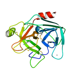
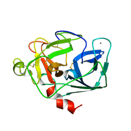
1ELT
DownloadVisualize
BU of 1elt by Molmil
STRUCTURE OF NATIVE PANCREATIC ELASTASE FROM NORTH ATLANTIC SALMON AT 1.61 ANGSTROMS RESOLUTION
Descriptor: CALCIUM ION, ELASTASE
Authors:Berglund, G.I, Smalaas, A.O.
Deposit date:1995-01-02
Release date:1996-01-01
Last modified:2018-04-18
Method:X-RAY DIFFRACTION (1.61 Å)
Cite:Structure of native pancreatic elastase from North Atlantic salmon at 1.61 A resolution.
Acta Crystallogr.,Sect.D, 51, 1995
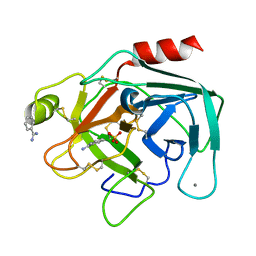
1BIT
DownloadVisualize
BU of 1bit by Molmil
THE CRYSTAL STRUCTURE OF ANIONIC SALMON TRYPSIN IN A SECOND CRYSTAL FORM
Descriptor: BENZAMIDINE, CALCIUM ION, SULFATE ION, ...
Authors:Berglund, G.I.
Deposit date:1994-08-26
Release date:1994-11-01
Last modified:2019-08-14
Method:X-RAY DIFFRACTION (1.83 Å)
Cite:Structure of anionic salmon trypsin in a second crystal form.
Acta Crystallogr.,Sect.D, 51, 1995
2TBS
DownloadVisualize
BU of 2tbs by Molmil
COLD-ADAPTION OF ENZYMES: STRUCTURAL COMPARISON BETWEEN SALMON AND BOVINE TRYPSINS
Descriptor: BENZAMIDINE, CALCIUM ION, TRYPSIN
Authors:Smalas, A.O.
Deposit date:1994-01-14
Release date:1994-04-30
Last modified:2011-07-13
Method:X-RAY DIFFRACTION (1.8 Å)
Cite:Cold adaption of enzymes: structural comparison between salmon and bovine trypsins.
Proteins, 20, 1994
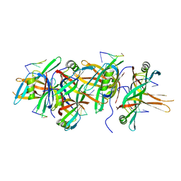
5ODN
DownloadVisualize
BU of 5odn by Molmil
Salinibacter ruber Single-Strand Binding protein
Descriptor: DNA (5'-D(*TP*TP*T)-3'), DNA (5'-D(P*TP*TP*TP*T)-3'), Single-stranded DNA-binding protein
Authors:Pierechod, M, Rothweiler, U.
Deposit date:2017-07-06
Release date:2018-08-01
Last modified:2024-01-17
Method:X-RAY DIFFRACTION (2.598 Å)
Cite:Salinibacter ruber Single-Strand Binding protein
To Be Published
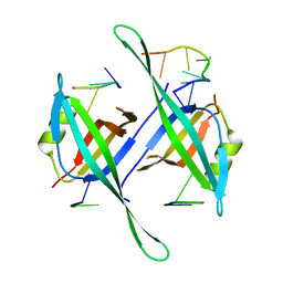
5ODP
DownloadVisualize
BU of 5odp by Molmil
Salinibacter ruber Single-Strand Binding protein D17K D71K mutant
Descriptor: DNA (5'-D(*TP*TP*TP*TP*TP*TP*TP*TP*T)-3'), Single-stranded DNA-binding protein
Authors:Pierechod, M, Rothweiler, U.
Deposit date:2017-07-06
Release date:2018-08-01
Last modified:2024-01-17
Method:X-RAY DIFFRACTION (2.535 Å)
Cite:Salinibacter ruber Single-Strand Binding protein D17K D71K mutant
To Be Published
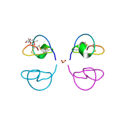
1LU0
DownloadVisualize
BU of 1lu0 by Molmil
Atomic Resolution Structure of Squash Trypsin Inhibitor: Unexpected Metal Coordination
Descriptor: (4R)-2-METHYLPENTANE-2,4-DIOL, GLYCEROL, SULFATE ION, ...
Authors:Thaimattam, R, Tykarska, E, Bierzynski, A, Sheldrick, G.M, Jaskolski, M.
Deposit date:2002-05-21
Release date:2002-08-28
Last modified:2021-10-27
Method:X-RAY DIFFRACTION (1.03 Å)
Cite:Atomic resolution structure of squash trypsin inhibitor: unexpected metal coordination.
Acta Crystallogr.,Sect.D, 58, 2002
<12


219869

건을2024-05-15부터공개중

PDB statisticsPDBj update infoContact PDBjnumon