Loading
PDBj
メニューPDBj@FacebookPDBj@TwitterPDBj@YouTubewwPDB FoundationwwPDB
RCSB PDBPDBeBMRBAdv. SearchSearch help
Search by PDB author
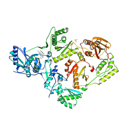
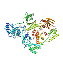
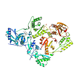
6X4A
DownloadVisualize
BU of 6x4a by Molmil
Crystal Structure of HIV-1 Reverse Transcriptase (Y181C) Variant in Complex with 5-chloro-7-(2-(2-(2,4-dioxo-3,4-dihydropyrimidin-1(2H)-yl)ethoxy)phenoxy)-8-methyl-2-naphthonitrile (JLJ651), a Non-nucleoside Inhibitor
分子名称: 5-chloro-7-(2-(2-(2,4-dioxo-3,4-dihydropyrimidin-1(2H)-yl)ethoxy)phenoxy)-8-methyl-2-naphthonitrile, Reverse transcriptase/ribonuclease H, SULFATE ION, ...
著者Chan, A.H, Duong, V.N, Ippolito, J.A, Jorgensen, W.L, Anderson, K.S.
登録日2020-05-22
公開日2020-07-22
最終更新日2023-10-18
実験手法X-RAY DIFFRACTION (2.537 Å)
主引用文献Structural investigation of 2-naphthyl phenyl ether inhibitors bound to WT and Y181C reverse transcriptase highlights key features of the NNRTI binding site.
Protein Sci., 29, 2020
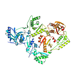
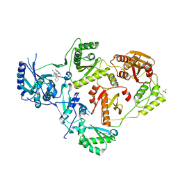
6X4D
DownloadVisualize
BU of 6x4d by Molmil
Crystal Structure of HIV-1 Reverse Transcriptase in Complex with 5-(cyclopropylmethyl)-7-(2-(2-(2,4-dioxo-3,4-dihydropyrimidin-1(2H)-yl)ethoxy)phenoxy)-8-methyl-2-naphthonitrile (JLJ678), a Non-nucleoside Inhibitor
分子名称: 5-(cyclopropylmethyl)-7-{2-[2-(2,4-dioxo-3,4-dihydropyrimidin-1(2H)-yl)ethoxy]phenoxy}-8-methylnaphthalene-2-carbonitrile, Reverse transcriptase/ribonuclease H, SULFATE ION, ...
著者Chan, A.H, Duong, V.N, Ippolito, J.A, Jorgensen, W.L, Anderson, K.S.
登録日2020-05-22
公開日2020-07-22
最終更新日2023-10-18
実験手法X-RAY DIFFRACTION (2.65 Å)
主引用文献Structural investigation of 2-naphthyl phenyl ether inhibitors bound to WT and Y181C reverse transcriptase highlights key features of the NNRTI binding site.
Protein Sci., 29, 2020
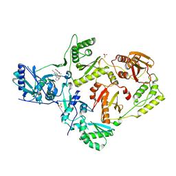
6X47
DownloadVisualize
BU of 6x47 by Molmil
Crystal Structure of HIV-1 Reverse Transcriptase in Complex with 7-(2-(2-(2,4-dioxo-3,4-dihydropyrimidin-1(2H)-yl)ethoxy)phenoxy)-2-naphthonitrile (JLJ649), a Non-nucleoside Inhibitor
分子名称: 7-(2-(2-(2,4-dioxo-3,4-dihydropyrimidin-1(2H)-yl)ethoxy)phenoxy)-2-naphthonitrile, Reverse transcriptase/ribonuclease H, p51 RT
著者Chan, A.H, Duong, V.N, Ippolito, J.A, Jorgensen, W.L, Anderson, K.S.
登録日2020-05-22
公開日2020-07-22
最終更新日2023-10-18
実験手法X-RAY DIFFRACTION (2.767 Å)
主引用文献Structural investigation of 2-naphthyl phenyl ether inhibitors bound to WT and Y181C reverse transcriptase highlights key features of the NNRTI binding site.
Protein Sci., 29, 2020
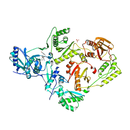
6X4E
DownloadVisualize
BU of 6x4e by Molmil
Crystal Structure of HIV-1 Reverse Transcriptase in Complex with methyl 2-(6-cyano-3-(2-(2-(2,4-dioxo-3,4-dihydropyrimidin-1(2H)-yl)ethoxy)phenoxy)-4-methylnaphthalen-1-yl)acetate (JLJ681), a Non-nucleoside Inhibitor
分子名称: Reverse transcriptase/ribonuclease H, SULFATE ION, methyl (6-cyano-3-{2-[2-(2,4-dioxo-3,4-dihydropyrimidin-1(2H)-yl)ethoxy]phenoxy}-4-methylnaphthalen-1-yl)acetate, ...
著者Chan, A.H, Duong, V.N, Ippolito, J.A, Jorgensen, W.L, Anderson, K.S.
登録日2020-05-22
公開日2020-07-22
最終更新日2023-10-18
実験手法X-RAY DIFFRACTION (2.6 Å)
主引用文献Structural investigation of 2-naphthyl phenyl ether inhibitors bound to WT and Y181C reverse transcriptase highlights key features of the NNRTI binding site.
Protein Sci., 29, 2020
6X49
DownloadVisualize
BU of 6x49 by Molmil
Crystal Structure of HIV-1 Reverse Transcriptase (Y181C) Variant in Complex with 7-(2-(2-(2,4-dioxo-3,4-dihydropyrimidin-1(2H)-yl)ethoxy)phenoxy)-2-naphthonitrile (JLJ649), a Non-nucleoside Inhibitor
分子名称: 7-(2-(2-(2,4-dioxo-3,4-dihydropyrimidin-1(2H)-yl)ethoxy)phenoxy)-2-naphthonitrile, Reverse transcriptase/ribonuclease H, SULFATE ION, ...
著者Chan, A.H, Duong, V.N, Ippolito, J.A, Jorgensen, W.L, Anderson, K.S.
登録日2020-05-22
公開日2020-07-22
最終更新日2023-10-18
実験手法X-RAY DIFFRACTION (2.745 Å)
主引用文献Structural investigation of 2-naphthyl phenyl ether inhibitors bound to WT and Y181C reverse transcriptase highlights key features of the NNRTI binding site.
Protein Sci., 29, 2020
6X4F
DownloadVisualize
BU of 6x4f by Molmil
Crystal Structure of HIV-1 Reverse Transcriptase (Y181C) Variant in Complex with methyl 2-(6-cyano-3-(2-(2-(2,4-dioxo-3,4-dihydropyrimidin-1(2H)-yl)ethoxy)phenoxy)-4-methylnaphthalen-1-yl)acetate (JLJ681), a Non-nucleoside Inhibitor
分子名称: Reverse transcriptase/ribonuclease H, SULFATE ION, methyl (6-cyano-3-{2-[2-(2,4-dioxo-3,4-dihydropyrimidin-1(2H)-yl)ethoxy]phenoxy}-4-methylnaphthalen-1-yl)acetate, ...
著者Chan, A.H, Duong, V.N, Ippolito, J.A, Jorgensen, W.L, Anderson, K.S.
登録日2020-05-22
公開日2020-07-22
最終更新日2023-10-18
実験手法X-RAY DIFFRACTION (2.72 Å)
主引用文献Structural investigation of 2-naphthyl phenyl ether inhibitors bound to WT and Y181C reverse transcriptase highlights key features of the NNRTI binding site.
Protein Sci., 29, 2020
6X4B
DownloadVisualize
BU of 6x4b by Molmil
Crystal Structure of HIV-1 Reverse Transcriptase in Complex with 7-(2-(2-(2,4-dioxo-3,4-dihydropyrimidin-1(2H)-yl)ethoxy)phenoxy)-5-fluoro-8-methyl-2-naphthonitrile (JLJ655), a Non-nucleoside Inhibitor
分子名称: 7-{2-[2-(2,4-dioxo-3,4-dihydropyrimidin-1(2H)-yl)ethoxy]phenoxy}-5-fluoro-8-methylnaphthalene-2-carbonitrile, MAGNESIUM ION, Reverse transcriptase/ribonuclease H, ...
著者Chan, A.H, Duong, V.N, Ippolito, J.A, Jorgensen, W.L, Anderson, K.S.
登録日2020-05-22
公開日2020-07-22
最終更新日2023-10-18
実験手法X-RAY DIFFRACTION (2.5 Å)
主引用文献Structural investigation of 2-naphthyl phenyl ether inhibitors bound to WT and Y181C reverse transcriptase highlights key features of the NNRTI binding site.
Protein Sci., 29, 2020
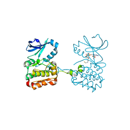
6XKA
DownloadVisualize
BU of 6xka by Molmil
TPX2 residues 7-20 fused to Aurora A residues 116-389 dephosphorylated, and CoAlated on C290
分子名称: COENZYME A, TPX2 fragment - Aurora A kinase domain fusion
著者Lim, D.C, Yaffe, M.B.
登録日2020-06-26
公開日2020-08-05
最終更新日2023-10-18
実験手法X-RAY DIFFRACTION (2.65 Å)
主引用文献Redox priming promotes Aurora A activation during mitosis.
Sci.Signal., 13, 2020
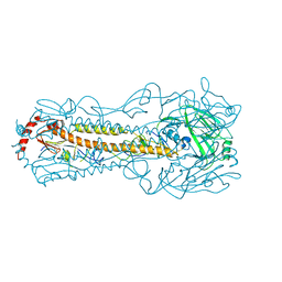
6IN5
DownloadVisualize
BU of 6in5 by Molmil
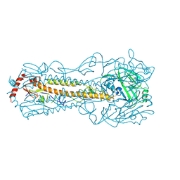
Crystal structure of H5N2 hemagglutinin G228S Q226L mutant with 3SLN from A/chicken/Taiwan/0502/2012
分子名称: Hemagglutinin, N-acetyl-alpha-neuraminic acid-(2-3)-beta-D-galactopyranose
著者Lin, T.H, Lee, M.S, Liu, J.S.
登録日2018-10-24
公開日2019-10-30
最終更新日2023-11-22
実験手法X-RAY DIFFRACTION (2.916 Å)
主引用文献crystal structure of H5 hemagglutinin from A/chicken/Taiwan/0502/2012
To Be Published
6IJT
DownloadVisualize
BU of 6ijt by Molmil
Crystal structure of H5N2 hemagglutinin G228S Q226L mutant with 6SLN from A/chicken/Taiwan/0502/2012
分子名称: Hemagglutinin, N-acetyl-alpha-neuraminic acid
著者Lin, T.H, Lee, M.S, Liu, J.S.
登録日2018-10-12
公開日2019-10-16
最終更新日2023-11-22
実験手法X-RAY DIFFRACTION (2.9 Å)
主引用文献crystal structure of H5 hemagglutinin from A/chicken/Taiwan/0502/2012
To Be Published
6JYS
DownloadVisualize
BU of 6jys by Molmil
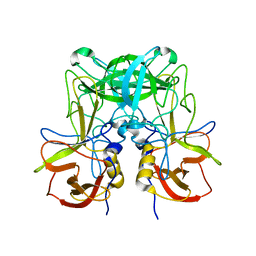
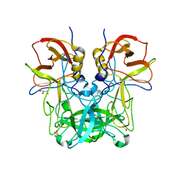
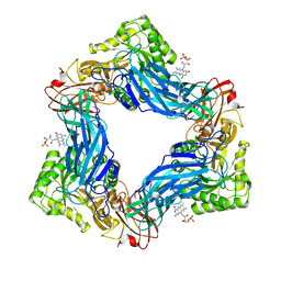
GII.13/21 noroviruses recognize glycans with a terminal beta-galactose via an unconventional glycan binding site
分子名称: THREONINE, beta-D-galactopyranose-(1-3)-2-acetamido-2-deoxy-alpha-D-galactopyranose, beta-D-galactopyranose-(1-3)-[2-acetamido-2-deoxy-beta-D-glucopyranose-(1-6)]2-acetamido-2-deoxy-alpha-D-galactopyranose, ...
著者Duan, Z, Xin, C.
登録日2019-04-27
公開日2019-05-22
最終更新日2024-05-01
実験手法X-RAY DIFFRACTION (1.697 Å)
主引用文献GII.13/21 Noroviruses Recognize Glycans with a Terminal beta-Galactose via an Unconventional Glycan Binding Site.
J.Virol., 93, 2019
6JYO
DownloadVisualize
BU of 6jyo by Molmil
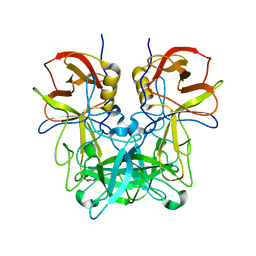
GII.13/21 noroviruses recognize glycans with a terminal beta-galactose via an unconventional glycan binding site
分子名称: norovirus P domain protein
著者Duan, Z, Xin, C.
登録日2019-04-27
公開日2019-05-22
最終更新日2024-03-27
実験手法X-RAY DIFFRACTION (1.502 Å)
主引用文献GII.13/21 Noroviruses Recognize Glycans with a Terminal beta-Galactose via an Unconventional Glycan Binding Site.
J.Virol., 93, 2019
6JYN
DownloadVisualize
BU of 6jyn by Molmil
GII.13/21 noroviruses recognize glycans with a terminal beta-galactose via an unconventional glycan binding site
分子名称: beta-D-galactopyranose-(1-3)-2-acetamido-2-deoxy-beta-D-glucopyranose, human norovirus P domain protein
著者Duan, Z, Xin, C.
登録日2019-04-26
公開日2019-05-22
最終更新日2024-03-27
実験手法X-RAY DIFFRACTION (1.599 Å)
主引用文献GII.13/21 Noroviruses Recognize Glycans with a Terminal beta-Galactose via an Unconventional Glycan Binding Site.
J.Virol., 93, 2019
2X0N
DownloadVisualize
BU of 2x0n by Molmil
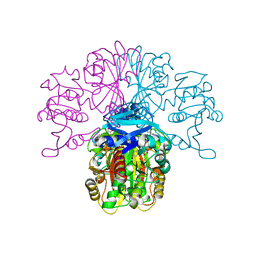
Structure of glycosomal glyceraldehyde-3-phosphate dehydrogenase from Trypanosoma brucei determined from Laue data
分子名称: GLYCERALDEHYDE-3-PHOSPHATE DEHYDROGENASE, GLYCOSOMAL, NICOTINAMIDE-ADENINE-DINUCLEOTIDE, ...
著者Vellieux, F.M.D, Hajdu, J, Hol, W.G.J.
登録日2009-12-16
公開日2009-12-22
最終更新日2023-12-20
実験手法X-RAY DIFFRACTION (3.2 Å)
主引用文献Structure of Glycosomal Glyceraldehyde-3-Phosphate Dehydrogenase from Trypanosoma Brucei Determined from Laue Data.
Proc.Natl.Acad.Sci.USA, 90, 1993
6JYR
DownloadVisualize
BU of 6jyr by Molmil
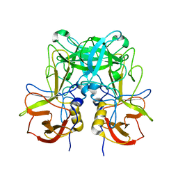
GII.13/21 noroviruses recognize glycans with a terminal beta-galactose via an unconventional glycan binding site
分子名称: GLYCEROL, norovirus P domain protein
著者Duan, Z, Xin, C.
登録日2019-04-27
公開日2019-05-22
最終更新日2024-03-27
実験手法X-RAY DIFFRACTION (1.501 Å)
主引用文献GII.13/21 Noroviruses Recognize Glycans with a Terminal beta-Galactose via an Unconventional Glycan Binding Site.
J.Virol., 93, 2019
6TIM
DownloadVisualize
BU of 6tim by Molmil
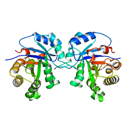
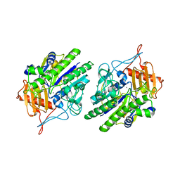
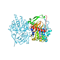
THE ADAPTABILITY OF THE ACTIVE SITE OF TRYPANOSOMAL TRIOSEPHOSPHATE ISOMERASE AS OBSERVED IN THE CRYSTAL STRUCTURES OF THREE DIFFERENT COMPLEXES
分子名称: SN-GLYCEROL-3-PHOSPHATE, TRIOSEPHOSPHATE ISOMERASE
著者Noble, M.E.M, Wierenga, R.K, Hol, W.G.J.
登録日1991-04-23
公開日1992-10-15
最終更新日2024-03-13
実験手法X-RAY DIFFRACTION (2.2 Å)
主引用文献The adaptability of the active site of trypanosomal triosephosphate isomerase as observed in the crystal structures of three different complexes.
Proteins, 10, 1991
6UFX
DownloadVisualize
BU of 6ufx by Molmil
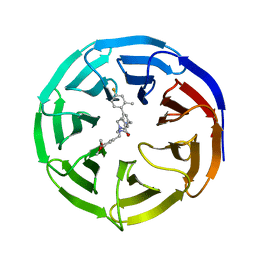
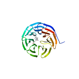
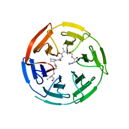
WD repeat-containing protein 5 complexed with N-[(3,5-dimethoxyphenyl)methyl]-4'-fluoro-5-{[(2E)-2-imino-3-methyl-2,3-dihydro-1H-imidazol-1-yl]methyl}-2'-methyl[1,1'-biphenyl]-3-carboxamide (compound 13)
分子名称: N-[(3,5-dimethoxyphenyl)methyl]-4'-fluoro-5-{[(2E)-2-imino-3-methyl-2,3-dihydro-1H-imidazol-1-yl]methyl}-2'-methyl[1,1'-biphenyl]-3-carboxamide, WD repeat-containing protein 5
著者Rietz, T.A, Fesik, S.W, Zhao, B.
登録日2019-09-25
公開日2020-01-01
最終更新日2023-10-11
実験手法X-RAY DIFFRACTION (2.015 Å)
主引用文献Discovery and Structure-Based Optimization of Potent and Selective WD Repeat Domain 5 (WDR5) Inhibitors Containing a Dihydroisoquinolinone Bicyclic Core.
J.Med.Chem., 63, 2020
6UCS
DownloadVisualize
BU of 6ucs by Molmil
Discovery and Structure-Based optimization of potent and selective WDR5 inhibitors containing a dihydroisoquinolinone bicyclic core
分子名称: 2-amino-3-{[(5P)-2-[(3,5-dimethoxyphenyl)methyl]-5-(4-fluoro-2-methylphenyl)-1-oxo-1,2,3,4-tetrahydroisoquinolin-7-yl]methyl}-1-methyl-1H-imidazol-3-ium, WD repeat-containing protein 5
著者Zhao, B.
登録日2019-09-17
公開日2020-01-01
最終更新日2024-05-15
実験手法X-RAY DIFFRACTION (1.85 Å)
主引用文献Discovery and Structure-Based Optimization of Potent and Selective WD Repeat Domain 5 (WDR5) Inhibitors Containing a Dihydroisoquinolinone Bicyclic Core.
J.Med.Chem., 63, 2020
6MVH
DownloadVisualize
BU of 6mvh by Molmil
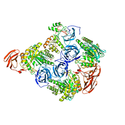
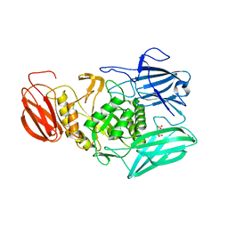
Crystal structure of FMN-binding beta-glucuronidase from Roseburia hominis
分子名称: Beta-galactosidase, CALCIUM ION, FLAVIN MONONUCLEOTIDE
著者Pellock, S.J, Redinbo, M.R.
登録日2018-10-25
公開日2019-01-30
最終更新日2024-03-13
実験手法X-RAY DIFFRACTION (2.4 Å)
主引用文献Discovery and Characterization of FMN-Binding beta-Glucuronidases in the Human Gut Microbiome.
J. Mol. Biol., 431, 2019
6USS
DownloadVisualize
BU of 6uss by Molmil
Catalytic S88C mutant of gut microbial sulfatase from Bacteroides fragilis CAG:558
分子名称: CALCIUM ION, Sulfatase
著者Ervin, S.M, Redinbo, M.R.
登録日2019-10-28
公開日2020-11-11
最終更新日2023-10-11
実験手法X-RAY DIFFRACTION (2.5 Å)
主引用文献Structural Insights into Endobiotic Reactivation by Human Gut Microbiome-Encoded Sulfatases.
Biochemistry, 59, 2020
6MVF
DownloadVisualize
BU of 6mvf by Molmil
Crystal structure of FMN-binding beta-glucuronidase from Facaelibacterium prausnitzii L2-6
分子名称: Beta-galactosidase/beta-glucuronidase, FLAVIN MONONUCLEOTIDE
著者Pellock, S.J, Redinbo, M.R.
登録日2018-10-25
公開日2019-01-30
最終更新日2023-10-11
実験手法X-RAY DIFFRACTION (2.55 Å)
主引用文献Discovery and Characterization of FMN-Binding beta-Glucuronidases in the Human Gut Microbiome.
J. Mol. Biol., 431, 2019
6V7O
DownloadVisualize
BU of 6v7o by Molmil
Structural Elucidation of Peptide Binding to KLHL-12, a Substrate Specific Adapter Protein in a Cul3-Ring E3 Ligase Complex
分子名称: Dvl3-peptide, Kelch-like protein 12
著者Zhao, B.
登録日2019-12-09
公開日2020-02-19
最終更新日2023-10-11
実験手法X-RAY DIFFRACTION (2.9 Å)
主引用文献Structural Elucidation of Peptide Binding to KLHL-12, a Substrate Specific Adapter Protein in a Cul3-Ring E3 Ligase Complex.
Biochemistry, 59, 2020
6MVG
DownloadVisualize
BU of 6mvg by Molmil
Crystal structure of FMN-binding beta-glucuronidase from Ruminococcus gnavus
分子名称: CALCIUM ION, FLAVIN MONONUCLEOTIDE, beta-glucuronidase
著者Pellock, S.J, Redinbo, M.R.
登録日2018-10-25
公開日2019-01-30
最終更新日2024-03-13
実験手法X-RAY DIFFRACTION (2.8 Å)
主引用文献Discovery and Characterization of FMN-Binding beta-Glucuronidases in the Human Gut Microbiome.
J. Mol. Biol., 431, 2019
6UST
DownloadVisualize
BU of 6ust by Molmil
Gut microbial sulfatase from Hungatella hathewayi
分子名称: CALCIUM ION, N-acetylgalactosamine 6-sulfate sulfatase
著者Ervin, S.M, Redinbo, M.R.
登録日2019-10-28
公開日2020-11-11
最終更新日2023-10-11
実験手法X-RAY DIFFRACTION (2.6 Å)
主引用文献Structural Insights into Endobiotic Reactivation by Human Gut Microbiome-Encoded Sulfatases.
Biochemistry, 59, 2020
6VPG
DownloadVisualize
BU of 6vpg by Molmil
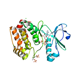
TPX2 residues 7-20 fused to Aurora A residues 116-389 in complex with AMP-PNP
分子名称: CHLORIDE ION, GLYCEROL, MAGNESIUM ION, ...
著者Lim, D.C, Yaffe, M.B.
登録日2020-02-03
公開日2020-08-05
最終更新日2023-10-11
実験手法X-RAY DIFFRACTION (2.64 Å)
主引用文献Redox priming promotes Aurora A activation during mitosis.
Sci.Signal., 13, 2020

219869

件を2024-05-15に公開中

PDB statisticsPDBj update infoContact PDBjnumon