8S2W
 
 | |
8S2U
 | |
8S2V
 | |
8S2X
 | |
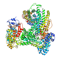
1B6S
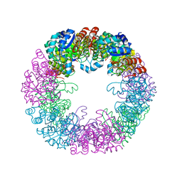
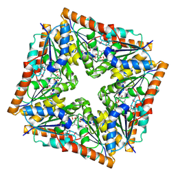
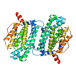
 | | STRUCTURE OF N5-CARBOXYAMINOIMIDAZOLE RIBONUCLEOTIDE SYNTHETASE | | 分子名称: | ADENOSINE-5'-DIPHOSPHATE, MAGNESIUM ION, PROTEIN (N5-CARBOXYAMINOIMIDAZOLE RIBONUCLEOTIDE SYNTHETASE) | | 著者 | Thoden, J.B, Kappock, T.J, Stubbe, J, Holden, H.M. | | 登録日 | 1999-01-18 | | 公開日 | 1999-11-30 | | 最終更新日 | 2023-08-09 | | 実験手法 | X-RAY DIFFRACTION (2.5 Å) | | 主引用文献 | Three-dimensional structure of N5-carboxyaminoimidazole ribonucleotide synthetase: a member of the ATP grasp protein superfamily.
Biochemistry, 38, 1999
|
|
1B6R
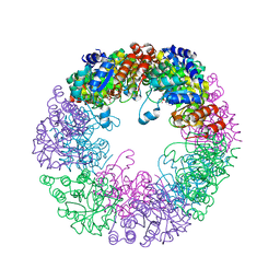
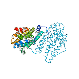
 | | N5-CARBOXYAMINOIMIDAZOLE RIBONUCLEOTIDE SYNTHETASE FROM E. COLI | | 分子名称: | PROTEIN (N5-CARBOXYAMINOIMIDAZOLE RIBONUCLEOTIDE SYNTHETASE), SULFATE ION | | 著者 | Thoden, J.B, Kappock, T.J, Stubbe, J, Holden, H.M. | | 登録日 | 1999-01-17 | | 公開日 | 1999-11-30 | | 最終更新日 | 2023-12-27 | | 実験手法 | X-RAY DIFFRACTION (2.1 Å) | | 主引用文献 | Three-dimensional structure of N5-carboxyaminoimidazole ribonucleotide synthetase: a member of the ATP grasp protein superfamily.
Biochemistry, 38, 1999
|
|
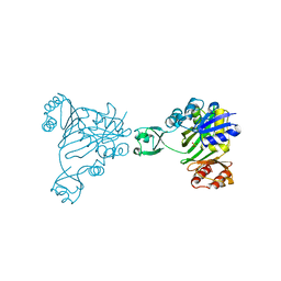
2ATE
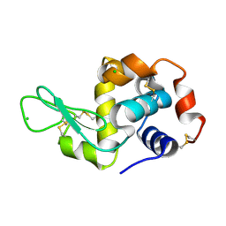
 | | Structure of the complex of PurE with NitroAIR | | 分子名称: | ((2R,3S,4R,5R)-5-(5-AMINO-4-NITRO-1H-IMIDAZOL-1-YL)-3,4-DIHYDROXYTETRAHYDROFURAN-2-YL)METHYL DIHYDROGEN PHOSPHATE, Phosphoribosylaminoimidazole carboxylase catalytic subunit | | 著者 | Kappock, T.J, Mathews, I.I, Zaugg, J.B, Peng, P, Hoskins, A.A, Okamoto, A, Ealick, S.E, Stubbe, J. | | 登録日 | 2005-08-24 | | 公開日 | 2006-08-29 | | 最終更新日 | 2023-11-15 | | 実験手法 | X-RAY DIFFRACTION (1.8 Å) | | 主引用文献 | N5-CAIR mutase: role of a CO2 binding site and substrate movement in catalysis.
Biochemistry, 46, 2007
|
|
6W4X
 | | Holocomplex of E. coli class Ia ribonucleotide reductase with GDP and TTP | | 分子名称: | GUANOSINE-5'-DIPHOSPHATE, MAGNESIUM ION, MU-OXO-DIIRON, ... | | 著者 | Kang, G, Taguchi, A, Stubbe, J, Drennan, C. | | 登録日 | 2020-03-11 | | 公開日 | 2020-04-08 | | 最終更新日 | 2020-05-06 | | 実験手法 | ELECTRON MICROSCOPY (3.6 Å) | | 主引用文献 | Structure of a trapped radical transfer pathway within a ribonucleotide reductase holocomplex.
Science, 368, 2020
|
|
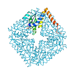
1D7A
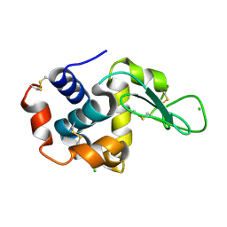
 | | CRYSTAL STRUCTURE OF E. COLI PURE-MONONUCLEOTIDE COMPLEX. | | 分子名称: | 5-AMINOIMIDAZOLE RIBONUCLEOTIDE, PHOSPHORIBOSYLAMINOIMIDAZOLE CARBOXYLASE | | 著者 | Mathews, I.I, Kappock, T.J, Stubbe, J, Ealick, S.E. | | 登録日 | 1999-10-16 | | 公開日 | 1999-12-03 | | 最終更新日 | 2021-11-03 | | 実験手法 | X-RAY DIFFRACTION (2.5 Å) | | 主引用文献 | Crystal structure of Escherichia coli PurE, an unusual mutase in the purine biosynthetic pathway.
Structure Fold.Des., 7, 1999
|
|
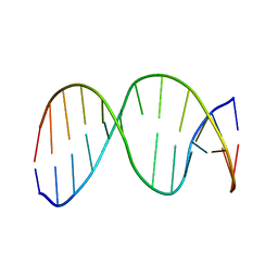
2O80
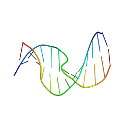 | | Duplex DNA containing an abasic site with an opposite dC (alpha anomer) in 5'-G_AC-3' (10 structure ensemble and averaged structure) | | 分子名称: | 5'-D(*CP*CP*AP*AP*AP*GP*(ORP)P*AP*CP*CP*GP*GP*G)-3', 5'-D(*CP*CP*CP*GP*GP*TP*CP*CP*TP*TP*TP*GP*G)-3' | | 著者 | Chen, J, Dupradeau, F.Y, Case, D.A, Turner, C.J, Stubbe, J. | | 登録日 | 2006-12-11 | | 公開日 | 2007-11-27 | | 最終更新日 | 2023-11-29 | | 実験手法 | SOLUTION NMR | | 主引用文献 | DNA oligonucleotides with A, T, G or C opposite an abasic site: structure and dynamics.
Nucleic Acids Res., 36, 2008
|
|
2O7W
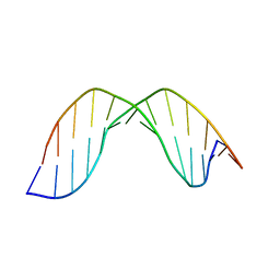 | | Duplex DNA containing an abasic site with an opposite G (alpha anomer) in 5'-G_AC-3' (10 structure ensemble and averaged structure) | | 分子名称: | 5'-D(*CP*CP*AP*AP*AP*GP*(ORP)P*AP*CP*CP*GP*GP*G)-3', 5'-D(*CP*CP*CP*GP*GP*TP*GP*CP*TP*TP*TP*GP*G)-3' | | 著者 | Chen, J, Dupradeau, F.Y, Case, D.A, Turner, C.J, Stubbe, J. | | 登録日 | 2006-12-11 | | 公開日 | 2007-11-27 | | 最終更新日 | 2023-11-29 | | 実験手法 | SOLUTION NMR | | 主引用文献 | DNA oligonucleotides with A, T, G or C opposite an abasic site: structure and dynamics.
Nucleic Acids Res., 36, 2008
|
|
2O7Z
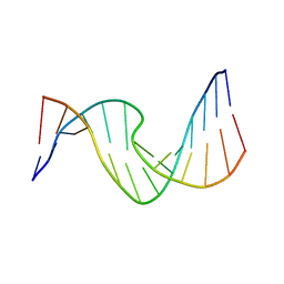 | | Duplex DNA containing an abasic site with an opposite T (beta anomer) in 5'-G_AC-3' (10 structure ensemble and averaged structure) | | 分子名称: | 5'-D(*CP*CP*AP*AP*AP*GP*(AAB)P*AP*CP*CP*GP*GP*G)-3', 5'-D(*CP*CP*CP*GP*GP*TP*TP*CP*TP*TP*TP*GP*G)-3' | | 著者 | Chen, J, Dupradeau, F.Y, Case, D.A, Turner, C.J, Stubbe, J. | | 登録日 | 2006-12-11 | | 公開日 | 2007-11-27 | | 最終更新日 | 2023-11-29 | | 実験手法 | SOLUTION NMR | | 主引用文献 | DNA oligonucleotides with A, T, G or C opposite an abasic site: structure and dynamics.
Nucleic Acids Res., 36, 2008
|
|
2O82
 | | Duplex DNA containing an abasic site with an opposite dC (beta anomer) in 5'-G_AC-3' (10 structure ensemble and averaged structure) | | 分子名称: | 5'-D(*CP*CP*AP*AP*AP*GP*(AAB)P*AP*CP*CP*GP*GP*G)-3', 5'-D(*CP*CP*CP*GP*GP*TP*CP*CP*TP*TP*TP*GP*G)-3' | | 著者 | Chen, J, Dupradeau, F.Y, Case, D.A, Turner, C.J, Stubbe, J. | | 登録日 | 2006-12-11 | | 公開日 | 2007-11-27 | | 最終更新日 | 2023-11-29 | | 実験手法 | SOLUTION NMR | | 主引用文献 | DNA oligonucleotides with A, T, G or C opposite an abasic site: structure and dynamics.
Nucleic Acids Res., 36, 2008
|
|
2O7X
 | | Duplex DNA containing an abasic site with an opposite G (beta anomer) in 5'-G_AC-3' (10 structure ensemble and averaged structure) | | 分子名称: | 5'-D(*CP*CP*AP*AP*AP*GP*(AAB)P*AP*CP*CP*GP*GP*G)-3', 5'-D(*CP*CP*CP*GP*GP*TP*GP*CP*TP*TP*TP*GP*G)-3' | | 著者 | Chen, J, Dupradeau, F.Y, Case, D.A, Turner, C.J, Stubbe, J. | | 登録日 | 2006-12-11 | | 公開日 | 2007-11-27 | | 最終更新日 | 2023-11-29 | | 実験手法 | SOLUTION NMR | | 主引用文献 | DNA oligonucleotides with A, T, G or C opposite an abasic site: structure and dynamics.
Nucleic Acids Res., 36, 2008
|
|
2O7Y
 | | Duplex DNA containing an abasic site with an opposite T (alpha anomer) in 5'-G_AC-3' (10 structure ensemble and averaged structure) | | 分子名称: | 5'-D(*CP*CP*AP*AP*AP*GP*(ORP)P*AP*CP*CP*GP*GP*G)-3', 5'-D(*CP*CP*CP*GP*GP*TP*TP*CP*TP*TP*TP*GP*G)-3' | | 著者 | Dupradeau, F.Y, Case, D.A, Turner, C.J, Stubbe, J. | | 登録日 | 2006-12-11 | | 公開日 | 2007-11-27 | | 最終更新日 | 2023-11-29 | | 実験手法 | SOLUTION NMR | | 主引用文献 | DNA oligonucleotides with A, T, G or C opposite an abasic site: structure and dynamics.
Nucleic Acids Res., 36, 2008
|
|
2HSL
 | | NMR structure of 13mer duplex DNA containing an abasic site, averaged structure (alpha anomer) | | 分子名称: | 5'-D(*CP*CP*AP*AP*AP*GP*(D1P)P*AP*CP*CP*GP*GP*G)-3', 5'-D(*CP*CP*CP*GP*GP*TP*AP*CP*TP*TP*TP*GP*G)-3' | | 著者 | Chen, J, Dupradeau, F.Y, Case, D.A, Turner, C.J, Stubbe, J. | | 登録日 | 2006-07-22 | | 公開日 | 2007-05-29 | | 最終更新日 | 2018-08-08 | | 実験手法 | SOLUTION NMR | | 主引用文献 | Nuclear magnetic resonance structural studies and molecular modeling of duplex DNA containing normal and 4'-oxidized abasic sites.
Biochemistry, 46, 2007
|
|
2HSS
 | | 13mer duplex DNA containg an abasic site with beta anomer, averaged structure | | 分子名称: | 5'-D(*CP*CP*AP*AP*AP*GP*(AAB)P*AP*CP*CP*GP*GP*G)-3', 5'-D(*CP*CP*CP*GP*GP*TP*AP*CP*TP*TP*TP*GP*G)-3' | | 著者 | Chen, J, Dupradeau, F.Y, Case, D.A, Turner, C.J, Stubbe, J. | | 登録日 | 2006-07-22 | | 公開日 | 2007-05-29 | | 最終更新日 | 2018-08-08 | | 実験手法 | SOLUTION NMR | | 主引用文献 | Nuclear magnetic resonance structural studies and molecular modeling of duplex DNA containing normal and 4'-oxidized abasic sites.
Biochemistry, 46, 2007
|
|
2HOU
 | | Structure ensembles of duplex DNA containing a 4'-oxidized abasic site. | | 分子名称: | 5'-D(*CP*CP*AP*AP*AP*GP*(X4A)P*AP*CP*CP*GP*GP*G)-3', 5'-D(*CP*CP*CP*GP*GP*TP*AP*CP*TP*TP*TP*GP*G)-3' | | 著者 | Chen, J, Dupradeau, F.Y, Case, D.A, Turner, C.J, Stubbe, J. | | 登録日 | 2006-07-16 | | 公開日 | 2007-05-29 | | 最終更新日 | 2018-08-08 | | 実験手法 | SOLUTION NMR | | 主引用文献 | Nuclear magnetic resonance structural studies and molecular modeling of duplex DNA containing normal and 4'-oxidized abasic sites.
Biochemistry, 46, 2007
|
|
2HSK
 | | NMR Structure of 13mer Duplex DNA containing an abasic site (Y) in 5'-CCAAAGYACCGGG-3' (10 structures, alpha anomer) | | 分子名称: | 5'-D(*CP*CP*AP*AP*AP*GP*(D1P)P*AP*CP*CP*GP*GP*G)-3', 5'-D(*CP*CP*CP*GP*GP*TP*AP*CP*TP*TP*TP*GP*G)-3' | | 著者 | Chen, J, Dupradeau, F.Y, Case, D.A, Turner, C.J, Stubbe, J. | | 登録日 | 2006-07-21 | | 公開日 | 2007-05-29 | | 最終更新日 | 2018-08-08 | | 実験手法 | SOLUTION NMR | | 主引用文献 | Nuclear magnetic resonance structural studies and molecular modeling of duplex DNA containing normal and 4'-oxidized abasic sites.
Biochemistry, 46, 2007
|
|
2HPX
 | | 13mer Duplex DNA containing a 4'-oxidized abasic site, averaged structure | | 分子名称: | 5'-D(*CP*CP*AP*AP*AP*GP*(X4A)P*AP*CP*CP*GP*GP*G)-3', 5'-D(*CP*CP*CP*GP*GP*TP*AP*CP*TP*TP*TP*GP*G)-3' | | 著者 | Chen, J, Dupradeau, F.Y, Case, D.A, Turner, C.J, Stubbe, J. | | 登録日 | 2006-07-17 | | 公開日 | 2007-05-29 | | 最終更新日 | 2018-08-08 | | 実験手法 | SOLUTION NMR | | 主引用文献 | Nuclear magnetic resonance structural studies and molecular modeling of duplex DNA containing normal and 4'-oxidized abasic sites.
Biochemistry, 46, 2007
|
|
2HSR
 | | 13mer duplex DNA containing an abasic site with beta anomer | | 分子名称: | 5'-D(*CP*CP*AP*AP*AP*GP*(AAB)P*AP*CP*CP*GP*GP*G)-3', 5'-D(*CP*CP*CP*GP*GP*TP*AP*CP*TP*TP*TP*GP*G)-3' | | 著者 | Chen, J, Dupradeau, F.Y, Case, D.A, Turner, C.J, Stubbe, J. | | 登録日 | 2006-07-22 | | 公開日 | 2007-05-29 | | 最終更新日 | 2018-08-08 | | 実験手法 | SOLUTION NMR | | 主引用文献 | Nuclear magnetic resonance structural studies and molecular modeling of duplex DNA containing normal and 4'-oxidized abasic sites.
Biochemistry, 46, 2007
|
|
1SMS
 | | Structure of the Ribonucleotide Reductase Rnr4 Homodimer from Saccharomyces cerevisiae | | 分子名称: | MERCURY (II) ION, Ribonucleoside-diphosphate reductase small chain 2 | | 著者 | Sommerhalter, M, Voegtli, W.C, Perlstein, D.L, Ge, J, Stubbe, J, Rosenzweig, A.C. | | 登録日 | 2004-03-09 | | 公開日 | 2004-08-10 | | 最終更新日 | 2023-08-23 | | 実験手法 | X-RAY DIFFRACTION (3.1 Å) | | 主引用文献 | Structures of the yeast ribonucleotide reductase Rnr2 and Rnr4 homodimers.
Biochemistry, 43, 2004
|
|
4M1F
 | |
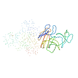
1GJ2
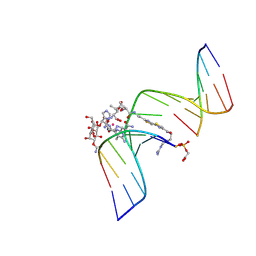 | | CO(III)-BLEOMYCIN-OOH BOUND TO AN OLIGONUCLEOTIDE CONTAINING A PHOSPHOGLYCOLATE LESION | | 分子名称: | 2-PHOSPHOGLYCOLIC ACID, 5'-D(*CP*CP*AP*AP*AP*G)-3', 5'-D(*CP*CP*CP*AP*GP*TP*AP*CP*TP*TP*TP*GP*G)-3', ... | | 著者 | Hoehn, S.T, Junker, H.-D, Bunt, R.C, Turner, C.J, Stubbe, J. | | 登録日 | 2000-11-01 | | 公開日 | 2001-06-06 | | 最終更新日 | 2023-12-27 | | 実験手法 | SOLUTION NMR | | 主引用文献 | Solution structure of Co(III)-bleomycin-OOH bound to a phosphoglycolate lesion containing oligonucleotide: implications for bleomycin-induced double-strand DNA cleavage.
Biochemistry, 40, 2001
|
|
1G5L
 | | CO(III)-BLEOMYCIN-OOH BOUND TO AN OLIGONUCLEOTIDE CONTAINING A PHOSPHOGLYCOLATE LESION | | 分子名称: | 2-PHOSPHOGLYCOLIC ACID, 5'-D(*CP*CP*AP*AP*AP*G)-3', 5'-D(*CP*CP*CP*AP*GP*TP*AP*CP*TP*TP*TP*GP*G)-3', ... | | 著者 | Hoehn, S.T, Junker, H.-D, Bunt, R.C, Turner, C.J, Stubbe, J. | | 登録日 | 2000-11-01 | | 公開日 | 2001-06-06 | | 最終更新日 | 2022-02-23 | | 実験手法 | SOLUTION NMR | | 主引用文献 | Solution structure of Co(III)-bleomycin-OOH bound to a phosphoglycolate lesion containing oligonucleotide: implications for bleomycin-induced double-strand DNA cleavage.
Biochemistry, 40, 2001
|
|


