Loading
PDBj
MenuPDBj@FacebookPDBj@TwitterPDBj@YouTubewwPDB FoundationwwPDB
RCSB PDBPDBeBMRBAdv. SearchSearch help
Search by PDB author
3EDD
DownloadVisualize
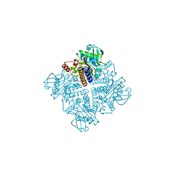
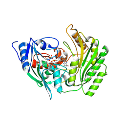
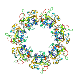
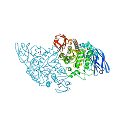
BU of 3edd by Molmil
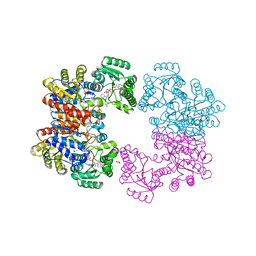
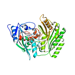
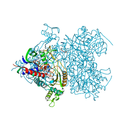
Structural base for cyclodextrin hydrolysis
Descriptor: CALCIUM ION, Cyclohexakis-(1-4)-(alpha-D-glucopyranose), Cyclomaltodextrinase
Authors:Buedenbender, S, Schulz, G.E.
Deposit date:2008-09-03
Release date:2009-03-03
Last modified:2021-11-10
Method:X-RAY DIFFRACTION (2.65 Å)
Cite:Structural base for enzymatic cyclodextrin hydrolysis
J.Mol.Biol., 385, 2009
2V7G
DownloadVisualize
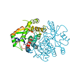
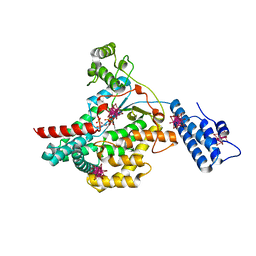
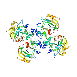
BU of 2v7g by Molmil
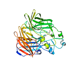
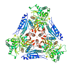
Crystal Structure of an Engineered Urocanase Tetramer
Descriptor: ACETATE ION, GLYCEROL, NICOTINAMIDE-ADENINE-DINUCLEOTIDE, ...
Authors:Treiber, N, Schulz, G.E.
Deposit date:2007-07-30
Release date:2008-01-15
Last modified:2023-12-13
Method:X-RAY DIFFRACTION (2 Å)
Cite:Designed Protein-Protein Association.
Science, 319, 2008
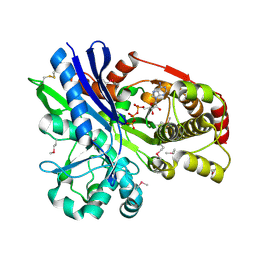
2PBG
DownloadVisualize
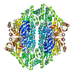
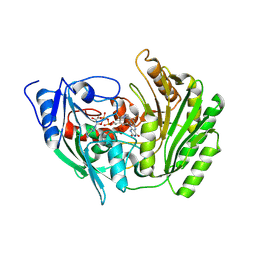
BU of 2pbg by Molmil
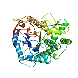
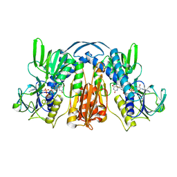
6-PHOSPHO-BETA-D-GALACTOSIDASE FORM-B
Descriptor: 6-PHOSPHO-BETA-D-GALACTOSIDASE, SULFATE ION
Authors:Wiesmann, C, Schulz, G.E.
Deposit date:1997-02-21
Release date:1997-07-23
Last modified:2023-08-09
Method:X-RAY DIFFRACTION (2.5 Å)
Cite:Crystal structures and mechanism of 6-phospho-beta-galactosidase from Lactococcus lactis.
J.Mol.Biol., 269, 1997
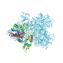
2UYT
DownloadVisualize
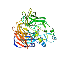
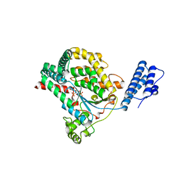
BU of 2uyt by Molmil
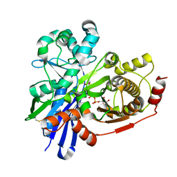
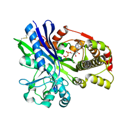
Structure of L-rhamnulose kinase in complex with ADP and beta-L- rhamnulose.
Descriptor: 6-deoxy-beta-L-fructofuranose, ADENOSINE-5'-DIPHOSPHATE, RHAMNULOKINASE
Authors:Grueninger, D, Schulz, G.E.
Deposit date:2007-04-13
Release date:2007-06-26
Last modified:2023-12-13
Method:X-RAY DIFFRACTION (1.55 Å)
Cite:Substrate Spectrum of L-Rhamnulose Kinase Related to Models Derived from Two Ternary Complex Structures.
FEBS Lett., 581, 2007
2V2A
DownloadVisualize
BU of 2v2a by Molmil
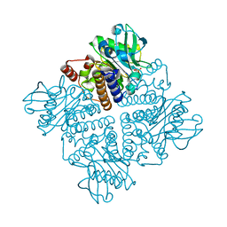
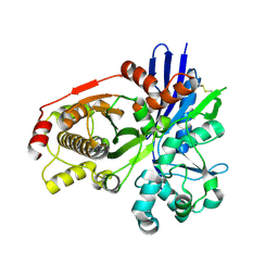
L-RHAMNULOSE-1-PHOSPHATE ALDOLASE FROM ESCHERICHIA COLI (MUTANT E192A- K248G-R253A-E254A)
Descriptor: 1,3-DIHYDROXYACETONEPHOSPHATE, RHAMNULOSE-1-PHOSPHATE ALDOLASE, ZINC ION
Authors:Grueninger, D, Schulz, G.E.
Deposit date:2007-06-04
Release date:2008-01-08
Last modified:2023-12-13
Method:X-RAY DIFFRACTION (1.75 Å)
Cite:Antenna Domain Mobility and Enzymatic Reaction of L-Rhamnulose-1-Phosphate Aldolase.
Biochemistry, 47, 2008
2V29
DownloadVisualize
BU of 2v29 by Molmil
L-RHAMNULOSE-1-PHOSPHATE ALDOLASE FROM ESCHERICHIA COLI (MUTANT K15W)
Descriptor: 1,2-ETHANEDIOL, ACETATE ION, RHAMNULOSE-1-PHOSPHATE ALDOLASE, ...
Authors:Grueninger, D, Schulz, G.E.
Deposit date:2007-06-04
Release date:2008-01-08
Last modified:2023-12-13
Method:X-RAY DIFFRACTION (2 Å)
Cite:Antenna Domain Mobility and Enzymatic Reaction of L-Rhamnulose-1-Phosphate Aldolase.
Biochemistry, 47, 2008
2VTK
DownloadVisualize
BU of 2vtk by Molmil
THYMIDINE KINASE FROM HERPES SIMPLEX VIRUS TYPE 1 IN COMPLEX WITH ADP AND DEOXYTHYMIDINE
Descriptor: ADENOSINE-5'-DIPHOSPHATE, THYMIDINE, THYMIDINE KINASE
Authors:Wild, K, Schulz, G.E.
Deposit date:1997-04-01
Release date:1997-10-22
Last modified:2024-02-21
Method:X-RAY DIFFRACTION (2.8 Å)
Cite:The structures of thymidine kinase from herpes simplex virus type 1 in complex with substrates and a substrate analogue.
Protein Sci., 6, 1997
2AG0
DownloadVisualize
BU of 2ag0 by Molmil
Crystal structure of Benzaldehyde lyase (BAL)- native
Descriptor: MAGNESIUM ION, THIAMINE DIPHOSPHATE, benzaldehyde lyase
Authors:Mosbacher, T.G, Mueller, M, Schulz, G.E.
Deposit date:2005-07-26
Release date:2006-01-24
Last modified:2024-03-13
Method:X-RAY DIFFRACTION (2.58 Å)
Cite:Structure and mechanism of the ThDP-dependent benzaldehyde lyase from Pseudomonas fluorescens
Febs J., 272, 2005
2BIX
DownloadVisualize
BU of 2bix by Molmil
Crystal structure of apocarotenoid cleavage oxygenase from Synechocystis, Fe-free apoenzyme
Descriptor: (HYDROXYETHYLOXY)TRI(ETHYLOXY)OCTANE, APOCAROTENOID-CLEAVING OXYGENASE, GLYCEROL
Authors:Kloer, D.P, Ruch, S, Al-Babili, S, Beyer, P, Schulz, G.E.
Deposit date:2005-01-26
Release date:2005-04-14
Last modified:2024-05-08
Method:X-RAY DIFFRACTION (2.68 Å)
Cite:The Structure of a Retinal-Forming Carotenoid Oxygenase
Science, 308, 2005
2BVF
DownloadVisualize
BU of 2bvf by Molmil
Crystal structure of 6-hydoxy-D-nicotine oxidase from Arthrobacter nicotinovorans. Crystal Form 3 (P1)
Descriptor: 6-HYDROXY-D-NICOTINE OXIDASE, FLAVIN-ADENINE DINUCLEOTIDE
Authors:Koetter, J.W.A, Schulz, G.E.
Deposit date:2005-06-28
Release date:2005-08-17
Last modified:2011-07-13
Method:X-RAY DIFFRACTION (1.92 Å)
Cite:Crystal Structure of 6-Hydroxy-D-Nicotine Oxidase from Arthrobacter Nicotinovorans.
J.Mol.Biol., 352, 2005
2BVL
DownloadVisualize
BU of 2bvl by Molmil
Crystal structure of the catalytic domain of toxin B from Clostridium difficile in complex with UDP, Glc and manganese ion
Descriptor: HEXATANTALUM DODECABROMIDE, MANGANESE (II) ION, SULFATE ION, ...
Authors:Reinert, D.J, Jank, T, Aktories, K, Schulz, G.E.
Deposit date:2005-06-30
Release date:2005-08-03
Last modified:2024-05-08
Method:X-RAY DIFFRACTION (2.2 Å)
Cite:Structural Basis for the Function of Clostridium Difficile Toxin B.
J.Mol.Biol., 351, 2005
2BVH
DownloadVisualize
BU of 2bvh by Molmil
Crystal structure of 6-hydoxy-D-nicotine oxidase from Arthrobacter nicotinovorans. Crystal Form 2 (P21)
Descriptor: 6-HYDROXY-D-NICOTINE OXIDASE, FLAVIN-ADENINE DINUCLEOTIDE
Authors:Koetter, J.W.A, Schulz, G.E.
Deposit date:2005-06-28
Release date:2005-08-17
Last modified:2011-07-13
Method:X-RAY DIFFRACTION (2.9 Å)
Cite:Crystal Structure of 6-Hydroxy-D-Nicotine Oxidase from Arthrobacter Nicotinovorans.
J.Mol.Biol., 352, 2005
2BVM
DownloadVisualize
BU of 2bvm by Molmil
Crystal structure of the catalytic domain of toxin B from Clostridium difficile in complex with UDP, Glc and manganese ion
Descriptor: MANGANESE (II) ION, SULFATE ION, TOXIN B, ...
Authors:Reinert, D.J, Jank, T, Aktories, K, Schulz, G.E.
Deposit date:2005-06-30
Release date:2005-08-03
Last modified:2023-12-13
Method:X-RAY DIFFRACTION (2.55 Å)
Cite:Structural Basis for the Function of Clostridium Difficile Toxin B.
J.Mol.Biol., 351, 2005
2BVG
DownloadVisualize
BU of 2bvg by Molmil
Crystal structure of 6-hydoxy-D-nicotine oxidase from Arthrobacter nicotinovorans. Crystal Form 1 (P21)
Descriptor: 6-HYDROXY-D-NICOTINE OXIDASE, FLAVIN-ADENINE DINUCLEOTIDE
Authors:Koetter, J.W.A, Schulz, G.E.
Deposit date:2005-06-28
Release date:2005-08-17
Last modified:2011-07-13
Method:X-RAY DIFFRACTION (3.18 Å)
Cite:Crystal Structure of 6-Hydroxy-D-Nicotine Oxidase from Arthrobacter Nicotinovorans.
J.Mol.Biol., 352, 2005
2BIW
DownloadVisualize
BU of 2biw by Molmil
Crystal structure of apocarotenoid cleavage oxygenase from Synechocystis, native enzyme
Descriptor: (3R)-3-HYDROXY-8'-APOCAROTENOL, APOCAROTENOID-CLEAVING OXYGENASE, FE (III) ION
Authors:Kloer, D.P, Ruch, S, Al-Babili, S, Beyer, P, Schulz, G.E.
Deposit date:2005-01-26
Release date:2005-04-14
Last modified:2024-05-01
Method:X-RAY DIFFRACTION (2.39 Å)
Cite:The Structure of a Retinal-Forming Carotenoid Oxygenase
Science, 308, 2005
2CGJ
DownloadVisualize
BU of 2cgj by Molmil
Crystal Structure of L-rhamnulose kinase from Escherichia coli in complex with L-fructose and ADP.
Descriptor: ADENOSINE-5'-DIPHOSPHATE, L-RHAMNULOSE KINASE, beta-L-fructofuranose
Authors:Grueninger, D, Schulz, G.E.
Deposit date:2006-03-09
Release date:2006-05-31
Last modified:2020-07-29
Method:X-RAY DIFFRACTION (2.26 Å)
Cite:Structure and Reaction Mechanism of L-Rhamnulose Kinase from Escherichia Coli.
J.Mol.Biol., 359, 2006
2CGK
DownloadVisualize
BU of 2cgk by Molmil
Crystal Structure of L-rhamnulose kinase from Escherichia coli in an open uncomplexed conformation.
Descriptor: L-RHAMNULOSE KINASE
Authors:Grueninger, D, Schulz, G.E.
Deposit date:2006-03-09
Release date:2006-05-31
Last modified:2023-12-13
Method:X-RAY DIFFRACTION (2.46 Å)
Cite:Structure and Reaction Mechanism of L-Rhamnulose Kinase from Escherichia Coli.
J.Mol.Biol., 359, 2006
2CB4
DownloadVisualize
BU of 2cb4 by Molmil
Crystal structure of the catalytic domain of the mosquitocidal toxin from Bacillus sphaericus, mutant E197Q
Descriptor: MOSQUITOCIDAL TOXIN
Authors:Reinert, D.J, Carpusca, I, Aktories, K, Schulz, G.E.
Deposit date:2005-12-29
Release date:2006-02-22
Last modified:2011-07-13
Method:X-RAY DIFFRACTION (2.5 Å)
Cite:Structure of the Mosquitocidal Toxin from Bacillus Sphaericus.
J.Mol.Biol., 357, 2006
2CGL
DownloadVisualize
BU of 2cgl by Molmil
Crystal Structure of L-rhamnulose kinase from Escherichia coli in complex with L-fructose, ADP and a modeled ATP gamma phosphate.
Descriptor: ADENOSINE-5'-DIPHOSPHATE, PHOSPHITE ION, RHAMNULOKINASE, ...
Authors:Grueninger, D, Schulz, G.E.
Deposit date:2006-03-09
Release date:2006-05-31
Last modified:2020-07-29
Method:X-RAY DIFFRACTION (1.88 Å)
Cite:Structure and Reaction Mechanism of L-Rhamnulose Kinase from Escherichia Coli.
J.Mol.Biol., 359, 2006
2CB6
DownloadVisualize
BU of 2cb6 by Molmil
Crystal structure of the catalytic domain of the mosquitocidal toxin from Bacillus sphaericus, mutant E195Q
Descriptor: MOSQUITOCIDAL TOXIN
Authors:Reinert, D.J, Carpusca, I, Aktories, K, Schulz, G.E.
Deposit date:2005-12-29
Release date:2006-02-22
Last modified:2023-12-13
Method:X-RAY DIFFRACTION (3 Å)
Cite:Structure of the Mosquitocidal Toxin from Bacillus Sphaericus.
J.Mol.Biol., 357, 2006
2F6C
DownloadVisualize
BU of 2f6c by Molmil
Reaction geometry and thermostability of pyranose 2-oxidase from the white-rot fungus Peniophora sp., Thermostability mutant E542K
Descriptor: DI(HYDROXYETHYL)ETHER, FLAVIN-ADENINE DINUCLEOTIDE, Pyranose 2-oxidase, ...
Authors:Bannwarth, M, Heckmann-Pohl, D.M, Bastian, S, Giffhorn, F, Schulz, G.E.
Deposit date:2005-11-29
Release date:2006-06-13
Last modified:2021-10-20
Method:X-RAY DIFFRACTION (1.84 Å)
Cite:Reaction Geometry and Thermostable Variant of Pyranose 2-Oxidase from the White-Rot Fungus Peniophora sp.
Biochemistry, 45, 2006
2F5V
DownloadVisualize
BU of 2f5v by Molmil
Reaction geometry and thermostability mutant of pyranose 2-oxidase from the white-rot fungus Peniophora sp.
Descriptor: DI(HYDROXYETHYL)ETHER, FLAVIN-ADENINE DINUCLEOTIDE, Pyranose 2-oxidase, ...
Authors:Bannwarth, M, Bastian, S, Heckmann-Pohl, D, Giffhorn, F, Schulz, G.E.
Deposit date:2005-11-28
Release date:2006-06-13
Last modified:2020-07-29
Method:X-RAY DIFFRACTION (1.41 Å)
Cite:Reaction Geometry and Thermostable Variant of Pyranose 2-Oxidase from the White-Rot Fungus Peniophora sp.
Biochemistry, 45, 2006
1FUI
DownloadVisualize
BU of 1fui by Molmil
L-FUCOSE ISOMERASE FROM ESCHERICHIA COLI
Descriptor: FUCITOL, L-FUCOSE ISOMERASE, MANGANESE (II) ION, ...
Authors:Seemann, J.E, Schulz, G.E.
Deposit date:1997-04-14
Release date:1997-10-15
Last modified:2024-02-07
Method:X-RAY DIFFRACTION (2.5 Å)
Cite:Structure and mechanism of L-fucose isomerase from Escherichia coli.
J.Mol.Biol., 273, 1997
1GER
DownloadVisualize
BU of 1ger by Molmil
THE STRUCTURE OF GLUTATHIONE REDUCTASE FROM ESCHERICHIA COLI AT 1.86 ANGSTROMS RESOLUTION: COMPARISON WITH THE ENZYME FROM HUMAN ERYTHROCYTES
Descriptor: FLAVIN-ADENINE DINUCLEOTIDE, GLUTATHIONE REDUCTASE
Authors:Mittl, P.R.E, Schulz, G.E.
Deposit date:1994-01-18
Release date:1994-11-01
Last modified:2011-07-13
Method:X-RAY DIFFRACTION (1.86 Å)
Cite:Structure of glutathione reductase from Escherichia coli at 1.86 A resolution: comparison with the enzyme from human erythrocytes.
Protein Sci., 3, 1994
1GXY
DownloadVisualize
BU of 1gxy by Molmil
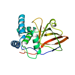
crystal structure of the eucaryotic mono-ADP-ribosyltransferase ART2.2; CRYSTAL FORM A (P21)
Descriptor: GLYCEROL, T-CELL ECTO-ADP-RIBOSYLTRANSFERASE 2
Authors:Mueller-Dieckmann, C, Schulz, G.E.
Deposit date:2002-04-15
Release date:2002-09-26
Last modified:2019-07-24
Method:X-RAY DIFFRACTION (1.71 Å)
Cite:Structure of the Ecto-Adp-Ribosyl Transferase Art2.2 From Rat
J.Mol.Biol., 322, 2002

219869

數據於2024-05-15公開中

PDB statisticsPDBj update infoContact PDBjnumon