3BW6
 
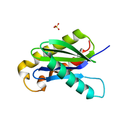 | | Crystal structure of the longin domain of yeast Ykt6 | | 分子名称: | SULFATE ION, Synaptobrevin homolog YKT6 | | 著者 | Pylypenko, O, Schonichen, A, Ludwig, D, Ungermann, C, Goody, R.S, Rak, A, Geyer, M. | | 登録日 | 2008-01-08 | | 公開日 | 2008-04-01 | | 最終更新日 | 2024-02-21 | | 実験手法 | X-RAY DIFFRACTION (2.5 Å) | | 主引用文献 | Farnesylation of the SNARE protein Ykt6 increases its stability and helical folding.
J.Mol.Biol., 377, 2008
|
|
3DAD
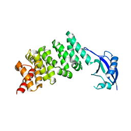 | | Crystal structure of the N-terminal regulatory domains of the formin FHOD1 | | 分子名称: | FH1/FH2 domain-containing protein 1 | | 著者 | Schulte, A, Stolp, B, Schonichen, A, Pylypenko, O, Rak, A, Fackler, O.T, Geyer, M. | | 登録日 | 2008-05-29 | | 公開日 | 2008-09-16 | | 最終更新日 | 2024-02-21 | | 実験手法 | X-RAY DIFFRACTION (2.3 Å) | | 主引用文献 | The Human Formin FHOD1 Contains a Bipartite Structure of FH3 and GTPase-Binding Domains Required for Activation.
Structure, 16, 2008
|
|


