1S2J
 
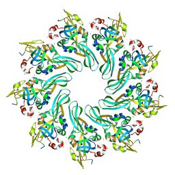
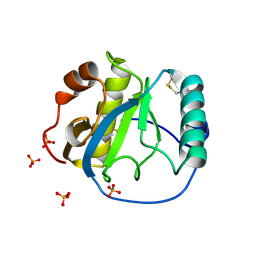 | | Crystal structure of the Drosophila pattern-recognition receptor PGRP-SA | | 分子名称: | PHOSPHATE ION, Peptidoglycan recognition protein SA CG11709-PA | | 著者 | Chang, C.-I, Pili-Floury, S, Chelliah, Y, Lemaitre, B, Mengin-Lecreulx, D, Deisenhofer, J. | | 登録日 | 2004-01-08 | | 公開日 | 2004-09-14 | | 最終更新日 | 2023-08-23 | | 実験手法 | X-RAY DIFFRACTION (2.2 Å) | | 主引用文献 | A Drosophila pattern recognition receptor contains a peptidoglycan docking groove and unusual l,d-carboxypeptidase activity.
PLOS BIOL., 2, 2004
|
|
2W3Y
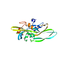
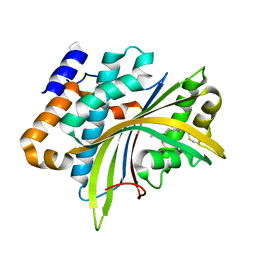 | | Structure of the Evf virulence factor | | 分子名称: | PALMITIC ACID, VIRULENCE FACTOR | | 著者 | Quevillon-Cheruel, S, Leulliot, N, Acosta Muniz C, C, Vincent, M, Gallay, J, Argentini, M, Cornu, D, Boccard, F, Lemaitre, B, van Tilbeurgh, H. | | 登録日 | 2008-11-17 | | 公開日 | 2008-12-30 | | 最終更新日 | 2011-07-13 | | 実験手法 | X-RAY DIFFRACTION (2 Å) | | 主引用文献 | Evf, a Virulence Factor Produced by the Drosophila Pathogen Erwinia Carotovora is a S-Palmitoylated Protein with a New Fold that Binds to Lipid Vesicles.
J.Biol.Chem., 284, 2009
|
|
8PBV
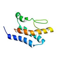 | |
8SMQ
 | | Crystal Structure of the N-terminal Domain of the Cryptic Surface Protein (CD630_25440) from Clostridium difficile. | | 分子名称: | 1,2-ETHANEDIOL, CHLORIDE ION, GLYCEROL, ... | | 著者 | Minasov, G, Shuvalova, L, Brunzelle, J.S, Kiryukhina, O, Wawrzak, Z, Satchell, K.J.F, Center for Structural Biology of Infectious Diseases (CSBID), Center for Structural Genomics of Infectious Diseases (CSGID) | | 登録日 | 2023-04-26 | | 公開日 | 2023-05-10 | | 最終更新日 | 2023-12-06 | | 実験手法 | X-RAY DIFFRACTION (2 Å) | | 主引用文献 | Protein target highlights in CASP15: Analysis of models by structure providers.
Proteins, 91, 2023
|
|
4MKQ
 | |
4MJT
 | |
4MKO
 | |


