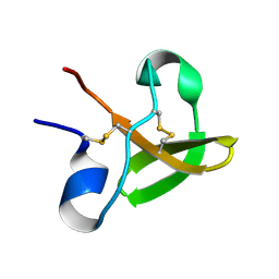
2XBD
 
 | | INTERNAL XYLAN BINDING DOMAIN FROM CELLULOMONAS FIMI XYLANASE D, NMR, MINIMIZED AVERAGE STRUCTURE | | 分子名称: | XYLANASE D | | 著者 | Simpson, P.J, Bolam, D.N, Cooper, A, Ciruela, A, Hazlewood, G.P, Gilbert, H.J, Williamson, M.P. | | 登録日 | 1998-10-27 | | 公開日 | 1999-07-21 | | 最終更新日 | 2022-03-16 | | 実験手法 | SOLUTION NMR | | 主引用文献 | A family IIb xylan-binding domain has a similar secondary structure to a homologous family IIa cellulose-binding domain but different ligand specificity.
Structure Fold.Des., 7, 1999
|
|
1XBD
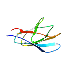 | | INTERNAL XYLAN BINDING DOMAIN FROM CELLULOMONAS FIMI XYLANASE D, NMR, 5 STRUCTURES | | 分子名称: | XYLANASE D | | 著者 | Simpson, P.J, Bolam, D.N, Cooper, A, Ciruela, A, Hazlewood, G.P, Gilbert, H.J, Williamson, M.P. | | 登録日 | 1998-10-16 | | 公開日 | 1999-07-21 | | 最終更新日 | 2022-03-02 | | 実験手法 | SOLUTION NMR | | 主引用文献 | A family IIb xylan-binding domain has a similar secondary structure to a homologous family IIa cellulose-binding domain but different ligand specificity.
Structure Fold.Des., 7, 1999
|
|
1E8P
 | | Characterisation of the cellulose docking domain from Piromyces equi | | 分子名称: | Endoglucanase 45A | | 著者 | Raghothama, S, Eberhardt, R.Y, White, P, Hazlewood, G.P, Gilbert, H.J, Simpson, P.J, Williamson, M.P. | | 登録日 | 2000-09-28 | | 公開日 | 2001-09-07 | | 最終更新日 | 2018-06-20 | | 実験手法 | SOLUTION NMR | | 主引用文献 | Characterization of a cellulosome dockerin domain from the anaerobic fungus Piromyces equi.
Nat. Struct. Biol., 8, 2001
|
|
1E8Q
 | | Characterisation of the cellulose docking domain from Piromyces equi | | 分子名称: | Endoglucanase 45A | | 著者 | Raghothama, S, Eberhardt, R.Y, White, P, Hazlewood, G.P, Gilbert, H.J, Simpson, P.J, Williamson, M.P. | | 登録日 | 2000-09-28 | | 公開日 | 2001-09-07 | | 最終更新日 | 2018-06-20 | | 実験手法 | SOLUTION NMR | | 主引用文献 | Characterization of a cellulosome dockerin domain from the anaerobic fungus Piromyces equi.
Nat. Struct. Biol., 8, 2001
|
|
1XYS
 | | CATALYTIC CORE OF XYLANASE A E246C MUTANT | | 分子名称: | CALCIUM ION, XYLANASE A | | 著者 | Harris, G.W, Jenkins, J.A, Connerton, I, Pickersgill, R.W. | | 登録日 | 1994-09-02 | | 公開日 | 1995-07-10 | | 最終更新日 | 2024-02-14 | | 実験手法 | X-RAY DIFFRACTION (2.5 Å) | | 主引用文献 | Structure of the catalytic core of the family F xylanase from Pseudomonas fluorescens and identification of the xylopentaose-binding sites.
Structure, 2, 1994
|
|
1CLX
 | | CATALYTIC CORE OF XYLANASE A | | 分子名称: | CALCIUM ION, XYLANASE A | | 著者 | Harris, G.W, Jenkins, J.A, Connerton, I, Pickersgill, R.W. | | 登録日 | 1995-08-31 | | 公開日 | 1996-06-20 | | 最終更新日 | 2011-07-13 | | 実験手法 | X-RAY DIFFRACTION (1.8 Å) | | 主引用文献 | Refined crystal structure of the catalytic domain of xylanase A from Pseudomonas fluorescens at 1.8 A resolution.
Acta Crystallogr.,Sect.D, 52, 1996
|
|
1E5N
 | | E246C mutant of P fluorescens subsp. cellulosa xylanase A in complex with xylopentaose | | 分子名称: | CALCIUM ION, ENDO-1,4-BETA-XYLANASE A, beta-D-xylopyranose-(1-4)-beta-D-xylopyranose-(1-4)-beta-D-xylopyranose-(1-4)-beta-D-xylopyranose-(1-4)-beta-D-xylopyranose | | 著者 | Lo Leggio, L, Jenkins, J.A, Harris, G.W, Pickersgill, R.W. | | 登録日 | 2000-07-27 | | 公開日 | 2000-12-08 | | 最終更新日 | 2023-12-13 | | 実験手法 | X-RAY DIFFRACTION (3.2 Å) | | 主引用文献 | X-ray crystallographic study of xylopentaose binding to Pseudomonas fluorescens xylanase A.
Proteins, 41, 2000
|
|


