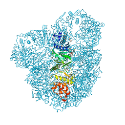
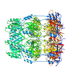
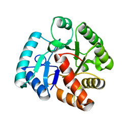
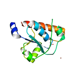
4UW0
 
 | |
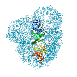
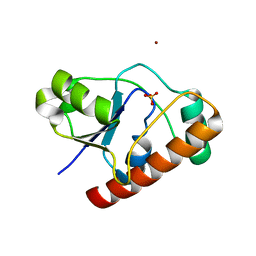
5FH9
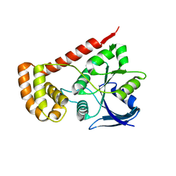 | |
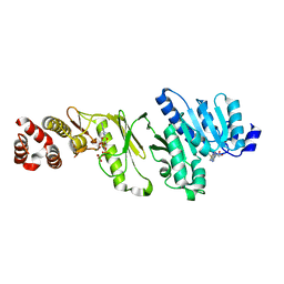
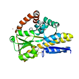
2CG3
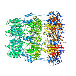
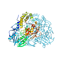 | | Crystal structure of SdsA1, an alkylsulfatase from Pseudomonas aeruginosa. | | 分子名称: | SDSA1, ZINC ION | | 著者 | Hagelueken, G, Adams, T.M, Wiehlmann, L, Widow, U, Kolmar, H, Tuemmler, B, Heinz, D.W, Schubert, W.-D. | | 登録日 | 2006-02-27 | | 公開日 | 2006-04-26 | | 最終更新日 | 2019-07-24 | | 実験手法 | X-RAY DIFFRACTION (2.6 Å) | | 主引用文献 | The Crystal Structure of Sdsa1, an Alkylsulfatase from Pseudomonas Aeruginosa, Defines a Third Third Class of Sulfatases
Proc.Natl.Acad.Sci.USA, 103, 2006
|
|
2CG2
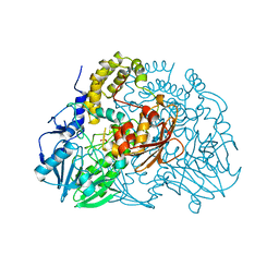 | | Crystal structure of SdsA1, an alkylsulfatase from Pseudomonas aeruginosa, in complex with sulfate | | 分子名称: | SDSA1, SULFATE ION, ZINC ION | | 著者 | Hagelueken, G, Adams, T.M, Wiehlmann, L, Widow, U, Kolmar, H, Tuemmler, B, Heinz, D.W, Schubert, W.-D. | | 登録日 | 2006-02-27 | | 公開日 | 2006-04-26 | | 最終更新日 | 2023-12-13 | | 実験手法 | X-RAY DIFFRACTION (2.1 Å) | | 主引用文献 | The Crystal Structure of Sdsa1, an Alkylsulfatase from Pseudomonas Aeruginosa, Defines a Third Class of Sulfatases.
Proc.Natl.Acad.Sci.USA, 103, 2006
|
|
2CFZ
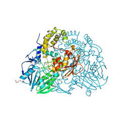 | | Crystal structure of SdsA1, an alkylsulfatase from Pseudomonas aeruginosa, in complex with 1-dodecanol | | 分子名称: | 1-DODECANOL, DI(HYDROXYETHYL)ETHER, SDS HYDROLASE SDSA1, ... | | 著者 | Hagelueken, G, Adams, T.M, Wiehlmann, L, Widow, U, Kolmar, H, Tuemmler, B, Heinz, D.W, Schubert, W.-D. | | 登録日 | 2006-02-26 | | 公開日 | 2006-04-26 | | 最終更新日 | 2023-12-13 | | 実験手法 | X-RAY DIFFRACTION (2.05 Å) | | 主引用文献 | The Crystal Structure of Sdsa1, an Alkylsulfatase from Pseudomonas Aeruginosa, Defines a Third Class of Sulfatases.
Proc.Natl.Acad.Sci.USA, 103, 2006
|
|
2CFU
 | | Crystal structure of SdsA1, an alkylsulfatase from Pseudomonas aeruginosa, in complex with 1-decane-sulfonic-acid. | | 分子名称: | 1-DECANE-SULFONIC-ACID, DI(HYDROXYETHYL)ETHER, ISOPROPYL ALCOHOL, ... | | 著者 | Hagelueken, G, Adams, T.M, Wiehlmann, L, Widow, U, Kolmar, H, Tuemmler, B, Heinz, D.W, Schubert, W.-D. | | 登録日 | 2006-02-23 | | 公開日 | 2006-04-26 | | 最終更新日 | 2023-12-13 | | 実験手法 | X-RAY DIFFRACTION (1.9 Å) | | 主引用文献 | The Crystal Structure of Sdsa1, an Alkylsulfatase from Pseudomonas Aeruginosa, Defines a Third Class of Sulfatases.
Proc.Natl.Acad.Sci.USA, 103, 2006
|
|
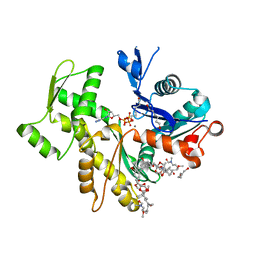
4AZS
 | | High resolution (2.2 A) crystal structure of WbdD. | | 分子名称: | ADENOSINE MONOPHOSPHATE, CHLORIDE ION, METHYLTRANSFERASE WBDD, ... | | 著者 | Hagelueken, G, Huang, H, Naismith, J.H. | | 登録日 | 2012-06-26 | | 公開日 | 2012-09-26 | | 最終更新日 | 2012-11-14 | | 実験手法 | X-RAY DIFFRACTION (2.15 Å) | | 主引用文献 | Structure of Wbdd; a Bifunctional Kinase and Methyltransferase that Regulates the Chain Length of the O Antigen in Escherichia Coli O9A.
Mol.Microbiol., 86, 2012
|
|
4AZT
 | | Co-crystal structure of WbdD and kinase inhibitor LY294002. | | 分子名称: | 2-MORPHOLIN-4-YL-7-PHENYL-4H-CHROMEN-4-ONE, CHLORIDE ION, METHYLTRANSFERASE WBDD, ... | | 著者 | Hagelueken, G, Huang, H, Naismith, J.H. | | 登録日 | 2012-06-26 | | 公開日 | 2012-09-26 | | 最終更新日 | 2012-11-14 | | 実験手法 | X-RAY DIFFRACTION (2.34 Å) | | 主引用文献 | Structure of Wbdd; a Bifunctional Kinase and Methyltransferase that Regulates the Chain Length of the O Antigen in Escherichia Coli O9A.
Mol.Microbiol., 86, 2012
|
|
4AZW
 | | Crystal structure of monomeric WbdD. | | 分子名称: | ADENOSINE-5'-TRIPHOSPHATE, MAGNESIUM ION, S-ADENOSYLMETHIONINE, ... | | 著者 | Hagelueken, G, Huang, H, Naismith, J.H. | | 登録日 | 2012-06-26 | | 公開日 | 2012-09-26 | | 最終更新日 | 2012-11-14 | | 実験手法 | X-RAY DIFFRACTION (2.47 Å) | | 主引用文献 | Structure of Wbdd; a Bifunctional Kinase and Methyltransferase that Regulates the Chain Length of the O Antigen in Escherichia Coli O9A.
Mol.Microbiol., 86, 2012
|
|
4AX8
 | | Medium resolution structure of the bifunctional kinase- methyltransferase WbdD | | 分子名称: | ADENOSINE-5'-DIPHOSPHATE, CHLORIDE ION, S-ADENOSYLMETHIONINE, ... | | 著者 | Hagelueken, G, Huang, H, Naismith, J.H. | | 登録日 | 2012-06-11 | | 公開日 | 2012-10-03 | | 実験手法 | X-RAY DIFFRACTION (3 Å) | | 主引用文献 | Crystallization, Dehydration and Experimental Phasing of Wbdd, a Bifunctional Kinase and Methyltransferase from Escherichia Coli O9A.
Acta Crystallogr.,Sect.D, 68, 2012
|
|
4AZV
 | | Co-crystal structure of WbdD and kinase inhibitor GW435821x. | | 分子名称: | CHLORIDE ION, S-ADENOSYLMETHIONINE, SULFATE ION, ... | | 著者 | Hagelueken, G, Huang, H, Naismith, J.H. | | 登録日 | 2012-06-26 | | 公開日 | 2012-09-26 | | 最終更新日 | 2013-02-06 | | 実験手法 | X-RAY DIFFRACTION (3.291 Å) | | 主引用文献 | Structure of Wbdd; a Bifunctional Kinase and Methyltransferase that Regulates the Chain Length of the O Antigen in Escherichia Coli O9A.
Mol.Microbiol., 86, 2012
|
|
2V3A
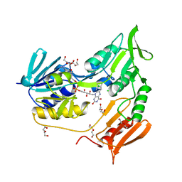 | | Crystal structure of rubredoxin reductase from Pseudomonas aeruginosa. | | 分子名称: | CHLORIDE ION, DI(HYDROXYETHYL)ETHER, FLAVIN-ADENINE DINUCLEOTIDE, ... | | 著者 | Hagelueken, G, Wiehlmann, L, Adams, T.M, Kolmar, H, Heinz, D.W, Tuemmler, B, Schubert, W.-D. | | 登録日 | 2007-06-14 | | 公開日 | 2007-08-14 | | 最終更新日 | 2023-12-13 | | 実験手法 | X-RAY DIFFRACTION (2.4 Å) | | 主引用文献 | Crystal Structure of the Electron Transfer Complex Rubredoxin - Rubredoxin Reductase from Pseudomonas Aeruginosa.
Proc.Natl.Acad.Sci.USA, 104, 2007
|
|
2V3B
 | | Crystal structure of the electron transfer complex rubredoxin - rubredoxin reductase from Pseudomonas aeruginosa. | | 分子名称: | FE (III) ION, FLAVIN-ADENINE DINUCLEOTIDE, RUBREDOXIN 2, ... | | 著者 | Hagelueken, G, Wiehlmann, L, Adams, T.M, Kolmar, H, Heinz, D.W, Tuemmler, B, Schubert, W.-D. | | 登録日 | 2007-06-14 | | 公開日 | 2007-08-14 | | 最終更新日 | 2023-12-13 | | 実験手法 | X-RAY DIFFRACTION (2.45 Å) | | 主引用文献 | Crystal Structure of the Electron Transfer Complex Rubredoxin - Rubredoxin Reductase from Pseudomonas Aeruginosa.
Proc.Natl.Acad.Sci.USA, 104, 2007
|
|
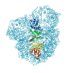
2W8I
 | | Crystal structure of Wza24-345. | | 分子名称: | PUTATIVE OUTER MEMBRANE LIPOPROTEIN WZA | | 著者 | Hagelueken, G, Ingledew, W.J, Huang, H, Petrovic-Stojanovska, B, Whitfield, C, ElMkami, H, Schiemann, O, Naismith, J.H. | | 登録日 | 2009-01-16 | | 公開日 | 2009-02-10 | | 最終更新日 | 2023-12-13 | | 実験手法 | X-RAY DIFFRACTION (3 Å) | | 主引用文献 | Peldor Distance Fingerprinting of the Octameric Outer-Membrane Protein Wza from Escherichia Coli.
Angew.Chem.Int.Ed.Engl., 48, 2009
|
|
2W8H
 | | Crystal structure of spin labeled Wza24-345. | | 分子名称: | CHLORIDE ION, PUTATIVE OUTER MEMBRANE LIPOPROTEIN WZA, S-[(1-oxyl-2,2,5,5-tetramethyl-2,5-dihydro-1H-pyrrol-3-yl)methyl] methanesulfonothioate | | 著者 | Hagelueken, G, Ingledew, W.J, Huang, H, Petrovic-Stojanovska, B, Whitfield, C, ElMkami, H, Schiemann, O, Naismith, J.H. | | 登録日 | 2009-01-16 | | 公開日 | 2009-02-10 | | 最終更新日 | 2023-12-13 | | 実験手法 | X-RAY DIFFRACTION (2.76 Å) | | 主引用文献 | Peldor Distance Fingerprinting of the Octameric Outer-Membrane Protein Wza from Escherichia Coli.
Angew.Chem.Int.Ed.Engl., 48, 2009
|
|
2VYP
 | | Rabbit-muscle G-actin in complex with myxobacterial rhizopodin | | 分子名称: | ACTIN, ALPHA SKELETAL MUSCLE, ADENOSINE-5'-TRIPHOSPHATE, ... | | 著者 | Hagelueken, G, Albrecht, S.C, Steinmetz, H, Jansen, R, Heinz, D.W, Kalesse, M, Schubert, W.-D. | | 登録日 | 2008-07-25 | | 公開日 | 2009-02-24 | | 最終更新日 | 2023-12-13 | | 実験手法 | X-RAY DIFFRACTION (2.35 Å) | | 主引用文献 | The Absolute Configuration of Rhizopodin and its Inhibition of Actin Polymerization by Dimerization.
Angew.Chem.Int.Ed.Engl., 48, 2009
|
|
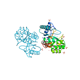
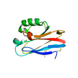
2WJF
 | | Crystal structure of the tyrosine phosphatase Cps4B from Steptococcus pneumoniae TIGR4 in complex with phosphate. | | 分子名称: | MANGANESE (II) ION, PHOSPHATE ION, TYROSINE-PROTEIN PHOSPHATASE CPSB | | 著者 | Hagelueken, G, Huang, H, Naismith, J.H. | | 登録日 | 2009-05-25 | | 公開日 | 2009-07-14 | | 最終更新日 | 2011-07-13 | | 実験手法 | X-RAY DIFFRACTION (2.22 Å) | | 主引用文献 | Crystal Structures of Wzb of Escherichia Coli and Cpsb of Streptococcus Pneumoniae, Representatives of Two Families of Tyrosine Phosphatases that Regulate Capsule Assembly.
J.Mol.Biol., 392, 2009
|
|
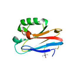
2WJD
 | | Crystal structure of the tyrosine phosphatase Cps4B from Steptococcus pneumoniae TIGR4. | | 分子名称: | MANGANESE (II) ION, SAMARIUM (III) ION, SULFATE ION, ... | | 著者 | Hagelueken, G, Huang, H, Naismith, J.H. | | 登録日 | 2009-05-25 | | 公開日 | 2009-07-14 | | 最終更新日 | 2017-07-12 | | 実験手法 | X-RAY DIFFRACTION (2.799 Å) | | 主引用文献 | Crystal Structures of Wzb of Escherichia Coli and Cpsb of Streptococcus Pneumoniae, Representatives of Two Families of Tyrosine Phosphatases that Regulate Capsule Assembly.
J.Mol.Biol., 392, 2009
|
|
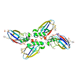
2WJE
 | |
5I26
 | | Azurin T30R1, crystal form I | | 分子名称: | Azurin, COPPER (II) ION | | 著者 | Hagelueken, G. | | 登録日 | 2016-02-08 | | 公開日 | 2016-04-13 | | 最終更新日 | 2024-01-10 | | 実験手法 | X-RAY DIFFRACTION (1.888 Å) | | 主引用文献 | Determination of nitroxide spin label conformations via PELDOR and X-ray crystallography.
Phys Chem Chem Phys, 18, 2016
|
|
5I28
 | | Azurin T30R1, crystal form II | | 分子名称: | Azurin, COPPER (II) ION, GLYCEROL | | 著者 | Hagelueken, G. | | 登録日 | 2016-02-08 | | 公開日 | 2016-04-13 | | 最終更新日 | 2024-01-10 | | 実験手法 | X-RAY DIFFRACTION (1.95 Å) | | 主引用文献 | Determination of nitroxide spin label conformations via PELDOR and X-ray crystallography.
Phys Chem Chem Phys, 18, 2016
|
|
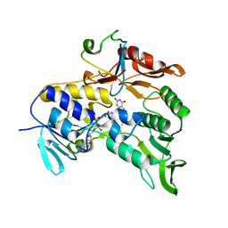
4BWW
 | | Crystal structure of spin labelled azurin T21R1. | | 分子名称: | AZURIN, COPPER (II) ION, GLYCEROL, ... | | 著者 | Hagelueken, G. | | 登録日 | 2013-07-04 | | 公開日 | 2014-06-11 | | 最終更新日 | 2023-12-20 | | 実験手法 | X-RAY DIFFRACTION (1.48 Å) | | 主引用文献 | High-Resolution Crystal Structure of Spin Labelled (T21R1) Azurin from Pseudomonas Aeruginosa: A Challenging Structural Benchmark for in Silico Spin Labelling Algorithms.
Bmc Struct.Biol., 14, 2014
|
|
2WJA
 | | Crystal structure of the tyrosine phosphatase Wzb from Escherichia coli K30 in complex with phosphate. | | 分子名称: | NICKEL (II) ION, PHOSPHATE ION, PUTATIVE ACID PHOSPHATASE WZB | | 著者 | Huang, H, Hagelueken, G, Whitfield, C, Naismith, J.H. | | 登録日 | 2009-05-25 | | 公開日 | 2009-07-14 | | 最終更新日 | 2023-12-13 | | 実験手法 | X-RAY DIFFRACTION (2.5 Å) | | 主引用文献 | Crystal Structures of Wzb of Escherichia Coli and Cpsb of Streptococcus Pneumoniae, Representatives of Two Families of Tyrosine Phosphatases that Regulate Capsule Assembly.
J.Mol.Biol., 392, 2009
|
|
2WMY
 | |
8CP7
 | |


