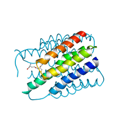
Loading
PDBj
メニューPDBj@FacebookPDBj@TwitterPDBj@YouTubewwPDB FoundationwwPDB
RCSB PDBPDBeBMRBAdv. SearchSearch help
Search by PDB author
2D57
DownloadVisualize
BU of 2d57 by Molmil
Double layered 2D crystal structure of AQUAPORIN-4 (AQP4M23) at 3.2 a resolution by electron crystallography
分子名称: Aquaporin-4
著者Hiroaki, Y, Tani, K, Kamegawa, A, Gyobu, N, Nishikawa, K, Suzuki, H, Walz, T, Sasaki, S, Mitsuoka, K, Kimura, K, Mizoguchi, A, Fujiyoshi, Y.
登録日2005-10-29
公開日2006-01-31
最終更新日2023-11-08
実験手法ELECTRON CRYSTALLOGRAPHY (3.2 Å)
主引用文献Implications of the Aquaporin-4 Structure on Array Formation and Cell Adhesion
J.Mol.Biol., 355, 2005
2H8A
DownloadVisualize
BU of 2h8a by Molmil
Structure of Microsomal Glutathione Transferase 1 in Complex with Glutathione
分子名称: GLUTATHIONE, Microsomal glutathione S-transferase 1
著者Hebert, H.
登録日2006-06-07
公開日2007-05-22
最終更新日2024-02-14
実験手法ELECTRON CRYSTALLOGRAPHY (3.2 Å)
主引用文献Structural basis for detoxification and oxidative stress protection in membranes.
J.Mol.Biol., 360, 2006
3DWW
DownloadVisualize
BU of 3dww by Molmil
Electron crystallographic structure of human microsomal prostaglandin E synthase 1
分子名称: GLUTATHIONE, Prostaglandin E synthase
著者Hebert, H, Jegerschold, C.
登録日2008-07-23
公開日2009-07-28
最終更新日2024-03-20
実験手法ELECTRON CRYSTALLOGRAPHY (3.5 Å)
主引用文献Structural basis for induced formation of the inflammatory mediator prostaglandin E2
Proc.Natl.Acad.Sci.USA, 105, 2008

225158

件を2024-09-18に公開中

PDB statisticsPDBj update infoContact PDBjnumon