Loading
PDBj
MenuPDBj@FacebookPDBj@TwitterPDBj@YouTubewwPDB FoundationwwPDB
RCSB PDBPDBeBMRBAdv. SearchSearch help
Search by PDB author
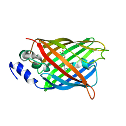
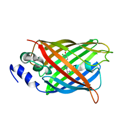
5HDS
DownloadVisualize
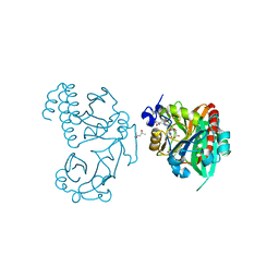
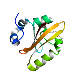
BU of 5hds by Molmil
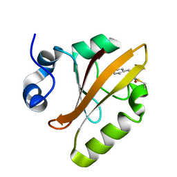
Femtosecond Structural Dynamics Drives the Trans/Cis Isomerization in Photoactive Yellow Protein: 3 ps Structure
Descriptor: Photoactive yellow protein
Authors:Pande, K, Tenboer, J, Schmidt, M.
Deposit date:2016-01-05
Release date:2016-05-25
Last modified:2023-11-15
Method:X-RAY DIFFRACTION (1.6 Å)
Cite:Femtosecond structural dynamics drives the trans/cis isomerization in photoactive yellow protein.
Science, 352, 2016
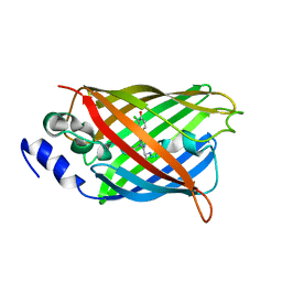
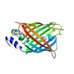
5HDC
DownloadVisualize
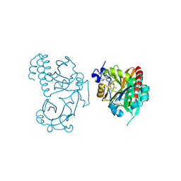
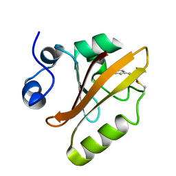
BU of 5hdc by Molmil
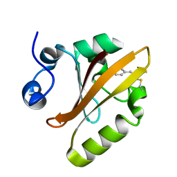
Femtosecond Structural Dynamics Drives the Trans/Cis Isomerization in Photoactive Yellow Protein: 100 fs to 400 fs Structure
Descriptor: Photoactive yellow protein
Authors:Pande, K, Tenboer, J, Schmidt, M.
Deposit date:2016-01-05
Release date:2016-05-25
Last modified:2023-11-15
Method:X-RAY DIFFRACTION (1.6 Å)
Cite:Femtosecond structural dynamics drives the trans/cis isomerization in photoactive yellow protein.
Science, 352, 2016
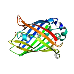
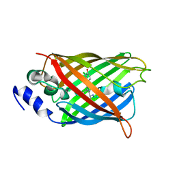
5HDD
DownloadVisualize
BU of 5hdd by Molmil
Femtosecond Structural Dynamics Drives the Trans/Cis Isomerization in Photoactive Yellow Protein: 800 fs to 1200 fs Structure
Descriptor: Photoactive yellow protein
Authors:Pande, K, Tenboer, J, Schmidt, M.
Deposit date:2016-01-05
Release date:2016-05-25
Last modified:2023-11-15
Method:X-RAY DIFFRACTION (1.6 Å)
Cite:Femtosecond structural dynamics drives the trans/cis isomerization in photoactive yellow protein.
Science, 352, 2016
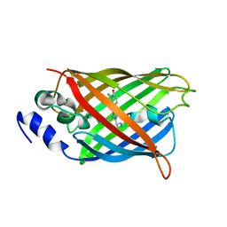
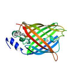
5HD5
DownloadVisualize
BU of 5hd5 by Molmil
Femtosecond Structural Dynamics Drives the Trans/Cis Isomerization in Photoactive Yellow Protein: 200 ns time delay photo-activated (light) structure
Descriptor: Photoactive yellow protein
Authors:Pande, K, Tenboer, J, Schmidt, M.
Deposit date:2016-01-04
Release date:2016-05-25
Last modified:2023-11-15
Method:X-RAY DIFFRACTION (1.6 Å)
Cite:Femtosecond structural dynamics drives the trans/cis isomerization in photoactive yellow protein.
Science, 352, 2016
6T3L
DownloadVisualize
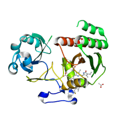
BU of 6t3l by Molmil
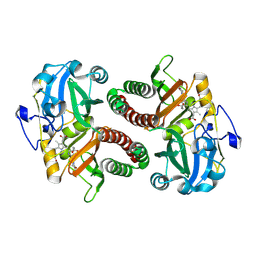
PAS-GAF fragment from Deinococcus radiodurans phytochrome in dark state
Descriptor: 3-[2-[(Z)-[3-(2-carboxyethyl)-5-[(Z)-(4-ethenyl-3-methyl-5-oxidanylidene-pyrrol-2-ylidene)methyl]-4-methyl-pyrrol-1-ium -2-ylidene]methyl]-5-[(Z)-[(3E)-3-ethylidene-4-methyl-5-oxidanylidene-pyrrolidin-2-ylidene]methyl]-4-methyl-1H-pyrrol-3- yl]propanoic acid, Bacteriophytochrome
Authors:Claesson, E, Takala, H, Yuan Wahlgren, W, Pandey, S, Schmidt, M, Westenhoff, S.
Deposit date:2019-10-11
Release date:2020-04-08
Last modified:2024-01-24
Method:X-RAY DIFFRACTION (2.07 Å)
Cite:The primary structural photoresponse of phytochrome proteins captured by a femtosecond X-ray laser.
Elife, 9, 2020
6T3U
DownloadVisualize
BU of 6t3u by Molmil
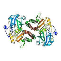
PAS-GAF fragment from Deinococcus radiodurans phytochrome 1ps after photoexcitation
Descriptor: 3-[2-[(Z)-[3-(2-carboxyethyl)-5-[(Z)-(4-ethenyl-3-methyl-5-oxidanylidene-pyrrol-2-ylidene)methyl]-4-methyl-pyrrol-1-ium -2-ylidene]methyl]-5-[(Z)-[(3E)-3-ethylidene-4-methyl-5-oxidanylidene-pyrrolidin-2-ylidene]methyl]-4-methyl-1H-pyrrol-3- yl]propanoic acid, Bacteriophytochrome
Authors:Claesson, E, Takala, H, Yuan Wahlgren, W, Pandey, S, Schmidt, M, Westenhoff, S.
Deposit date:2019-10-11
Release date:2020-04-08
Last modified:2024-01-24
Method:X-RAY DIFFRACTION (2.21 Å)
Cite:The primary structural photoresponse of phytochrome proteins captured by a femtosecond X-ray laser.
Elife, 9, 2020
5K5B
DownloadVisualize
BU of 5k5b by Molmil
Wild-type PAS-GAF fragment from Deinococcus radiodurans BphP
Descriptor: (4S)-2-METHYL-2,4-PENTANEDIOL, 3-[2-[(Z)-[3-(2-carboxyethyl)-5-[(Z)-(4-ethenyl-3-methyl-5-oxidanylidene-pyrrol-2-ylidene)methyl]-4-methyl-pyrrol-1-ium -2-ylidene]methyl]-5-[(Z)-[(3E)-3-ethylidene-4-methyl-5-oxidanylidene-pyrrolidin-2-ylidene]methyl]-4-methyl-1H-pyrrol-3- yl]propanoic acid, ACETATE ION, ...
Authors:Takala, H, Edlund, P, Claesson, E, Ihalainen, J.A, Westenhoff, S.
Deposit date:2016-05-23
Release date:2016-10-26
Last modified:2024-01-10
Method:X-RAY DIFFRACTION (1.35 Å)
Cite:The room temperature crystal structure of a bacterial phytochrome determined by serial femtosecond crystallography.
Sci Rep, 6, 2016
5LBR
DownloadVisualize
BU of 5lbr by Molmil
Wild-type PAS-GAF fragment from Deinococcus radiodurans Bphp collected at SACLA
Descriptor: 3-[2-[(Z)-[3-(2-carboxyethyl)-5-[(Z)-(4-ethenyl-3-methyl-5-oxidanylidene-pyrrol-2-ylidene)methyl]-4-methyl-pyrrol-1-ium -2-ylidene]methyl]-5-[(Z)-[(3E)-3-ethylidene-4-methyl-5-oxidanylidene-pyrrolidin-2-ylidene]methyl]-4-methyl-1H-pyrrol-3- yl]propanoic acid, ACETATE ION, Bacteriophytochrome
Authors:Edlund, P, Claesson, E, Nakane, T, Takala, H, Dods, R, Schmidt, M, Westenhoff, S.
Deposit date:2016-06-17
Release date:2016-10-26
Last modified:2024-01-10
Method:X-RAY DIFFRACTION (2.2 Å)
Cite:The room temperature crystal structure of a bacterial phytochrome determined by serial femtosecond crystallography.
Sci Rep, 6, 2016
5L8M
DownloadVisualize
BU of 5l8m by Molmil
Wild-type PAS-GAF fragment from Deinococcus radiodurans Bphp collected at LCLS
Descriptor: 3-[2-[(Z)-[3-(2-carboxyethyl)-5-[(Z)-(4-ethenyl-3-methyl-5-oxidanylidene-pyrrol-2-ylidene)methyl]-4-methyl-pyrrol-1-ium -2-ylidene]methyl]-5-[(Z)-[(3E)-3-ethylidene-4-methyl-5-oxidanylidene-pyrrolidin-2-ylidene]methyl]-4-methyl-1H-pyrrol-3- yl]propanoic acid, ACETATE ION, Bacteriophytochrome
Authors:Claesson, E, Takala, H, Edlund, P, Henry, L, Dods, R, Schmidt, M, Westenhoff, S.
Deposit date:2016-06-08
Release date:2016-10-26
Last modified:2024-01-10
Method:X-RAY DIFFRACTION (2.1 Å)
Cite:The room temperature crystal structure of a bacterial phytochrome determined by serial femtosecond crystallography.
Sci Rep, 6, 2016
8A6O
DownloadVisualize
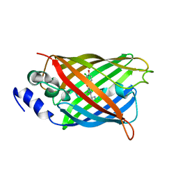
BU of 8a6o by Molmil
Room temperature rsEGFP2 with a chlorinated chromophore 600 fs after Photoexcitation
Descriptor: Green fluorescent protein
Authors:Fadini, A, van Thor, J.
Deposit date:2022-06-18
Release date:2023-07-19
Last modified:2024-02-07
Method:X-RAY DIFFRACTION (1.63 Å)
Cite:Serial Femtosecond Crystallography Reveals that Photoactivation in a Fluorescent Protein Proceeds via the Hula Twist Mechanism.
J.Am.Chem.Soc., 145, 2023
8A6S
DownloadVisualize
BU of 8a6s by Molmil
Room temperature rsEGFP2 with a chlorinated chromophore 1 microsecond after Photoexcitation
Descriptor: Green fluorescent protein
Authors:Fadini, A, van Thor, J.
Deposit date:2022-06-19
Release date:2023-07-19
Last modified:2024-02-07
Method:X-RAY DIFFRACTION (1.63 Å)
Cite:Serial Femtosecond Crystallography Reveals that Photoactivation in a Fluorescent Protein Proceeds via the Hula Twist Mechanism.
J.Am.Chem.Soc., 145, 2023
8A6N
DownloadVisualize
BU of 8a6n by Molmil
Room temperature rsEGFP2 with a chlorinated chromophore 300 fs after Photoexcitation
Descriptor: Green fluorescent protein
Authors:Fadini, A, van Thor, J.
Deposit date:2022-06-18
Release date:2023-07-19
Last modified:2024-02-07
Method:X-RAY DIFFRACTION (1.63 Å)
Cite:Serial Femtosecond Crystallography Reveals that Photoactivation in a Fluorescent Protein Proceeds via the Hula Twist Mechanism.
J.Am.Chem.Soc., 145, 2023
8A83
DownloadVisualize
BU of 8a83 by Molmil
rsEGFP2 with a chlorinated chromophore in the fluorescent ON-state in a crystal dehydrated after illumination
Descriptor: Green fluorescent protein, SULFATE ION
Authors:Fadini, A, van Thor, J, Chang, J.
Deposit date:2022-06-21
Release date:2023-07-19
Last modified:2024-02-07
Method:X-RAY DIFFRACTION (1.81 Å)
Cite:Serial Femtosecond Crystallography Reveals that Photoactivation in a Fluorescent Protein Proceeds via the Hula Twist Mechanism.
J.Am.Chem.Soc., 145, 2023
8A6R
DownloadVisualize
BU of 8a6r by Molmil
Room temperature rsEGFP2 with a chlorinated chromophore 100 ps after Photoexcitation
Descriptor: Green fluorescent protein
Authors:Fadini, A, van Thor, J.
Deposit date:2022-06-18
Release date:2023-07-19
Last modified:2024-02-07
Method:X-RAY DIFFRACTION (1.63 Å)
Cite:Serial Femtosecond Crystallography Reveals that Photoactivation in a Fluorescent Protein Proceeds via the Hula Twist Mechanism.
J.Am.Chem.Soc., 145, 2023
8A6G
DownloadVisualize
BU of 8a6g by Molmil
Room temperature rsEGFP2 with a chlorinated chromophore in the non-fluorescent OFF-state
Descriptor: Green fluorescent protein
Authors:Fadini, A, van Thor, J.
Deposit date:2022-06-17
Release date:2023-07-19
Last modified:2024-02-07
Method:X-RAY DIFFRACTION (1.63 Å)
Cite:Serial Femtosecond Crystallography Reveals that Photoactivation in a Fluorescent Protein Proceeds via the Hula Twist Mechanism.
J.Am.Chem.Soc., 145, 2023
8A6P
DownloadVisualize
BU of 8a6p by Molmil
Room temperature rsEGFP2 with a chlorinated chromophore 900 fs after photoexcitation
Descriptor: Green fluorescent protein
Authors:Fadini, A, van Thor, J.
Deposit date:2022-06-18
Release date:2023-07-19
Last modified:2024-02-07
Method:X-RAY DIFFRACTION (1.63 Å)
Cite:Serial Femtosecond Crystallography Reveals that Photoactivation in a Fluorescent Protein Proceeds via the Hula Twist Mechanism.
J.Am.Chem.Soc., 145, 2023
8A6Q
DownloadVisualize
BU of 8a6q by Molmil
Room temperature rsEGFP2 with a chlorinated chromophore 5 ps after photoexcitation
Descriptor: Green fluorescent protein
Authors:Fadini, A, van Thor, J.
Deposit date:2022-06-18
Release date:2023-07-19
Last modified:2024-02-07
Method:X-RAY DIFFRACTION (1.63 Å)
Cite:Serial Femtosecond Crystallography Reveals that Photoactivation in a Fluorescent Protein Proceeds via the Hula Twist Mechanism.
J.Am.Chem.Soc., 145, 2023
8A7V
DownloadVisualize
BU of 8a7v by Molmil
Room temperature rsEGFP2 in its OFF-state obtained with SFX
Descriptor: Green fluorescent protein
Authors:Fadini, A, van Thor, J.
Deposit date:2022-06-21
Release date:2023-07-19
Last modified:2024-02-07
Method:X-RAY DIFFRACTION (1.46 Å)
Cite:Serial Femtosecond Crystallography Reveals that Photoactivation in a Fluorescent Protein Proceeds via the Hula Twist Mechanism.
J.Am.Chem.Soc., 145, 2023
<12


219515

数据于2024-05-08公开中

PDB statisticsPDBj update infoContact PDBjnumon