6GLC
 
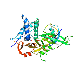
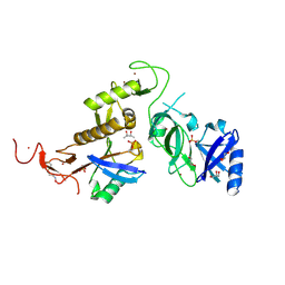 | | Structure of phospho-Parkin bound to phospho-ubiquitin | | 分子名称: | (4S)-2-METHYL-2,4-PENTANEDIOL, E3 ubiquitin-protein ligase parkin, GLYCEROL, ... | | 著者 | Gladkova, C, Maslen, S.L, Skehel, J.M, Komander, D. | | 登録日 | 2018-05-23 | | 公開日 | 2018-06-13 | | 最終更新日 | 2024-01-17 | | 実験手法 | X-RAY DIFFRACTION (1.8 Å) | | 主引用文献 | Mechanism of parkin activation by PINK1.
Nature, 559, 2018
|
|
5OXH
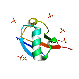 | | C-terminally retracted ubiquitin T66V/L67N mutant | | 分子名称: | SULFATE ION, Ubiquitin T66V/L67N mutant | | 著者 | Gladkova, C, Schubert, A.F, Wagstaff, J.L, Pruneda, J.P, Freund, S.M.V, Komander, D. | | 登録日 | 2017-09-06 | | 公開日 | 2017-11-22 | | 最終更新日 | 2024-01-17 | | 実験手法 | X-RAY DIFFRACTION (1.601 Å) | | 主引用文献 | An invisible ubiquitin conformation is required for efficient phosphorylation by PINK1.
EMBO J., 36, 2017
|
|
6EQI
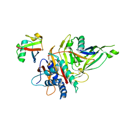
 | | Structure of PINK1 bound to ubiquitin | | 分子名称: | GLYCEROL, Nb696, Serine/threonine-protein kinase PINK1, ... | | 著者 | Schubert, A.F, Gladkova, C, Pardon, E, Wagstaff, J.L, Freund, S.M.V, Steyaert, J, Maslen, S, Komander, D. | | 登録日 | 2017-10-13 | | 公開日 | 2017-11-08 | | 最終更新日 | 2024-01-17 | | 実験手法 | X-RAY DIFFRACTION (3.1 Å) | | 主引用文献 | Structure of PINK1 in complex with its substrate ubiquitin.
Nature, 552, 2017
|
|
5OXI
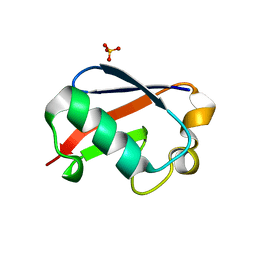 | | C-terminally retracted ubiquitin L67S mutant | | 分子名称: | SULFATE ION, Ubiquitin L67S mutant | | 著者 | Gladkova, C.G, Schubert, A.F, Wagstaff, J.L, Pruneda, J.N, Freund, S.M.V, Komander, D. | | 登録日 | 2017-09-06 | | 公開日 | 2017-11-22 | | 最終更新日 | 2024-01-17 | | 実験手法 | X-RAY DIFFRACTION (1.63 Å) | | 主引用文献 | An invisible ubiquitin conformation is required for efficient phosphorylation by PINK1.
EMBO J., 36, 2017
|
|
4WZP
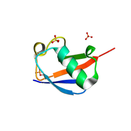 | | Ser65 phosphorylated ubiquitin, major conformation | | 分子名称: | SULFATE ION, ubiquitin | | 著者 | Wauer, T, Wagstaff, J, Freund, S.M.V, Komander, D. | | 登録日 | 2014-11-20 | | 公開日 | 2015-01-14 | | 最終更新日 | 2024-01-10 | | 実験手法 | X-RAY DIFFRACTION (1.9 Å) | | 主引用文献 | Ubiquitin Ser65 phosphorylation affects ubiquitin structure, chain assembly and hydrolysis.
Embo J., 34, 2015
|
|
5OHN
 | |
5OHP
 | |
5OHK
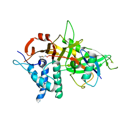 | | Crystal structure of USP30 in covalent complex with ubiquitin propargylamide (high resolution) | | 分子名称: | Polyubiquitin-B, Ubiquitin carboxyl-terminal hydrolase 30,Ubiquitin carboxyl-terminal hydrolase 30,Ubiquitin carboxyl-terminal hydrolase 30, ZINC ION, ... | | 著者 | Gersch, M, Komander, D. | | 登録日 | 2017-07-17 | | 公開日 | 2017-09-20 | | 最終更新日 | 2024-02-07 | | 実験手法 | X-RAY DIFFRACTION (2.34 Å) | | 主引用文献 | Mechanism and regulation of the Lys6-selective deubiquitinase USP30.
Nat. Struct. Mol. Biol., 24, 2017
|
|


