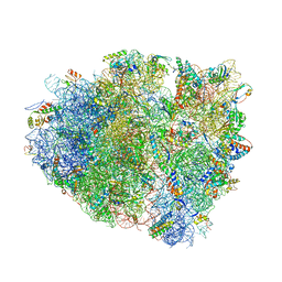
4V7J
 
 | | Structure of RelE nuclease bound to the 70S ribosome (precleavage state) | | 分子名称: | 30S ribosomal protein S10, 30S ribosomal protein S11, 30S ribosomal protein S12, ... | | 著者 | Neubauer, C, Gao, Y.-G, Andersen, K.R, Dunham, C.M, Kelley, A.C, Hentschel, J, Gerdes, K, Ramakrishnan, V, Brodersen, D.E. | | 登録日 | 2009-11-02 | | 公開日 | 2014-07-09 | | 最終更新日 | 2023-09-20 | | 実験手法 | X-RAY DIFFRACTION (3.3 Å) | | 主引用文献 | The structural basis for mRNA recognition and cleavage by the ribosome-dependent endonuclease RelE.
Cell(Cambridge,Mass.), 139, 2009
|
|
4V7K
 | | Structure of RelE nuclease bound to the 70S ribosome (postcleavage state) | | 分子名称: | 30S ribosomal protein S10, 30S ribosomal protein S11, 30S ribosomal protein S12, ... | | 著者 | Neubauer, C, Gao, Y.-G, Andersen, K.R, Dunham, C.M, Kelley, A.C, Hentschel, J, Gerdes, K, Ramakrishnan, V, Brodersen, D.E. | | 登録日 | 2009-11-02 | | 公開日 | 2014-07-09 | | 最終更新日 | 2014-12-10 | | 実験手法 | X-RAY DIFFRACTION (3.6 Å) | | 主引用文献 | The structural basis for mRNA recognition and cleavage by the ribosome-dependent endonuclease RelE.
Cell(Cambridge,Mass.), 139, 2009
|
|
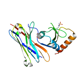
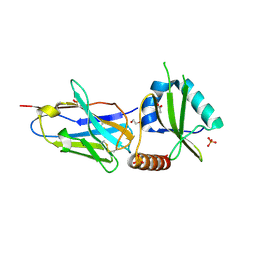
3TND
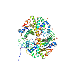 | | Crystal structure of Shigella flexneri VapBC toxin-antitoxin complex | | 分子名称: | Antitoxin VapB, SODIUM ION, SULFATE ION, ... | | 著者 | Dienemann, C, Boggild, A, Winther, K.S, Gerdes, K, Brodersen, D.E. | | 登録日 | 2011-09-01 | | 公開日 | 2011-11-02 | | 最終更新日 | 2024-02-28 | | 実験手法 | X-RAY DIFFRACTION (2.7 Å) | | 主引用文献 | Crystal Structure of the VapBC Toxin-Antitoxin Complex from Shigella flexneri Reveals a Hetero-Octameric DNA-Binding Assembly.
J.Mol.Biol., 414, 2011
|
|
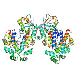
1MWM
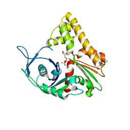 | | ParM from plasmid R1 ADP form | | 分子名称: | ADENOSINE-5'-DIPHOSPHATE, MAGNESIUM ION, ParM | | 著者 | Van den Ent, F, Moller-Jensen, J, Amos, L.A, Gerdes, K, Lowe, J. | | 登録日 | 2002-09-30 | | 公開日 | 2003-01-28 | | 最終更新日 | 2024-04-03 | | 実験手法 | X-RAY DIFFRACTION (2 Å) | | 主引用文献 | F-actin-like filaments formed by plasmid segregation protein ParM
EMBO J., 21, 2002
|
|
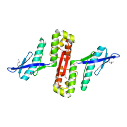
1MWK
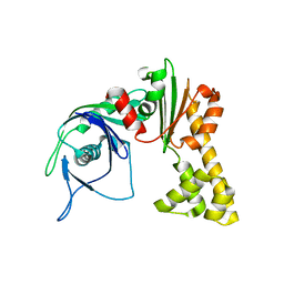 | | ParM from plasmid R1 APO form | | 分子名称: | ParM | | 著者 | Van den Ent, F, Moller-Jensen, J, Amos, L.A, Gerdes, K, Lowe, J. | | 登録日 | 2002-09-30 | | 公開日 | 2003-01-28 | | 最終更新日 | 2024-03-13 | | 実験手法 | X-RAY DIFFRACTION (2.3 Å) | | 主引用文献 | F-actin-like filaments formed by plasmid segregation protein ParM
EMBO J., 21, 2002
|
|
2JEE
 | |
6GFM
 | | Crystal structure of the Escherichia coli nucleosidase PpnN (pppGpp-form) | | 分子名称: | Pyrimidine/purine nucleotide 5'-monophosphate nucleosidase, guanosine 5'-(tetrahydrogen triphosphate) 3'-(trihydrogen diphosphate) | | 著者 | Zhang, Y, Baerentsen, R.L, Gerdes, K, Brodersen, D.E. | | 登録日 | 2018-05-01 | | 公開日 | 2019-04-24 | | 最終更新日 | 2024-01-17 | | 実験手法 | X-RAY DIFFRACTION (2.77 Å) | | 主引用文献 | (p)ppGpp Regulates a Bacterial Nucleosidase by an Allosteric Two-Domain Switch.
Mol.Cell, 74, 2019
|
|
6GFL
 | |
6HPB
 | |
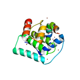
5MJE
 | | Crystal structure of the HigB2 toxin in complex with Nb8 | | 分子名称: | Cytotoxic translational repressor of toxin-antitoxin stability system, DI(HYDROXYETHYL)ETHER, Nanobody 8, ... | | 著者 | Hadzi, S, Loris, R. | | 登録日 | 2016-11-30 | | 公開日 | 2017-04-05 | | 最終更新日 | 2019-10-16 | | 実験手法 | X-RAY DIFFRACTION (2.599 Å) | | 主引用文献 | Ribosome-dependent Vibrio cholerae mRNAse HigB2 is regulated by a beta-strand sliding mechanism.
Nucleic Acids Res., 45, 2017
|
|
7AB5
 | |
7AB3
 | |
7AB4
 | |
2JD3
 | |
5J9I
 | |
5JA9
 | |
5JAA
 | |
5JA8
 | | Crystal structure of the HigB2 toxin in complex with Nb2 | | 分子名称: | 1,2-ETHANEDIOL, 1,3-PROPANDIOL, 4-(2-HYDROXYETHYL)-1-PIPERAZINE ETHANESULFONIC ACID, ... | | 著者 | Hadzi, S, Loris, R. | | 登録日 | 2016-04-12 | | 公開日 | 2017-04-05 | | 最終更新日 | 2019-02-20 | | 実験手法 | X-RAY DIFFRACTION (2.49 Å) | | 主引用文献 | Ribosome-dependent Vibrio cholerae mRNAse HigB2 is regulated by a beta-strand sliding mechanism.
Nucleic Acids Res., 45, 2017
|
|
6GW6
 | |
6HPC
 | |
3DD7
 | |
4FXI
 | |


