1IDY
 
 | |
1IDZ
 | |
5XFV
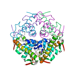
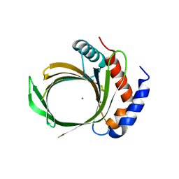
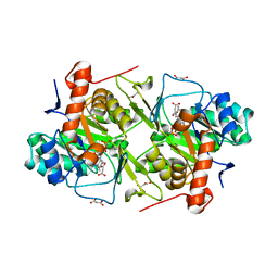 | | Crystal structures of FMN-bound form of dihydroorotate dehydrogenase from Trypanosoma brucei | | 分子名称: | Dihydroorotate dehydrogenase (fumarate), FLAVIN MONONUCLEOTIDE, MALONATE ION | | 著者 | Kubota, T, Tani, O, Yamaguchi, T, Namatame, I, Sakashita, H, Furukawa, K, Yamasaki, K. | | 登録日 | 2017-04-11 | | 公開日 | 2018-04-25 | | 最終更新日 | 2023-11-22 | | 実験手法 | X-RAY DIFFRACTION (1.79 Å) | | 主引用文献 | Crystal structures of FMN-bound and FMN-free forms of dihydroorotate dehydrogenase fromTrypanosoma brucei.
FEBS Open Bio, 8, 2018
|
|
5XFW
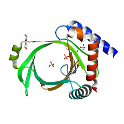
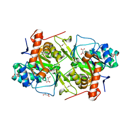 | | Crystal structures of FMN-free form of dihydroorotate dehydrogenase from Trypanosoma brucei | | 分子名称: | Dihydroorotate dehydrogenase (fumarate), MALONATE ION | | 著者 | Kubota, T, Tani, O, Yamaguchi, T, Namatame, I, Sakashita, H, Furukawa, K, Yamasaki, K. | | 登録日 | 2017-04-11 | | 公開日 | 2018-04-25 | | 最終更新日 | 2023-11-22 | | 実験手法 | X-RAY DIFFRACTION (1.6 Å) | | 主引用文献 | Crystal structures of FMN-bound and FMN-free forms of dihydroorotate dehydrogenase fromTrypanosoma brucei.
FEBS Open Bio, 8, 2018
|
|
5XDH
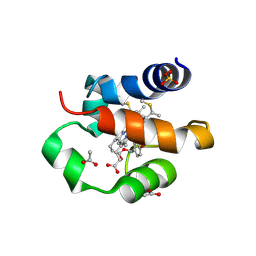 | | His/DOPA ligated cytochrome c from an anammox organism KSU-1 | | 分子名称: | ACETATE ION, HEME C, Putative cytochrome c, ... | | 著者 | Hira, D, Kitamura, R, Nakamura, T, Yamagata, Y, Furukawa, K, Fujii, T. | | 登録日 | 2017-03-28 | | 公開日 | 2018-03-28 | | 最終更新日 | 2019-10-02 | | 実験手法 | X-RAY DIFFRACTION (1.32 Å) | | 主引用文献 | Anammox Organism KSU-1 Expresses a Novel His/DOPA Ligated Cytochrome c.
J. Mol. Biol., 430, 2018
|
|
4RUM
 | |
8H3M
 | | Conformation 1 of SARS-CoV-2 Omicron BA.1 Variant Spike protein complexed with MO1 Fab | | 分子名称: | 2-acetamido-2-deoxy-beta-D-glucopyranose, MO1 heavy chain, Spike glycoprotein | | 著者 | Ishimaru, H, Nishimura, M, Sutandhio, S, Shigematsu, H, Kato, K, Hasegawa, N, Mori, Y. | | 登録日 | 2022-10-09 | | 公開日 | 2023-05-10 | | 最終更新日 | 2023-08-02 | | 実験手法 | ELECTRON MICROSCOPY (2.48 Å) | | 主引用文献 | Identification and Analysis of Monoclonal Antibodies with Neutralizing Activity against Diverse SARS-CoV-2 Variants.
J.Virol., 97, 2023
|
|
8H3N
 | | Conformation 2 of SARS-CoV-2 Omicron BA.1 Variant Spike protein complexed with MO1 Fab | | 分子名称: | 2-acetamido-2-deoxy-beta-D-glucopyranose, MO1 heavy-chain, MO1 light chain, ... | | 著者 | Ishimaru, H, Nishimura, M, Sutandhio, S, Shigematsu, H, Kato, K, Hasegawa, N, Mori, Y. | | 登録日 | 2022-10-09 | | 公開日 | 2023-05-10 | | 最終更新日 | 2023-08-02 | | 実験手法 | ELECTRON MICROSCOPY (2.73 Å) | | 主引用文献 | Identification and Analysis of Monoclonal Antibodies with Neutralizing Activity against Diverse SARS-CoV-2 Variants.
J.Virol., 97, 2023
|
|
7YDO
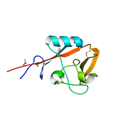
 | | Crystal structure of Atg44 | | 分子名称: | 1,2-Distearoyl-sn-glycerophosphoethanolamine, Uncharacterized protein C26A3.14c | | 著者 | Maruyama, T, Noda, N.N. | | 登録日 | 2022-07-04 | | 公開日 | 2023-05-17 | | 最終更新日 | 2023-06-28 | | 実験手法 | X-RAY DIFFRACTION (1.58 Å) | | 主引用文献 | The mitochondrial intermembrane space protein mitofissin drives mitochondrial fission required for mitophagy.
Mol.Cell, 83, 2023
|
|
5B1R
 | | Crystal structure of mouse CD72a CTLD | | 分子名称: | ACETATE ION, B-cell differentiation antigen CD72, GLYCEROL | | 著者 | Shinagawa, K, Numoto, N, Tsubata, T, Ito, N. | | 登録日 | 2015-12-15 | | 公開日 | 2016-10-19 | | 最終更新日 | 2023-11-08 | | 実験手法 | X-RAY DIFFRACTION (1.2 Å) | | 主引用文献 | CD72 negatively regulates B lymphocyte responses to the lupus-related endogenous toll-like receptor 7 ligand Sm/RNP
J.Exp.Med., 213, 2016
|
|
6L7V
 | |
6L7X
 | |
6L7W
 | |
6L7Y
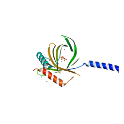
 | | Crystal structure of Cet1 from Trypanosoma cruzi in complex with #466 ligand. | | 分子名称: | 3,4,6,7-tetrahydroacridine-1,8(2H,5H)-dione, SULFATE ION, mRNA_triPase domain-containing protein | | 著者 | Kuwabara, N, Ho, K. | | 登録日 | 2019-11-03 | | 公開日 | 2020-06-03 | | 最終更新日 | 2023-11-22 | | 実験手法 | X-RAY DIFFRACTION (2.51 Å) | | 主引用文献 | Crystal structures of the RNA triphosphatase fromTrypanosoma cruziprovide insights into how it recognizes the 5'-end of the RNA substrate.
J.Biol.Chem., 295, 2020
|
|
3WJ2
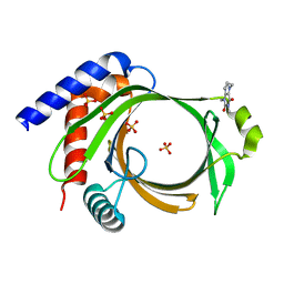
 | | Crystal structure of ESTFA (FE-lacking apo form) | | 分子名称: | Carboxylesterase | | 著者 | Ohara, K, Unno, H, Oshima, Y, Furukawa, K, Fujino, N, Hirooka, K, Hemmi, H, Takahashi, S, Nishino, T, Kusunoki, M, Nakayama, T. | | 登録日 | 2013-10-03 | | 公開日 | 2014-07-30 | | 最終更新日 | 2024-03-20 | | 実験手法 | X-RAY DIFFRACTION (1.61 Å) | | 主引用文献 | Structural insights into the low pH adaptation of a unique carboxylesterase from Ferroplasma: altering the pH optima of two carboxylesterases.
J.Biol.Chem., 289, 2014
|
|
3WJ1
 | | Crystal structure of SSHESTI | | 分子名称: | Carboxylesterase, octyl beta-D-glucopyranoside | | 著者 | Ohara, K, Unno, H, Oshima, Y, Furukawa, K, Fujino, N, Hirooka, K, Hemmi, H, Takahashi, S, Nishino, T, Kusunoki, M, Nakayama, T. | | 登録日 | 2013-10-03 | | 公開日 | 2014-07-30 | | 最終更新日 | 2020-07-29 | | 実験手法 | X-RAY DIFFRACTION (1.5 Å) | | 主引用文献 | Structural insights into the low pH adaptation of a unique carboxylesterase from Ferroplasma: altering the pH optima of two carboxylesterases.
J.Biol.Chem., 289, 2014
|
|


