5F6A
 
 | |
5F69
 | |
5F68
 | |
1R1C
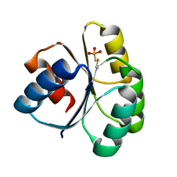
 | | PSEUDOMONAS AERUGINOSA W48F/Y72F/H83Q/Y108W-AZURIN RE(PHEN)(CO)3(HIS107) | | 分子名称: | (1,10 PHENANTHROLINE)-(TRI-CARBON MONOXIDE) RHENIUM (I), Azurin, COPPER (I) ION | | 著者 | Miller, J.E, Gradinaru, C, Crane, B.R, Di Bilio, A.J. | | 登録日 | 2003-09-23 | | 公開日 | 2003-09-30 | | 最終更新日 | 2023-08-23 | | 実験手法 | X-RAY DIFFRACTION (1.9 Å) | | 主引用文献 | Spectroscopy and reactivity of a photogenerated tryptophan radical in a structurally defined protein environment
J.Am.Chem.Soc., 125, 2003
|
|
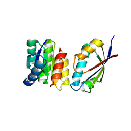
1TQG
 | | CheA phosphotransferase domain from Thermotoga maritima | | 分子名称: | Chemotaxis protein cheA | | 著者 | Quezada, C.M, Gradinaru, C, Simon, M.I, Bilwes, A.M, Crane, B.R. | | 登録日 | 2004-06-17 | | 公開日 | 2004-09-07 | | 最終更新日 | 2024-02-14 | | 実験手法 | X-RAY DIFFRACTION (0.98 Å) | | 主引用文献 | Helical Shifts Generate Two Distinct Conformers in the Atomic Resolution Structure of the CheA Phosphotransferase Domain from Thermotoga maritima.
J.Mol.Biol., 341, 2004
|
|
4QYW
 | |
1U0S
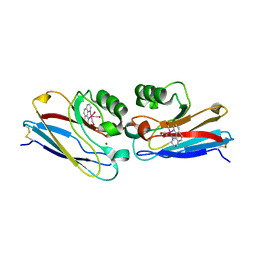
 | | Chemotaxis kinase CheA P2 domain in complex with response regulator CheY from the thermophile thermotoga maritima | | 分子名称: | Chemotaxis protein cheA, Chemotaxis protein cheY | | 著者 | Park, S.Y, Beel, B.D, Simon, M.I, Bilwes, A.M, Crane, B.R. | | 登録日 | 2004-07-14 | | 公開日 | 2004-08-10 | | 最終更新日 | 2023-10-25 | | 実験手法 | X-RAY DIFFRACTION (1.9 Å) | | 主引用文献 | In different organisms, the mode of interaction between two signaling proteins is not necessarily conserved
Proc.Natl.Acad.Sci.USA, 101, 2004
|
|
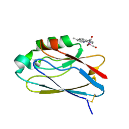
2I7S
 | | Crystal structure of Re(phen)(CO)3 (Thr124His)(His83Gln) Azurin Cu(II) from Pseudomonas aeruginosa | | 分子名称: | (1,10 PHENANTHROLINE)-(TRI-CARBON MONOXIDE) RHENIUM (I), Azurin, COBALT TETRAAMMINE ION, ... | | 著者 | Gradinaru, C, Crane, B.R. | | 登録日 | 2006-08-31 | | 公開日 | 2006-12-05 | | 最終更新日 | 2023-08-30 | | 実験手法 | X-RAY DIFFRACTION (1.35 Å) | | 主引用文献 | Relaxation dynamics of Pseudomonas aeruginosa Re(I)(CO)3(alpha-diimine)(HisX)+ (X = 83, 107, 109, 124, 126)Cu(II) azurins.
J.Am.Chem.Soc., 131, 2009
|
|
2I7O
 | | Structure of Re(4,7-dimethyl-phen)(Thr124His)(Lys122Trp)(His83Gln)AzCu(II), a Rhenium modified Azurin mutant | | 分子名称: | (1,10 PHENANTHROLINE)-(TRI-CARBON MONOXIDE) RHENIUM (I), Azurin, COPPER (II) ION | | 著者 | Sudhamsu, J, Crane, B.R. | | 登録日 | 2006-08-31 | | 公開日 | 2007-08-14 | | 最終更新日 | 2021-10-20 | | 実験手法 | X-RAY DIFFRACTION (1.5 Å) | | 主引用文献 | Tryptophan-accelerated electron flow through proteins.
Science, 320, 2008
|
|
2NOX
 | | Crystal structure of tryptophan 2,3-dioxygenase from Ralstonia metallidurans | | 分子名称: | PROTOPORPHYRIN IX CONTAINING FE, Tryptophan 2,3-dioxygenase | | 著者 | Zhang, Y, Kang, S.A, Mukherjee, T, Bale, S, Crane, B.R, Begley, T.P, Ealick, S.E. | | 登録日 | 2006-10-26 | | 公開日 | 2006-12-19 | | 最終更新日 | 2023-08-30 | | 実験手法 | X-RAY DIFFRACTION (2.4 Å) | | 主引用文献 | Crystal structure and mechanism of tryptophan 2,3-dioxygenase, a heme enzyme involved in tryptophan catabolism and in quinolinate biosynthesis.
Biochemistry, 46, 2007
|
|
4HI4
 | |
4I44
 | |
4I3M
 | | Aer2 poly-HAMP domains: L44H HAMP1 CW-lock mutant | | 分子名称: | Aerotaxis transducer Aer2, CHLORIDE ION, MAGNESIUM ION, ... | | 著者 | Airola, M.V, Sukomon, N, Crane, B.R. | | 登録日 | 2012-11-26 | | 公開日 | 2013-02-27 | | 最終更新日 | 2024-02-28 | | 実験手法 | X-RAY DIFFRACTION (1.95 Å) | | 主引用文献 | HAMP Domain Conformers That Propagate Opposite Signals in Bacterial Chemoreceptors.
Plos Biol., 11, 2013
|
|
4HZ1
 | | Crystal Structure of Pseudomonas aeruginosa azurin with iron(II) at the copper-binding site. | | 分子名称: | ACETATE ION, Azurin, FE (II) ION | | 著者 | McLaughlin, M.P, Retegan, M, Bill, E, Payne, T.M, Shafaat, H.S, Pea, S, Sudhamsu, J, Ensign, A.A, Crane, B.R, Neese, F, Holland, P.L. | | 登録日 | 2012-11-14 | | 公開日 | 2012-12-12 | | 最終更新日 | 2023-09-20 | | 実験手法 | X-RAY DIFFRACTION (2.2 Å) | | 主引用文献 | Azurin as a Protein Scaffold for a Low-coordinate Nonheme Iron Site with a Small-molecule Binding Pocket.
J.Am.Chem.Soc., 134, 2012
|
|
4JPB
 | | The structure of a ternary complex between CheA domains P4 and P5 with CheW and with an unzipped fragment of TM14, a chemoreceptor analog from Thermotoga maritima. | | 分子名称: | Chemotaxis protein CheA, Chemotaxis protein CheW, Methyl-accepting chemotaxis protein | | 著者 | Li, X, Bayas, C, Bilwes, A.M, Crane, B.R. | | 登録日 | 2013-03-19 | | 公開日 | 2013-08-28 | | 最終更新日 | 2024-02-28 | | 実験手法 | X-RAY DIFFRACTION (3.186 Å) | | 主引用文献 | The 3.2 angstrom resolution structure of a receptor: CheA:CheW signaling complex defines overlapping binding sites and key residue interactions within bacterial chemosensory arrays.
Biochemistry, 52, 2013
|
|
6NDT
 | |
6NDX
 | |
6NDV
 | | FlgE D2 domain K336A mutant | | 分子名称: | Flagellar hook protein FlgE | | 著者 | Lynch, M.J, Crane, B.R. | | 登録日 | 2018-12-14 | | 公開日 | 2019-08-21 | | 最終更新日 | 2024-03-13 | | 実験手法 | X-RAY DIFFRACTION (1.502 Å) | | 主引用文献 | Structure and chemistry of lysinoalanine crosslinking in the spirochaete flagella hook.
Nat.Chem.Biol., 15, 2019
|
|
6NDW
 | |
3RH8
 | |
3RTY
 | | Structure of an Enclosed Dimer Formed by The Drosophila Period Protein | | 分子名称: | 2,3-DIHYDROXY-1,4-DITHIOBUTANE, Period circadian protein | | 著者 | King, H.A, Hoelz, A, Crane, B.R, Young, M.W. | | 登録日 | 2011-05-04 | | 公開日 | 2011-12-21 | | 実験手法 | X-RAY DIFFRACTION (2.85 Å) | | 主引用文献 | Structure of an enclosed dimer formed by the Drosophila period protein.
J.Mol.Biol., 413, 2011
|
|
3SFT
 | |
3SOH
 | | Architecture of the Flagellar Rotor | | 分子名称: | Flagellar motor switch protein FliG, Flagellar motor switch protein FliM | | 著者 | Koushik, P, Gonzalez-Bonet, G, Bilwes, A.M, Crane, B.R, Blair, D. | | 登録日 | 2011-06-30 | | 公開日 | 2011-07-27 | | 最終更新日 | 2023-09-13 | | 実験手法 | X-RAY DIFFRACTION (3.5 Å) | | 主引用文献 | Architecture of the flagellar rotor.
Embo J., 30, 2011
|
|
3T8Y
 | |
2PD8
 | |


