2XSD
 
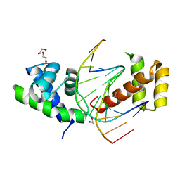 | | Crystal Structure of the dimeric Oct-6 (Pou3f1) POU domain bound to palindromic MORE DNA | | 分子名称: | 1,2-ETHANEDIOL, 5'-D(*AP*TP*GP*CP*AP*TP*GP*AP*GP*GP*AP)-3', 5'-D(*CP*CP*TP*CP*AP*TP*GP*CP*AP*TP*AP)-3', ... | | 著者 | Jauch, R, Choo, S.H, Ng, C.K.L, Kolatkar, P.R. | | 登録日 | 2010-09-28 | | 公開日 | 2010-10-20 | | 最終更新日 | 2023-12-20 | | 実験手法 | X-RAY DIFFRACTION (2.049 Å) | | 主引用文献 | Crystal Structure of the Dimeric Oct6 (POU3F1) POU Domain Bound to Palindromic More DNA.
Proteins, 79, 2011
|
|
4E6S
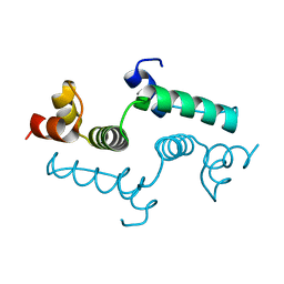 | | Crystal structure of the SCAN domain from mouse Zfp206 | | 分子名称: | Zinc finger and SCAN domain-containing protein 10 | | 著者 | Liang, Y, Choo, S.H, Rossbach, M, Baburajendran, N, Palasingam, P, Kolatkar, P.R. | | 登録日 | 2012-03-15 | | 公開日 | 2012-05-02 | | 最終更新日 | 2024-03-20 | | 実験手法 | X-RAY DIFFRACTION (1.85 Å) | | 主引用文献 | Crystal optimization and preliminary diffraction data analysis of the SCAN domain of Zfp206.
Acta Crystallogr.,Sect.F, 68, 2012
|
|


