Loading
PDBj
メニューPDBj@FacebookPDBj@TwitterPDBj@YouTubewwPDB FoundationwwPDB
RCSB PDBPDBeBMRBAdv. SearchSearch help
Search by PDB author
6WEE
DownloadVisualize
BU of 6wee by Molmil
Copper-bound M88I variant of Campylobacter jejuni P19
分子名称: 2-AMINO-2-HYDROXYMETHYL-PROPANE-1,3-DIOL, COPPER (II) ION, GLYCEROL, ...
著者Chan, A.C, Murphy, M.E.
登録日2020-04-02
公開日2021-02-10
最終更新日2023-10-18
実験手法X-RAY DIFFRACTION (2.3 Å)
主引用文献A copper site is required for iron transport by the periplasmic proteins P19 and FetP.
Metallomics, 12, 2020
6WEF
DownloadVisualize
BU of 6wef by Molmil
Copper-bound D92H variant of Campylobacter jejuni P19
分子名称: COPPER (II) ION, GLYCEROL, SULFATE ION, ...
著者Chan, A.C, Murphy, M.E.
登録日2020-04-02
公開日2021-02-10
最終更新日2023-10-18
実験手法X-RAY DIFFRACTION (2.501 Å)
主引用文献A copper site is required for iron transport by the periplasmic proteins P19 and FetP.
Metallomics, 12, 2020
8GKD
DownloadVisualize
BU of 8gkd by Molmil
Crystal structure of the peptidoglycan O-acetylesterase Ape1 (amino acids 41-392) from Campylobacter jejuni
分子名称: ACETATE ION, SGNH hydrolase-type esterase domain-containing protein
著者Lin, C.S, Murphy, M.E.
登録日2023-03-17
公開日2024-03-20
実験手法X-RAY DIFFRACTION (1.8 Å)
主引用文献Mechanism of the CBM35 domain in assisting catalysis by Ape1, a Campylobacter jejuni O-acetyl esterase
To be published
8GLD
DownloadVisualize
BU of 8gld by Molmil
Crystal structure of the peptidoglycan O-acetylesterase Ape1 (amino acids 22-392) from Campylobacter jejuni
分子名称: ACETATE ION, SGNH hydrolase-type esterase domain-containing protein
著者Lin, C.S, Murphy, M.E.
登録日2023-03-22
公開日2024-03-27
実験手法X-RAY DIFFRACTION (2.3 Å)
主引用文献Mechanism of the CBM35 domain in assisting catalysis by Ape1, a Campylobacter jejuni O-acetyl esterase
To be published
3NRP
DownloadVisualize
BU of 3nrp by Molmil
Crystal structure of 'as isolated' uropathogenic E. coli strain F11 FetP recombinantly expressed in the periplasm of E. coli BL21(DE3)
分子名称: Periplasmic protein-probably involved in high-affinity Fe2+ transport
著者Chan, A.C.K, Murphy, M.E.P.
登録日2010-06-30
公開日2011-05-18
最終更新日2023-12-27
実験手法X-RAY DIFFRACTION (1.6 Å)
主引用文献Characterization of a Dipartite Iron Uptake System from Uropathogenic Escherichia coli Strain F11.
J.Biol.Chem., 286, 2011
3NRQ
DownloadVisualize
BU of 3nrq by Molmil
Crystal structure of copper-reconstituted FetP from uropathogenic Escherichia coli strain F11
分子名称: COPPER (II) ION, Periplasmic protein-probably involved in high-affinity Fe2+ transport
著者Chan, A.C.K, Murphy, M.E.P.
登録日2010-06-30
公開日2011-05-18
最終更新日2023-12-27
実験手法X-RAY DIFFRACTION (1.7 Å)
主引用文献Characterization of a Dipartite Iron Uptake System from Uropathogenic Escherichia coli Strain F11.
J.Biol.Chem., 286, 2011
6E6I
DownloadVisualize
BU of 6e6i by Molmil
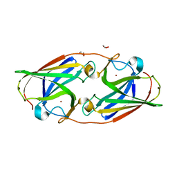
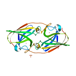
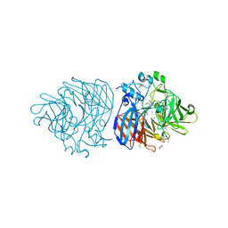
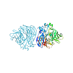
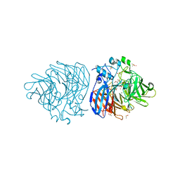
Crystal structure of 4-methyl HOPDA bound to LigY from Sphingobium sp. strain SYK-6
分子名称: (1Z,3E)-5-carboxy-3-methyl-5-oxo-1-phenylpenta-1,3-dien-1-olate, 2,2',3-trihydroxy-3'-methoxy-5,5'-dicarboxybiphenyl meta-cleavage compound hydrolase, ZINC ION
著者Kuatsjah, E, Chan, A.C, Hurst, T.E, Snieckus, V, Murphy, M.E, Eltis, L.D.
登録日2018-07-24
公開日2019-01-30
最終更新日2023-10-11
実験手法X-RAY DIFFRACTION (2.4 Å)
主引用文献Metal- and Serine-Dependent Meta-Cleavage Product Hydrolases Utilize Similar Nucleophile-Activation Strategies
Acs Catalysis, 8, 2018
1A81
DownloadVisualize
BU of 1a81 by Molmil
CRYSTAL STRUCTURE OF THE TANDEM SH2 DOMAIN OF THE SYK KINASE BOUND TO A DUALLY TYROSINE-PHOSPHORYLATED ITAM
分子名称: SYK KINASE, T-CELL SURFACE GLYCOPROTEIN CD3 EPSILON CHAIN
著者Fuetterer, K, Waksman, G.
登録日1998-03-31
公開日1998-10-21
最終更新日2016-05-25
実験手法X-RAY DIFFRACTION (3 Å)
主引用文献Structural basis for Syk tyrosine kinase ubiquity in signal transduction pathways revealed by the crystal structure of its regulatory SH2 domains bound to a dually phosphorylated ITAM peptide.
J.Mol.Biol., 281, 1998
6XJ6
DownloadVisualize
BU of 6xj6 by Molmil
Crystal structure of the helical cell shape determining protein Pgp2 from Campylobacter jejuni
分子名称: Pgp2
著者Lin, C.S, Chan, A.C, Murphy, M.E.
登録日2020-06-23
公開日2021-03-17
最終更新日2023-10-18
実験手法X-RAY DIFFRACTION (1.497 Å)
主引用文献Peptidoglycan binding by a pocket on the accessory NTF2-domain of Pgp2 directs helical cell shape of Campylobacter jejuni.
J.Biol.Chem., 296, 2021
6XJ7
DownloadVisualize
BU of 6xj7 by Molmil
Crystal structure of the helical cell shape determining protein Pgp2 (K307A mutant) from Campylobacter jejuni
分子名称: Pgp2
著者Lin, C.S, Chan, A.C, Murphy, M.E.
登録日2020-06-23
公開日2021-03-17
最終更新日2023-10-18
実験手法X-RAY DIFFRACTION (1.85 Å)
主引用文献Peptidoglycan binding by a pocket on the accessory NTF2-domain of Pgp2 directs helical cell shape of Campylobacter jejuni.
J.Biol.Chem., 296, 2021
6XM9
DownloadVisualize
BU of 6xm9 by Molmil
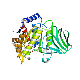
Crystal structure of vanillin bound to Co-LSD4 from Sphingobium sp. strain SYK-6
分子名称: 4-hydroxy-3-methoxybenzaldehyde, ACETATE ION, COBALT (II) ION, ...
著者Kuatsjah, E, Chan, A.C, Katahira, R, Beckham, G.T, Murphy, M.E, Eltis, L.D.
登録日2020-06-29
公開日2021-05-12
最終更新日2023-10-18
実験手法X-RAY DIFFRACTION (1.651 Å)
主引用文献Structural and functional analysis of lignostilbene dioxygenases from Sphingobium sp. SYK-6.
J.Biol.Chem., 296, 2021
6XM8
DownloadVisualize
BU of 6xm8 by Molmil
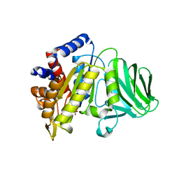
Crystal structure of lignostilbene bound to Co-LSD4 from Sphingobium sp. strain SYK-6
分子名称: COBALT (II) ION, DIMETHYL SULFOXIDE, Dioxygenase, ...
著者Kuatsjah, E, Chan, A.C, Katahira, R, Beckham, G.T, Murphy, M.E, Eltis, L.D.
登録日2020-06-29
公開日2021-05-12
最終更新日2023-10-18
実験手法X-RAY DIFFRACTION (1.85 Å)
主引用文献Structural and functional analysis of lignostilbene dioxygenases from Sphingobium sp. SYK-6.
J.Biol.Chem., 296, 2021
6XM7
DownloadVisualize
BU of 6xm7 by Molmil
Crystal structure of DCA-S bound to Co-LSD4 from Sphingobium sp. strain SYK-6
分子名称: (2E)-3-{4-hydroxy-3-[(E)-2-(4-hydroxy-3-methoxyphenyl)ethenyl]-5-methoxyphenyl}prop-2-enoic acid, COBALT (II) ION, DIMETHYL SULFOXIDE, ...
著者Kuatsjah, E, Chan, A.C, Katahira, R, Beckham, G.T, Murphy, M.E, Eltis, L.D.
登録日2020-06-29
公開日2021-05-12
最終更新日2023-10-18
実験手法X-RAY DIFFRACTION (1.45 Å)
主引用文献Structural and functional analysis of lignostilbene dioxygenases from Sphingobium sp. SYK-6.
J.Biol.Chem., 296, 2021
6XMA
DownloadVisualize
BU of 6xma by Molmil
Crystal structure of iron-bound LSD4 from Sphingobium sp. strain SYK-6
分子名称: Dioxygenase, FE (III) ION, SULFATE ION
著者Kuatsjah, E, Chan, A.C, Katahira, R, Beckham, G.T, Murphy, M.E, Eltis, L.D.
登録日2020-06-29
公開日2021-05-12
最終更新日2023-10-18
実験手法X-RAY DIFFRACTION (1.45 Å)
主引用文献Structural and functional analysis of lignostilbene dioxygenases from Sphingobium sp. SYK-6.
J.Biol.Chem., 296, 2021
6XM6
DownloadVisualize
BU of 6xm6 by Molmil
Crystal structure of cobalt-bound LSD4 from Sphingobium sp. strain SYK-6
分子名称: COBALT (II) ION, Dioxygenase
著者Kuatsjah, E, Chan, A.C, Katahira, R, Beckham, G.T, Murphy, M.E, Eltis, L.D.
登録日2020-06-29
公開日2021-05-12
最終更新日2023-10-18
実験手法X-RAY DIFFRACTION (1.45 Å)
主引用文献Structural and functional analysis of lignostilbene dioxygenases from Sphingobium sp. SYK-6.
J.Biol.Chem., 296, 2021
5VMM
DownloadVisualize
BU of 5vmm by Molmil
Staphylococcus aureus IsdB bound to human hemoglobin
分子名称: Hemoglobin subunit alpha, Hemoglobin subunit beta, Iron-regulated cell wall-anchored protein, ...
著者Bowden, C.F, Chan, A.C, Murphy, M.E.
登録日2017-04-27
公開日2017-11-15
最終更新日2023-10-04
実験手法X-RAY DIFFRACTION (3.6 Å)
主引用文献Structure-function analyses reveal key features in Staphylococcus aureus IsdB-associated unfolding of the heme-binding pocket of human hemoglobin.
J. Biol. Chem., 293, 2018
<12


220113

件を2024-05-22に公開中

PDB statisticsPDBj update infoContact PDBjnumon