Loading
PDBj
MenuPDBj@FacebookPDBj@TwitterPDBj@YouTubewwPDB FoundationwwPDB
RCSB PDBPDBeBMRBAdv. SearchSearch help
Search by PDB author
8FI3
DownloadVisualize
BU of 8fi3 by Molmil
Bifunctional ligase/repressor BirA from Klebsiella pneumoniae complexed with biotin (Domain Swapped Dimer)
Descriptor: BIOTIN, Bifunctional ligase/repressor BirA
Authors:Seattle Structural Genomics Center for Infectious Disease, Seattle Structural Genomics Center for Infectious Disease (SSGCID)
Deposit date:2022-12-15
Release date:2022-12-28
Last modified:2023-11-22
Method:X-RAY DIFFRACTION (2.9 Å)
Cite:Bifunctional ligase/repressor BirA from Klebsiella pneumoniae complexed with biotin (Domain Swapped Dimer)
To be published
8FI4
DownloadVisualize
BU of 8fi4 by Molmil
Crystal Structure of Glycylpeptide N-tetradecanoyltransferase (N-myristoyl transferase) (NMT) from Leishmania major Friedlin bound to tetradecanoyl-CoA (Orthorhombic P Form 1)
Descriptor: Glycylpeptide N-tetradecanoyltransferase, TETRADECANOYL-COA
Authors:Seattle Structural Genomics Center for Infectious Disease, Seattle Structural Genomics Center for Infectious Disease (SSGCID)
Deposit date:2022-12-15
Release date:2022-12-28
Last modified:2023-11-22
Method:X-RAY DIFFRACTION (1.8 Å)
Cite:Crystal Structure of Glycylpeptide N-tetradecanoyltransferase (N-myristoyl transferase) (NMT) from Leishmania major Friedlin bound to tetradecanoyl-CoA (Orthorhombic P Form 1)
To be published
8FT7
DownloadVisualize
BU of 8ft7 by Molmil
Crystal structure of SAH bound protein arginine N-methyltransferase 1 (PRMT1) from Naegleria fowleri
Descriptor: PUA domain-containing protein, S-ADENOSYL-L-HOMOCYSTEINE
Authors:Seattle Structural Genomics Center for Infectious Disease (SSGCID)
Deposit date:2023-01-11
Release date:2023-01-25
Last modified:2023-11-22
Method:X-RAY DIFFRACTION (2.15 Å)
Cite:Crystal structure of SAH bound protein arginine N-methyltransferase 1 (PRMT1) from Naegleria fowleri
To be published
8FUY
DownloadVisualize
BU of 8fuy by Molmil
Glucose-6-phosphate 1-dehydrogenase (G6PDH) from Crithidia fasciculata (citrate bound)
Descriptor: CHLORIDE ION, CITRIC ACID, Glucose-6-phosphate 1-dehydrogenase
Authors:Seattle Structural Genomics Center for Infectious Disease (SSGCID), Seattle Structural Genomics Center for Infectious Disease (SSGCID)
Deposit date:2023-01-18
Release date:2023-02-08
Method:X-RAY DIFFRACTION (2.45 Å)
Cite:Glucose-6-phosphate 1-dehydrogenase (G6PDH) from Crithidia fasciculata (citrate bound)
To be published
8G0V
DownloadVisualize
BU of 8g0v by Molmil
Crystal Structure of Acetyl-CoA synthetase in complex with a propyne ester AMP inhibitor from Cryptococcus neoformans H99
Descriptor: 1,2-ETHANEDIOL, 5'-O-{(S)-hydroxy[(prop-2-yn-1-yl)oxy]phosphoryl}adenosine, Acetyl-coenzyme A synthetase, ...
Authors:Seattle Structural Genomics Center for Infectious Disease, Seattle Structural Genomics Center for Infectious Disease (SSGCID)
Deposit date:2023-02-01
Release date:2023-02-15
Last modified:2024-05-22
Method:X-RAY DIFFRACTION (1.9 Å)
Cite:Crystal Structure of Acetyl-CoA synthetase in complex with a propyne ester AMP inhibitor from Cryptococcus neoformans H99
To be published
8G0R
DownloadVisualize
BU of 8g0r by Molmil
Crystal Structure of Acetyl-CoA synthetase in complex with a cyclopentyl ester AMP inhibitor from Cryptococcus neoformans H99
Descriptor: 5'-O-[(S)-(cyclopentyloxy)(hydroxy)phosphoryl]adenosine, Acetyl-coenzyme A synthetase
Authors:Seattle Structural Genomics Center for Infectious Disease, Seattle Structural Genomics Center for Infectious Disease (SSGCID)
Deposit date:2023-02-01
Release date:2023-02-15
Last modified:2024-05-22
Method:X-RAY DIFFRACTION (2.6 Å)
Cite:Crystal Structure of Acetyl-CoA synthetase in complex with a cyclopentyl ester AMP inhibitor from Cryptococcus neoformans H99
To be published
8G0T
DownloadVisualize
BU of 8g0t by Molmil
Crystal Structure of Acetyl-CoA synthetase in complex with a cyclopropyl ester AMP inhibitor from Cryptococcus neoformans H99
Descriptor: 1,2-ETHANEDIOL, 5'-O-[(R)-(cyclopropyloxy)(hydroxy)phosphoryl]adenosine, Acetyl-coenzyme A synthetase
Authors:Seattle Structural Genomics Center for Infectious Disease, Seattle Structural Genomics Center for Infectious Disease (SSGCID)
Deposit date:2023-02-01
Release date:2023-02-15
Last modified:2024-05-22
Method:X-RAY DIFFRACTION (2.45 Å)
Cite:Crystal Structure of Acetyl-CoA synthetase in complex with a cyclopropyl ester AMP inhibitor from Cryptococcus neoformans H99
To be published
8G0U
DownloadVisualize
BU of 8g0u by Molmil
Crystal Structure of Acetyl-CoA synthetase in complex with an isopropyl ester AMP inhibitor from Cryptococcus neoformans H99
Descriptor: 1,2-ETHANEDIOL, 5'-O-{(R)-hydroxy[(propan-2-yl)oxy]phosphoryl}adenosine, Acetyl-coenzyme A synthetase
Authors:Seattle Structural Genomics Center for Infectious Disease, Seattle Structural Genomics Center for Infectious Disease (SSGCID)
Deposit date:2023-02-01
Release date:2023-02-15
Last modified:2024-05-22
Method:X-RAY DIFFRACTION (1.9 Å)
Cite:Crystal Structure of Acetyl-CoA synthetase in complex with an isopropyl ester AMP inhibitor from Cryptococcus neoformans H99
To be published
8G0S
DownloadVisualize
BU of 8g0s by Molmil
Crystal Structure of Acetyl-CoA synthetase in complex with a cyclopentyl ester AMP inhibitor from Cryptococcus neoformans H99 (tetragonal form)
Descriptor: 5'-O-[(S)-(cyclopentyloxy)(hydroxy)phosphoryl]adenosine, Acetyl-coenzyme A synthetase, CHLORIDE ION
Authors:Seattle Structural Genomics Center for Infectious Disease (SSGCID)
Deposit date:2023-02-01
Release date:2023-02-15
Last modified:2024-05-22
Method:X-RAY DIFFRACTION (2.9 Å)
Cite:Crystal Structure of Acetyl-CoA synthetase in complex with a cyclopentyl ester AMP inhibitor from Cryptococcus neoformans H99 (tetragonal form)
To Be Published
4DC5
DownloadVisualize
BU of 4dc5 by Molmil
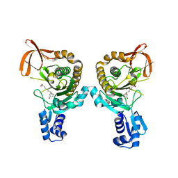
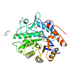
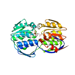
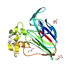
Crystal Structure of Thaumatin Unexposed to Excessive SONICC Imaging Laser Dose.
Descriptor: GLYCEROL, L(+)-TARTARIC ACID, Thaumatin I
Authors:Mulichak, A.M, Becker, M, Kissick, D.J, Keefe, L.J, Fischetti, R.F, Simpson, G.J.
Deposit date:2012-01-17
Release date:2013-01-23
Last modified:2023-09-13
Method:X-RAY DIFFRACTION (1.48 Å)
Cite:Towards protein-crystal centering using second-harmonic generation (SHG) microscopy.
Acta Crystallogr.,Sect.D, 69, 2013
4DC8
DownloadVisualize
BU of 4dc8 by Molmil
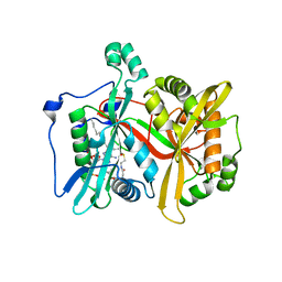
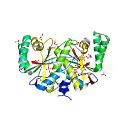
Crystal Structure of Myoglobin Unexposed to Excessive SONICC Imaging Laser Dose.
Descriptor: Myoglobin, PROTOPORPHYRIN IX CONTAINING FE, SULFATE ION
Authors:Becker, M, Mulichak, A.M, Kissick, D.J, Fischetti, R.F, Keefe, L.J, Simpson, D.J.
Deposit date:2012-01-17
Release date:2013-01-23
Last modified:2024-02-28
Method:X-RAY DIFFRACTION (1.5 Å)
Cite:Towards protein-crystal centering using second-harmonic generation (SHG) microscopy.
Acta Crystallogr.,Sect.D, 69, 2013
4DC6
DownloadVisualize
BU of 4dc6 by Molmil
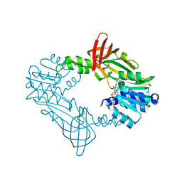
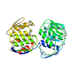
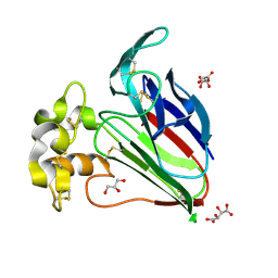
Crystal Structure of Thaumatin Exposed to Excessive SONICC Imaging Laser Dose.
Descriptor: GLYCEROL, L(+)-TARTARIC ACID, Thaumatin I
Authors:Mulichak, A.M, Becker, M, Kissick, D.J, Keefe, L.J, Fischetti, R.F, Simpson, G.J.
Deposit date:2012-01-17
Release date:2013-01-23
Last modified:2023-09-13
Method:X-RAY DIFFRACTION (1.48 Å)
Cite:Towards protein-crystal centering using second-harmonic generation (SHG) microscopy.
Acta Crystallogr.,Sect.D, 69, 2013
4DC7
DownloadVisualize
BU of 4dc7 by Molmil
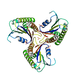
Crystal Structure of Myoglobin Exposed to Excessive SONICC Imaging Laser Dose.
Descriptor: Myoglobin, PROTOPORPHYRIN IX CONTAINING FE, SULFATE ION
Authors:Becker, M, Mulichak, A.M, Kissick, D.J, Fischetti, R.F, Keefe, L.J, Simpson, G.J.
Deposit date:2012-01-17
Release date:2013-01-23
Last modified:2024-02-28
Method:X-RAY DIFFRACTION (1.5 Å)
Cite:Towards protein-crystal centering using second-harmonic generation (SHG) microscopy.
Acta Crystallogr.,Sect.D, 69, 2013
7TMB
DownloadVisualize
BU of 7tmb by Molmil
Crystal Structure of N-ethylmaleimide reductase from Klebsiella pneumoniae
Descriptor: CITRATE ANION, FLAVIN MONONUCLEOTIDE, N-ethylmaleimide reductase, ...
Authors:Seattle Structural Genomics Center for Infectious Disease (SSGCID)
Deposit date:2022-01-19
Release date:2022-02-02
Last modified:2023-10-18
Method:X-RAY DIFFRACTION (2.1 Å)
Cite:Crystal Structure of N-ethylmaleimide reductase from Klebsiella pneumoniae
To be published
7TMF
DownloadVisualize
BU of 7tmf by Molmil
Crystal Structure of NAD(P)H nitroreductase from Klebsiella pneumoniae (short b-axis)
Descriptor: 2-[BIS-(2-HYDROXY-ETHYL)-AMINO]-2-HYDROXYMETHYL-PROPANE-1,3-DIOL, FLAVIN MONONUCLEOTIDE, GLYCEROL, ...
Authors:Seattle Structural Genomics Center for Infectious Disease (SSGCID)
Deposit date:2022-01-19
Release date:2022-02-02
Last modified:2023-10-18
Method:X-RAY DIFFRACTION (1.97 Å)
Cite:Crystal Structure of NAD(P)H nitroreductase from Klebsiella pneumoniae (short b-axis)
To be published
7TM4
DownloadVisualize
BU of 7tm4 by Molmil
Crystal structure of apo 3-phosphoshikimate 1-carboxyvinyltransferase from Klebsiella pneumoniae
Descriptor: 3-phosphoshikimate 1-carboxyvinyltransferase, NITRATE ION
Authors:Seattle Structural Genomics Center for Infectious Disease (SSGCID)
Deposit date:2022-01-19
Release date:2022-02-02
Last modified:2023-10-18
Method:X-RAY DIFFRACTION (1.7 Å)
Cite:Crystal structure of apo 3-phosphoshikimate 1-carboxyvinyltransferase from Klebsiella pneumoniae
To be published
7TM8
DownloadVisualize
BU of 7tm8 by Molmil
Crystal structure of Bifunctional adenosylcobalamin biosynthesis protein from Klebsiella pneumoniae
Descriptor: Bifunctional adenosylcobalamin biosynthesis protein, SULFATE ION
Authors:Seattle Structural Genomics Center for Infectious Disease (SSGCID)
Deposit date:2022-01-19
Release date:2022-02-02
Last modified:2023-10-18
Method:X-RAY DIFFRACTION (1.65 Å)
Cite:Crystal structure of Bifunctional adenosylcobalamin biosynthesis protein from Klebsiella pneumoniae
To be published
7TM7
DownloadVisualize
BU of 7tm7 by Molmil
Crystal structure of Alpha-1,4 glucan phosphorylase from Klebsiella pneumoniae
Descriptor: Alpha-1,4 glucan phosphorylase, PYRIDOXAL-5'-PHOSPHATE
Authors:Seattle Structural Genomics Center for Infectious Disease (SSGCID)
Deposit date:2022-01-19
Release date:2022-02-02
Last modified:2023-10-18
Method:X-RAY DIFFRACTION (1.91 Å)
Cite:Crystal structure of Alpha-1,4 glucan phosphorylase from Klebsiella pneumoniae
To be published
7TMV
DownloadVisualize
BU of 7tmv by Molmil
Crystal structure of a Putative structural protein from Klebsiella pneumoniae
Descriptor: Putative structural protein, ZINC ION
Authors:Seattle Structural Genomics Center for Infectious Disease (SSGCID)
Deposit date:2022-01-20
Release date:2022-02-02
Last modified:2023-10-18
Method:X-RAY DIFFRACTION (2.3 Å)
Cite:Crystal structure of a Putative structural protein from Klebsiella pneumoniae
To be published
7TM6
DownloadVisualize
BU of 7tm6 by Molmil
Crystal structure of shikimate-3-phosphate and glyphosate bound 3-phosphoshikimate 1-carboxyvinyltransferase from Klebsiella pneumoniae
Descriptor: 3-phosphoshikimate 1-carboxyvinyltransferase, GLYPHOSATE, PENTAETHYLENE GLYCOL, ...
Authors:Seattle Structural Genomics Center for Infectious Disease (SSGCID)
Deposit date:2022-01-19
Release date:2022-02-02
Last modified:2023-10-18
Method:X-RAY DIFFRACTION (1.26 Å)
Cite:Crystal structure of shikimate-3-phosphate and glyphosate bound 3-phosphoshikimate 1-carboxyvinyltransferase from Klebsiella pneumoniae
To be published
7TMG
DownloadVisualize
BU of 7tmg by Molmil
Crystal Structure of NAD(P)H nitroreductase from Klebsiella pneumoniae (long b-axis)
Descriptor: FLAVIN MONONUCLEOTIDE, NAD(P)H nitroreductase, PHOSPHATE ION
Authors:Seattle Structural Genomics Center for Infectious Disease (SSGCID)
Deposit date:2022-01-19
Release date:2022-02-02
Last modified:2023-10-18
Method:X-RAY DIFFRACTION (1.9 Å)
Cite:Crystal Structure of NAD(P)H nitroreductase from Klebsiella pneumoniae (long b-axis)
To be published
7TM9
DownloadVisualize
BU of 7tm9 by Molmil
Crystal structure of Bacterial alkaline phosphatase from Klebsiella pneumoniae
Descriptor: Bacterial alkaline phosphatase, MAGNESIUM ION, ZINC ION
Authors:Seattle Structural Genomics Center for Infectious Disease (SSGCID)
Deposit date:2022-01-19
Release date:2022-02-02
Last modified:2023-10-18
Method:X-RAY DIFFRACTION (1.95 Å)
Cite:Crystal structure of Bacterial alkaline phosphatase from Klebsiella pneumoniae
To be published
7TM5
DownloadVisualize
BU of 7tm5 by Molmil
Crystal structure of shikimate-3-phosphate bound 3-phosphoshikimate 1-carboxyvinyltransferase from Klebsiella pneumoniae
Descriptor: 3-phosphoshikimate 1-carboxyvinyltransferase, FORMIC ACID, GLYCEROL, ...
Authors:Seattle Structural Genomics Center for Infectious Disease (SSGCID)
Deposit date:2022-01-19
Release date:2022-02-02
Last modified:2023-10-18
Method:X-RAY DIFFRACTION (1.41 Å)
Cite:Crystal structure of shikimate-3-phosphate bound 3-phosphoshikimate 1-carboxyvinyltransferase from Klebsiella pneumoniae
To be published
7TMD
DownloadVisualize
BU of 7tmd by Molmil
Crystal structure of Ornithine carbamoyltransferase from Klebsiella pneumoniae
Descriptor: BROMIDE ION, IODIDE ION, Ornithine carbamoyltransferase
Authors:Seattle Structural Genomics Center for Infectious Disease (SSGCID)
Deposit date:2022-01-19
Release date:2022-02-02
Last modified:2023-10-18
Method:X-RAY DIFFRACTION (2.35 Å)
Cite:Crystal structure of Ornithine carbamoyltransferase from Klebsiella pneumoniae
To be published
3KOH
DownloadVisualize
BU of 3koh by Molmil
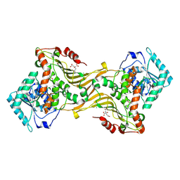
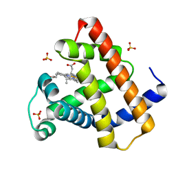
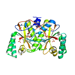
Cytochrome P450 2E1 with omega-imidazolyl octanoic acid
Descriptor: 8-(1H-imidazol-1-yl)octanoic acid, Cytochrome P450 2E1, PROTOPORPHYRIN IX CONTAINING FE
Authors:Scott, E.E, Porubsky, P.R.
Deposit date:2009-11-13
Release date:2010-05-12
Last modified:2023-09-06
Method:X-RAY DIFFRACTION (2.9 Å)
Cite:Human cytochrome P450 2E1 structures with fatty acid analogs reveal a previously unobserved binding mode.
J.Biol.Chem., 285, 2010

220113

数据于2024-05-22公开中

PDB statisticsPDBj update infoContact PDBjnumon