Loading
PDBj
MenuPDBj@FacebookPDBj@TwitterPDBj@YouTubewwPDB FoundationwwPDB
RCSB PDBPDBeBMRBAdv. SearchSearch help
Search by PDB author
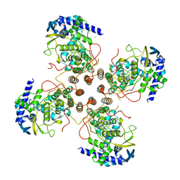
8UR4
DownloadVisualize
BU of 8ur4 by Molmil
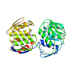
Crystal Structure of macrophage migration inhibitory factor (MIF) from Trichomonas vaginalis (I4122 form)
Descriptor: CHLORIDE ION, MACROPHAGE MIGRATION INHIBITORY FACTOR
Authors:Seattle Structural Genomics Center for Infectious Disease, Seattle Structural Genomics Center for Infectious Disease (SSGCID)
Deposit date:2023-10-25
Release date:2024-03-20
Method:X-RAY DIFFRACTION (2.55 Å)
Cite:Crystal Structure of macrophage migration inhibitory factor (MIF) from Trichomonas vaginalis (I4122 form)
To be published
9AV6
DownloadVisualize
BU of 9av6 by Molmil
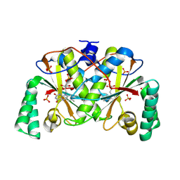
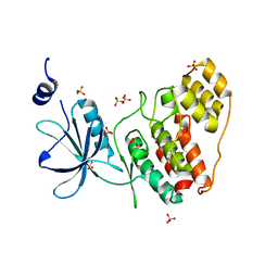
Crystal structure of CMGC family protein kinase from Trichomonas vaginalis (Apo)
Descriptor: SULFATE ION, non-specific serine/threonine protein kinase
Authors:Seattle Structural Genomics Center for Infectious Disease, Seattle Structural Genomics Center for Infectious Disease (SSGCID)
Deposit date:2024-03-01
Release date:2024-03-13
Method:X-RAY DIFFRACTION (2 Å)
Cite:Crystal structure of CMGC family protein kinase from Trichomonas vaginalis (Apo)
To be published
9AV7
DownloadVisualize
BU of 9av7 by Molmil
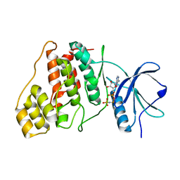
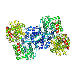
Crystal structure of CMGC family protein kinase from Trichomonas vaginalis (AMP-PNP)
Descriptor: PHOSPHOAMINOPHOSPHONIC ACID-ADENYLATE ESTER, non-specific serine/threonine protein kinase
Authors:Seattle Structural Genomics Center for Infectious Disease, Seattle Structural Genomics Center for Infectious Disease (SSGCID)
Deposit date:2024-03-01
Release date:2024-03-13
Method:X-RAY DIFFRACTION (2.1 Å)
Cite:Crystal structure of CMGC family protein kinase from Trichomonas vaginalis (AMP-PNP)
To be published
9B22
DownloadVisualize
BU of 9b22 by Molmil
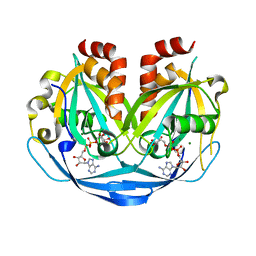
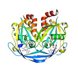
Crystal structure of ADP-ribose diphosphatase from Klebsiella pneumoniae (ADP Ribose and AMP bound)
Descriptor: ADENOSINE MONOPHOSPHATE, ADP-ribose pyrophosphatase, MAGNESIUM ION, ...
Authors:Seattle Structural Genomics Center for Infectious Disease, Seattle Structural Genomics Center for Infectious Disease (SSGCID)
Deposit date:2024-03-14
Release date:2024-03-27
Method:X-RAY DIFFRACTION (1.3 Å)
Cite:Crystal structure of ADP-ribose diphosphatase from Klebsiella pneumoniae (ADP Ribose and AMP bound)
To be published
9B1Z
DownloadVisualize
BU of 9b1z by Molmil
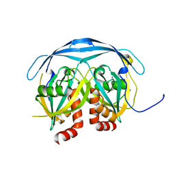
Crystal structure of ADP-ribose diphosphatase from Klebsiella pneumoniae (Apo)
Descriptor: ADP-ribose pyrophosphatase, SODIUM ION
Authors:Seattle Structural Genomics Center for Infectious Disease, Seattle Structural Genomics Center for Infectious Disease (SSGCID)
Deposit date:2024-03-14
Release date:2024-03-27
Method:X-RAY DIFFRACTION (1.25 Å)
Cite:Crystal structure of ADP-ribose diphosphatase from Klebsiella pneumoniae (Apo)
To be published
9AZZ
DownloadVisualize
BU of 9azz by Molmil
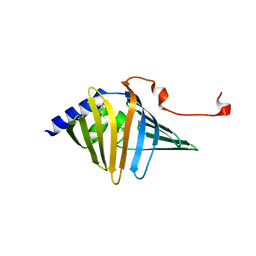
Crystal structure of outer membrane lipoprotein carrier protein (LolA) from Ehrlichia ruminantium
Descriptor: GLYCEROL, Outer membrane lipoprotein carrier protein LolA, TETRAETHYLENE GLYCOL
Authors:Seattle Structural Genomics Center for Infectious Disease (SSGCID)
Deposit date:2024-03-11
Release date:2024-03-20
Method:X-RAY DIFFRACTION (2 Å)
Cite:Crystal structure of outer membrane lipoprotein carrier protein (LolA) from Ehrlichia ruminantium
To be published
9B21
DownloadVisualize
BU of 9b21 by Molmil
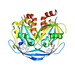
Crystal structure of ADP-ribose diphosphatase from Klebsiella pneumoniae (ADP Ribose bound, Orthorhombic P form)
Descriptor: ADP-ribose pyrophosphatase, MAGNESIUM ION, [(2R,3S,4R,5R)-5-(6-AMINOPURIN-9-YL)-3,4-DIHYDROXY-OXOLAN-2-YL]METHYL [HYDROXY-[[(2R,3S,4R,5S)-3,4,5-TRIHYDROXYOXOLAN-2-YL]METHOXY]PHOSPHORYL] HYDROGEN PHOSPHATE
Authors:Seattle Structural Genomics Center for Infectious Disease, Seattle Structural Genomics Center for Infectious Disease (SSGCID)
Deposit date:2024-03-14
Release date:2024-03-27
Method:X-RAY DIFFRACTION (1.6 Å)
Cite:Crystal structure of ADP-ribose diphosphatase from Klebsiella pneumoniae (ADP Ribose bound, Orthorhombic P form)
To be published
9B20
DownloadVisualize
BU of 9b20 by Molmil
Crystal structure of ADP-ribose diphosphatase from Klebsiella pneumoniae (AMP bound)
Descriptor: ADENOSINE MONOPHOSPHATE, ADP-ribose pyrophosphatase, MAGNESIUM ION
Authors:Seattle Structural Genomics Center for Infectious Disease, Seattle Structural Genomics Center for Infectious Disease (SSGCID)
Deposit date:2024-03-14
Release date:2024-03-27
Method:X-RAY DIFFRACTION (1.55 Å)
Cite:Crystal structure of ADP-ribose diphosphatase from Klebsiella pneumoniae (AMP bound)
To be published
9B0M
DownloadVisualize
BU of 9b0m by Molmil
Crystal structure of Macrophage migration inhibitory factor from Plasmodium vivax
Descriptor: DI(HYDROXYETHYL)ETHER, GLYCEROL, L-dopachrome isomerase
Authors:Seattle Structural Genomics Center for Infectious Disease, Seattle Structural Genomics Center for Infectious Disease (SSGCID)
Deposit date:2024-03-12
Release date:2024-03-20
Method:X-RAY DIFFRACTION (1.8 Å)
Cite:Crystal structure of Macrophage migration inhibitory factor from Plasmodium vivax
To be published
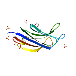
9BKW
DownloadVisualize
BU of 9bkw by Molmil
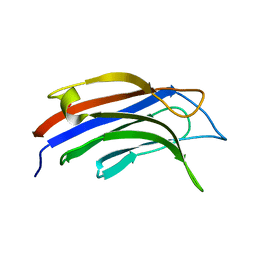
Crystal structure of a C2 domain from Trichomonas vaginalis
Descriptor: XYPPX repeat family protein
Authors:Seattle Structural Genomics Center for Infectious Disease, Seattle Structural Genomics Center for Infectious Disease (SSGCID)
Deposit date:2024-04-29
Release date:2024-05-08
Method:X-RAY DIFFRACTION (1.85 Å)
Cite:Crystal structure of a C2 domain from Trichomonas vaginalis
To be published
9BJL
DownloadVisualize
BU of 9bjl by Molmil
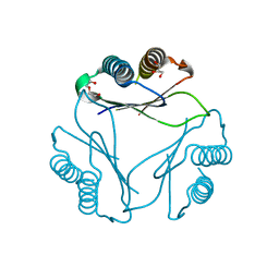
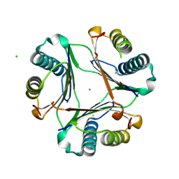
Crystal structure of Influenza D virus Nucleoprotein (Oklahoma)
Descriptor: CHLORIDE ION, Nucleoprotein
Authors:Seattle Structural Genomics Center for Infectious Disease (SSGCID)
Deposit date:2024-04-25
Release date:2024-05-08
Method:X-RAY DIFFRACTION (2.43 Å)
Cite:Crystal structure of Influenza D virus Nucleoprotein (Oklahoma)
To be published
9BKY
DownloadVisualize
BU of 9bky by Molmil
Crystal structure of a C2 domain from Trichomonas vaginalis (sulfate bound)
Descriptor: GLYCEROL, SULFATE ION, XYPPX repeat family protein
Authors:Seattle Structural Genomics Center for Infectious Disease, Seattle Structural Genomics Center for Infectious Disease (SSGCID)
Deposit date:2024-04-29
Release date:2024-05-08
Method:X-RAY DIFFRACTION (1.73 Å)
Cite:Crystal structure of a C2 domain from Trichomonas vaginalis (sulfate bound)
To be published
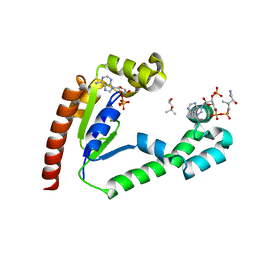
9BKZ
DownloadVisualize
BU of 9bkz by Molmil
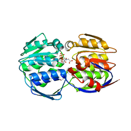
Crystal Structure of Dephospho-CoA kinase from Klebsiella aerogenes (CoA and ADP bound)
Descriptor: (4S)-2-METHYL-2,4-PENTANEDIOL, ADENOSINE-5'-DIPHOSPHATE, COENZYME A, ...
Authors:Seattle Structural Genomics Center for Infectious Disease, Seattle Structural Genomics Center for Infectious Disease (SSGCID)
Deposit date:2024-04-29
Release date:2024-05-08
Method:X-RAY DIFFRACTION (2.35 Å)
Cite:Crystal Structure of Dephospho-CoA kinase from Klebsiella aerogenes (CoA and ADP bound)
To be published
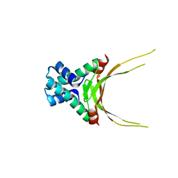
5FBM
DownloadVisualize
BU of 5fbm by Molmil
Crystal Structure of Histone Like Protein (HLP) from Streptococcus mutans Refined to 1.9 A Resolution
Descriptor: DNA-binding protein HU
Authors:Lovell, S, Battaile, K.P, Mehzabeen, N, O'Neil, P, Biswas, I.
Deposit date:2015-12-14
Release date:2016-04-06
Last modified:2023-09-27
Method:X-RAY DIFFRACTION (1.9 Å)
Cite:Crystal structure of histone-like protein from Streptococcus mutans refined to 1.9 angstrom resolution.
Acta Crystallogr F Struct Biol Commun, 72, 2016
4DC5
DownloadVisualize
BU of 4dc5 by Molmil
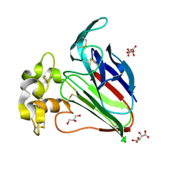
Crystal Structure of Thaumatin Unexposed to Excessive SONICC Imaging Laser Dose.
Descriptor: GLYCEROL, L(+)-TARTARIC ACID, Thaumatin I
Authors:Mulichak, A.M, Becker, M, Kissick, D.J, Keefe, L.J, Fischetti, R.F, Simpson, G.J.
Deposit date:2012-01-17
Release date:2013-01-23
Last modified:2023-09-13
Method:X-RAY DIFFRACTION (1.48 Å)
Cite:Towards protein-crystal centering using second-harmonic generation (SHG) microscopy.
Acta Crystallogr.,Sect.D, 69, 2013
4DC8
DownloadVisualize
BU of 4dc8 by Molmil
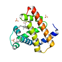
Crystal Structure of Myoglobin Unexposed to Excessive SONICC Imaging Laser Dose.
Descriptor: Myoglobin, PROTOPORPHYRIN IX CONTAINING FE, SULFATE ION
Authors:Becker, M, Mulichak, A.M, Kissick, D.J, Fischetti, R.F, Keefe, L.J, Simpson, D.J.
Deposit date:2012-01-17
Release date:2013-01-23
Last modified:2024-02-28
Method:X-RAY DIFFRACTION (1.5 Å)
Cite:Towards protein-crystal centering using second-harmonic generation (SHG) microscopy.
Acta Crystallogr.,Sect.D, 69, 2013
4DC6
DownloadVisualize
BU of 4dc6 by Molmil
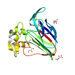
Crystal Structure of Thaumatin Exposed to Excessive SONICC Imaging Laser Dose.
Descriptor: GLYCEROL, L(+)-TARTARIC ACID, Thaumatin I
Authors:Mulichak, A.M, Becker, M, Kissick, D.J, Keefe, L.J, Fischetti, R.F, Simpson, G.J.
Deposit date:2012-01-17
Release date:2013-01-23
Last modified:2023-09-13
Method:X-RAY DIFFRACTION (1.48 Å)
Cite:Towards protein-crystal centering using second-harmonic generation (SHG) microscopy.
Acta Crystallogr.,Sect.D, 69, 2013
4DC7
DownloadVisualize
BU of 4dc7 by Molmil
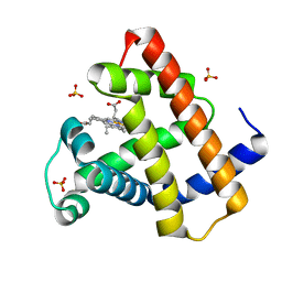
Crystal Structure of Myoglobin Exposed to Excessive SONICC Imaging Laser Dose.
Descriptor: Myoglobin, PROTOPORPHYRIN IX CONTAINING FE, SULFATE ION
Authors:Becker, M, Mulichak, A.M, Kissick, D.J, Fischetti, R.F, Keefe, L.J, Simpson, G.J.
Deposit date:2012-01-17
Release date:2013-01-23
Last modified:2024-02-28
Method:X-RAY DIFFRACTION (1.5 Å)
Cite:Towards protein-crystal centering using second-harmonic generation (SHG) microscopy.
Acta Crystallogr.,Sect.D, 69, 2013
6V02
DownloadVisualize
BU of 6v02 by Molmil
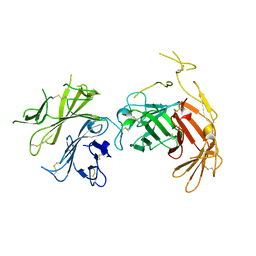
N-terminal 5 domains of CI-MPR
Descriptor: 2-acetamido-2-deoxy-beta-D-glucopyranose, Cation-independent mannose-6-phosphate receptor
Authors:Olson, L.J, Dahms, N.M, Kim, J.-J.P.
Deposit date:2019-11-18
Release date:2020-09-30
Last modified:2023-10-11
Method:X-RAY DIFFRACTION (2.46 Å)
Cite:Allosteric regulation of lysosomal enzyme recognition by the cation-independent mannose 6-phosphate receptor.
Commun Biol, 3, 2020
3KOH
DownloadVisualize
BU of 3koh by Molmil
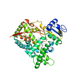
Cytochrome P450 2E1 with omega-imidazolyl octanoic acid
Descriptor: 8-(1H-imidazol-1-yl)octanoic acid, Cytochrome P450 2E1, PROTOPORPHYRIN IX CONTAINING FE
Authors:Scott, E.E, Porubsky, P.R.
Deposit date:2009-11-13
Release date:2010-05-12
Last modified:2023-09-06
Method:X-RAY DIFFRACTION (2.9 Å)
Cite:Human cytochrome P450 2E1 structures with fatty acid analogs reveal a previously unobserved binding mode.
J.Biol.Chem., 285, 2010
3LC4
DownloadVisualize
BU of 3lc4 by Molmil
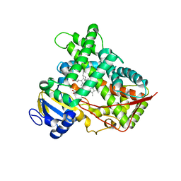
Human Cytochrome P450 2E1 in Complex with Omega-Imidazolyl-Dodecanoic Acid
Descriptor: 12-(1H-imidazol-1-yl)dodecanoic acid, Cytochrome P450 2E1, PROTOPORPHYRIN IX CONTAINING FE
Authors:Scott, E.E, Porubsky, P.R.
Deposit date:2010-01-09
Release date:2010-05-12
Last modified:2023-09-06
Method:X-RAY DIFFRACTION (3.1 Å)
Cite:Human cytochrome P450 2E1 structures with fatty acid analogs reveal a previously unobserved binding mode.
J.Biol.Chem., 285, 2010
7TM6
DownloadVisualize
BU of 7tm6 by Molmil
Crystal structure of shikimate-3-phosphate and glyphosate bound 3-phosphoshikimate 1-carboxyvinyltransferase from Klebsiella pneumoniae
Descriptor: 3-phosphoshikimate 1-carboxyvinyltransferase, GLYPHOSATE, PENTAETHYLENE GLYCOL, ...
Authors:Seattle Structural Genomics Center for Infectious Disease (SSGCID)
Deposit date:2022-01-19
Release date:2022-02-02
Last modified:2023-10-18
Method:X-RAY DIFFRACTION (1.26 Å)
Cite:Crystal structure of shikimate-3-phosphate and glyphosate bound 3-phosphoshikimate 1-carboxyvinyltransferase from Klebsiella pneumoniae
To be published
7TMG
DownloadVisualize
BU of 7tmg by Molmil
Crystal Structure of NAD(P)H nitroreductase from Klebsiella pneumoniae (long b-axis)
Descriptor: FLAVIN MONONUCLEOTIDE, NAD(P)H nitroreductase, PHOSPHATE ION
Authors:Seattle Structural Genomics Center for Infectious Disease (SSGCID)
Deposit date:2022-01-19
Release date:2022-02-02
Last modified:2023-10-18
Method:X-RAY DIFFRACTION (1.9 Å)
Cite:Crystal Structure of NAD(P)H nitroreductase from Klebsiella pneumoniae (long b-axis)
To be published
7TM7
DownloadVisualize
BU of 7tm7 by Molmil
Crystal structure of Alpha-1,4 glucan phosphorylase from Klebsiella pneumoniae
Descriptor: Alpha-1,4 glucan phosphorylase, PYRIDOXAL-5'-PHOSPHATE
Authors:Seattle Structural Genomics Center for Infectious Disease (SSGCID)
Deposit date:2022-01-19
Release date:2022-02-02
Last modified:2023-10-18
Method:X-RAY DIFFRACTION (1.91 Å)
Cite:Crystal structure of Alpha-1,4 glucan phosphorylase from Klebsiella pneumoniae
To be published
7TM4
DownloadVisualize
BU of 7tm4 by Molmil
Crystal structure of apo 3-phosphoshikimate 1-carboxyvinyltransferase from Klebsiella pneumoniae
Descriptor: 3-phosphoshikimate 1-carboxyvinyltransferase, NITRATE ION
Authors:Seattle Structural Genomics Center for Infectious Disease (SSGCID)
Deposit date:2022-01-19
Release date:2022-02-02
Last modified:2023-10-18
Method:X-RAY DIFFRACTION (1.7 Å)
Cite:Crystal structure of apo 3-phosphoshikimate 1-carboxyvinyltransferase from Klebsiella pneumoniae
To be published

219869

數據於2024-05-15公開中

PDB statisticsPDBj update infoContact PDBjnumon