1NYA
 
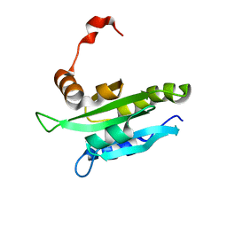
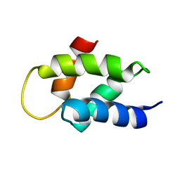
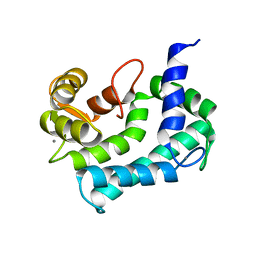 | | NMR SOLUTION STRUCTURE OF CALERYTHRIN, AN EF-HAND CALCIUM-BINDING PROTEIN | | 分子名称: | CALCIUM ION, Calerythrin | | 著者 | Tossavainen, H, Permi, P, Annila, A, Kilpelainen, I, Drakenberg, T. | | 登録日 | 2003-02-12 | | 公開日 | 2003-08-05 | | 最終更新日 | 2021-10-27 | | 実験手法 | SOLUTION NMR | | 主引用文献 | NMR solution structure of calerythrin, an EF-hand calcium-binding protein from Saccharopolyspora erythraea
Eur.J.Biochem., 270, 2003
|
|
4BMF
 | |
2HD7
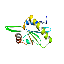 | | Solution structure of C-teminal domain of twinfilin-1. | | 分子名称: | Twinfilin-1 | | 著者 | Hellman, M.H, Paavilainen, V.O, Annila, A, Lappalainen, P, Permi, P.I. | | 登録日 | 2006-06-20 | | 公開日 | 2007-02-06 | | 最終更新日 | 2022-03-09 | | 実験手法 | SOLUTION NMR | | 主引用文献 | Structural basis and evolutionary origin of actin filament capping by twinfilin
Proc.Natl.Acad.Sci.Usa, 104, 2007
|
|
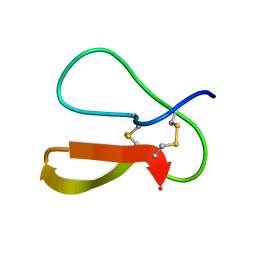
1Z6C
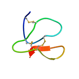
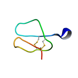
 | | Solution structure of an EGF pair (EGF34) from vitamin K-dependent protein S | | 分子名称: | CALCIUM ION, Vitamin K-dependent protein S | | 著者 | Drakenberg, T, Ghasriani, H, Thulin, E, Thamlitz, A.M, Muranyi, A, Annila, A, Stenflo, J. | | 登録日 | 2005-03-22 | | 公開日 | 2005-06-21 | | 最終更新日 | 2022-03-02 | | 実験手法 | SOLUTION NMR | | 主引用文献 | Solution Structure of the Ca(2+)-Binding EGF3-4 Pair from Vitamin K-Dependent Protein S: Identification of an Unusual Fold in EGF3.
Biochemistry, 44, 2005
|
|
1WM4
 | | Solution structure of mouse coactosin, an actin filament binding protein | | 分子名称: | Coactosin-like protein | | 著者 | Hellman, M, Paavilainen, V.O, Naumanen, P, Lappalainen, P, Annila, A, Permi, P. | | 登録日 | 2004-07-03 | | 公開日 | 2004-11-02 | | 最終更新日 | 2022-03-02 | | 実験手法 | SOLUTION NMR | | 主引用文献 | Solution structure of coactosin reveals structural homology to ADF/cofilin family proteins
Febs Lett., 576, 2004
|
|
1AY3
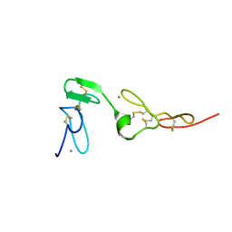
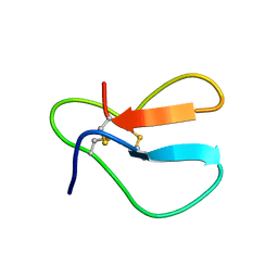
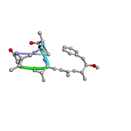 | | Nodularin from Nodularia spumigena | | 分子名称: | PEPTIDIC TOXIN NODULARIN | | 著者 | Annila, A.J. | | 登録日 | 1997-11-14 | | 公開日 | 1999-08-16 | | 最終更新日 | 2023-11-15 | | 実験手法 | SOLUTION NMR | | 主引用文献 | Solution structure of nodularin. An inhibitor of serine/threonine-specific protein phosphatases.
J.Biol.Chem., 271, 1996
|
|
1LCM
 | |
1AZ6
 | |
1AZH
 | |
1AZJ
 | |
1AZK
 | |
2V6Z
 | | Solution Structure of Amino-Terminal Domain of Human DNA Polymerase Epsilon Subunit B | | 分子名称: | DNA POLYMERASE EPSILON SUBUNIT 2 | | 著者 | Nuutinen, T, Fredriksson, K, Tossavainen, H, Pospiech, H, Pirila, P, Permi, P, Annila, A, Syvaoja, J.E. | | 登録日 | 2007-07-24 | | 公開日 | 2008-08-05 | | 最終更新日 | 2023-06-14 | | 実験手法 | SOLUTION NMR | | 主引用文献 | The Solution Structure of the Amino-Terminal Domain of Human DNA Polymerase Epsilon Subunit B is Homologous to C-Domains of Aaa+ Proteins.
Nucleic Acids Res., 36, 2008
|
|


