Loading
PDBj
MenuPDBj@FacebookPDBj@TwitterPDBj@YouTubewwPDB FoundationwwPDB
RCSB PDBPDBeBMRBAdv. SearchSearch help
Search by PDB author
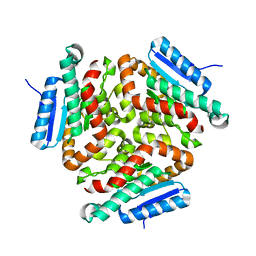
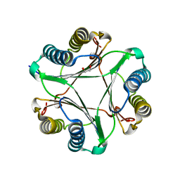
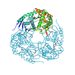
5J49
DownloadVisualize
BU of 5j49 by Molmil
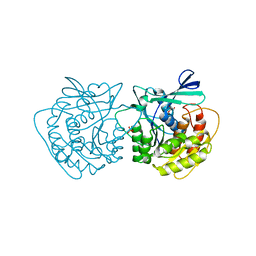
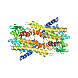
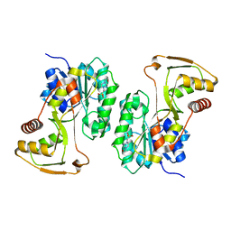
Crystal structure of UDP-glucose pyrophosporylase / UTP-glucose-1-phosphate uridylyltransferase from Burkholderia xenovorans
Descriptor: 1,2-ETHANEDIOL, SULFATE ION, UTP--glucose-1-phosphate uridylyltransferase
Authors:Seattle Structural Genomics Center for Infectious Disease (SSGCID)
Deposit date:2016-03-31
Release date:2016-05-04
Last modified:2023-09-27
Method:X-RAY DIFFRACTION (1.8 Å)
Cite:Crystal structure of UDP-glucose pyrophosporylase / UTP-glucose-1-phosphate uridylyltransferase from Burkholderia xenovorans
to be published
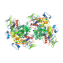
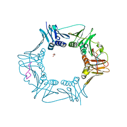
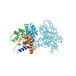
6C46
DownloadVisualize
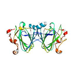
BU of 6c46 by Molmil
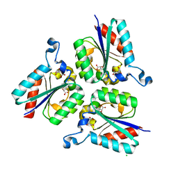
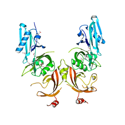
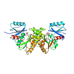
Crystal structure of dTDP-4-dehydrorhamnose 3,5-epimerase from Elizabethkingia anophelis NUHP1
Descriptor: CALCIUM ION, dTDP-4-dehydrorhamnose 3,5-epimerase
Authors:Seattle Structural Genomics Center for Infectious Disease (SSGCID)
Deposit date:2018-01-11
Release date:2018-02-14
Last modified:2023-10-04
Method:X-RAY DIFFRACTION (1.95 Å)
Cite:Crystal structure of dTDP-4-dehydrorhamnose 3,5-epimerase from Elizabethkingia anophelis NUHP1
To be Published
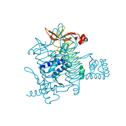
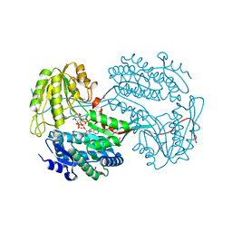
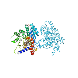
6C7C
DownloadVisualize
BU of 6c7c by Molmil
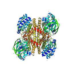
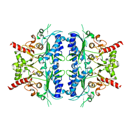
Crystal structure of Enoyl-CoA hydratase, EchA3, from Mycobacterium ulcerans Agy99
Descriptor: Enoyl-CoA hydratase, EchA3
Authors:Seattle Structural Genomics Center for Infectious Disease (SSGCID)
Deposit date:2018-01-22
Release date:2018-02-07
Last modified:2023-10-04
Method:X-RAY DIFFRACTION (2.1 Å)
Cite:Crystal structure of Enoyl-CoA hydratase, EchA3, from Mycobacterium ulcerans Agy99
to be published
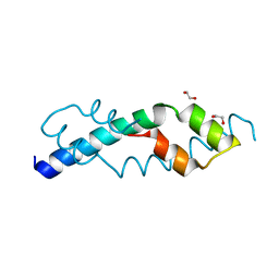
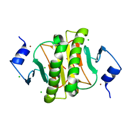
6C87
DownloadVisualize
BU of 6c87 by Molmil
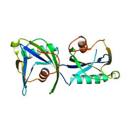
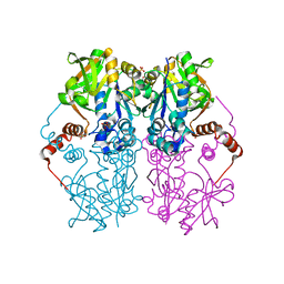
Crystal structure of rab gdp dissociation inhibitor alpha from Naegleria fowleri
Descriptor: Rab GDP dissociation inhibitor alpha, SULFATE ION
Authors:Seattle Structural Genomics Center for Infectious Disease (SSGCID)
Deposit date:2018-01-24
Release date:2018-02-14
Last modified:2023-10-04
Method:X-RAY DIFFRACTION (2.6 Å)
Cite:Crystal structure of rab gdp dissociation inhibitor alpha from Naegleria fowleri
TO BE PUBLISHED
6CKT
DownloadVisualize
BU of 6ckt by Molmil
Crystal structure of 2,3,4,5-tetrahydropyridine-2,6-dicarboxylate N-succinyltransferase from Legionella pneumophila Philadelphia 1
Descriptor: 1,2-ETHANEDIOL, 2,3,4,5-tetrahydropyridine-2,6-dicarboxylate N-succinyltransferase
Authors:Seattle Structural Genomics Center for Infectious Disease (SSGCID)
Deposit date:2018-02-28
Release date:2018-03-21
Last modified:2023-10-04
Method:X-RAY DIFFRACTION (1.8 Å)
Cite:Crystal structure of 2,3,4,5-tetrahydropyridine-2,6-dicarboxylate N-succinyltransferase from Legionella pneumophila Philadelphia 1
to be published
6CUM
DownloadVisualize
BU of 6cum by Molmil
Crystal structure of a C-terminal proteolytic fragment of a protein annotated as an LAO/AO transport system ATPase but likely MeaB and MMAA-like GTPase from Mycobacterium smegmatis
Descriptor: 1,2-ETHANEDIOL, LAO/AO transport system ATPase
Authors:Seattle Structural Genomics Center for Infectious Disease (SSGCID)
Deposit date:2018-03-26
Release date:2018-04-11
Last modified:2024-04-03
Method:X-RAY DIFFRACTION (1.6 Å)
Cite:Ab initio structure solution of a proteolytic fragment using ARCIMBOLDO.
Acta Crystallogr F Struct Biol Commun, 74, 2018
6CW5
DownloadVisualize
BU of 6cw5 by Molmil
Crystal structure of Ribokinase from Cryptococcus neoformans var. grubii serotype A
Descriptor: GLYCEROL, PHOSPHATE ION, Ribokinase
Authors:Seattle Structural Genomics Center for Infectious Disease (SSGCID)
Deposit date:2018-03-29
Release date:2018-04-18
Last modified:2023-10-04
Method:X-RAY DIFFRACTION (1.8 Å)
Cite:Crystal structure of Ribokinase from Cryptococcus neoformans var. grubii serotype A
to be published
6CV6
DownloadVisualize
BU of 6cv6 by Molmil
Crystal structure of 3-dehydroquinate dehydratase, type II, from Burkholderia phymatum STM815
Descriptor: 3-dehydroquinate dehydratase, CHLORIDE ION, D(-)-TARTARIC ACID, ...
Authors:Seattle Structural Genomics Center for Infectious Disease (SSGCID)
Deposit date:2018-03-27
Release date:2018-04-11
Last modified:2023-10-04
Method:X-RAY DIFFRACTION (2.6 Å)
Cite:Crystal structure of 3-dehydroquinate dehydratase, type II, from Burkholderia phymatum STM815
to be published
6CJA
DownloadVisualize
BU of 6cja by Molmil
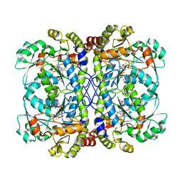
Crystal structure of Cystathionine beta-lyase from Legionella pneumophila Philadelphia 1 in complex with Alanyl-PLP and Serine
Descriptor: (E)-N-({3-hydroxy-2-methyl-5-[(phosphonooxy)methyl]pyridin-4-yl}methylidene)-L-alanine, Cystathionine beta-lyase, GLYCEROL, ...
Authors:Seattle Structural Genomics Center for Infectious Disease (SSGCID)
Deposit date:2018-02-26
Release date:2018-03-21
Last modified:2024-04-03
Method:X-RAY DIFFRACTION (1.7 Å)
Cite:Crystal structure of Cystathionine beta-lyase from Legionella pneumophila Philadelphia 1 in complex with Alanyl-PLP and Serine
to be published
6CJB
DownloadVisualize
BU of 6cjb by Molmil
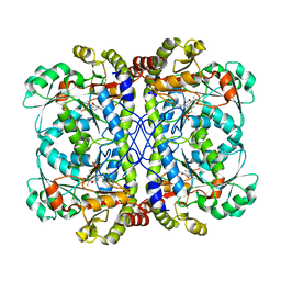
Crystal structure of Cystathionine beta-lyase from Legionella pneumophila Philadelphia 1 covalently bound to Pyridoxal phosphate
Descriptor: Cystathionine beta-lyase, FORMIC ACID, GLYCEROL
Authors:Seattle Structural Genomics Center for Infectious Disease (SSGCID)
Deposit date:2018-02-26
Release date:2018-03-21
Last modified:2023-11-15
Method:X-RAY DIFFRACTION (1.75 Å)
Cite:Crystal structure of Cystathionine beta-lyase from Legionella pneumophila Philadelphia 1 covalently bound to Pyridoxal phosphate
to be published
6CUQ
DownloadVisualize
BU of 6cuq by Molmil
Crystal structure of Macrophage migration inhibitory factor-like protein (EhMIF) from Entamoeba histolytica
Descriptor: 1,2-ETHANEDIOL, Macrophage migration inhibitory factor-like protein
Authors:Seattle Structural Genomics Center for Infectious Disease (SSGCID)
Deposit date:2018-03-26
Release date:2018-04-11
Last modified:2023-10-04
Method:X-RAY DIFFRACTION (2.45 Å)
Cite:Crystal structure of Macrophage migration inhibitory factor-like protein (EhMIF) from Entamoeba histolytica
to be published
6DJK
DownloadVisualize
BU of 6djk by Molmil
Structure of DNA polymerase III subunit beta from Rickettsia typhi in complex with a natural product
Descriptor: 1,2-ETHANEDIOL, ACE-MVA-MP8-NZC-LEU-MP8-LEU-MVA-PRO-MLU-GLY, Beta sliding clamp, ...
Authors:Seattle Structural Genomics Center for Infectious Disease (SSGCID)
Deposit date:2018-05-25
Release date:2019-05-29
Last modified:2023-11-15
Method:X-RAY DIFFRACTION (1.85 Å)
Cite:Structure of DNA polymerase III subunit beta from Rickettsia typhi in complex with a natural product
to be published
6DBB
DownloadVisualize
BU of 6dbb by Molmil
Crystal structure of a Putative aldehyde dehydrogenase family protein Burkholderia cenocepacia J2315 in complex with partially reduced NADH
Descriptor: 1,2-ETHANEDIOL, BETA-6-HYDROXY-1,4,5,6-TETRHYDRONICOTINAMIDE ADENINE DINUCLEOTIDE, NICOTINAMIDE-ADENINE-DINUCLEOTIDE, ...
Authors:Seattle Structural Genomics Center for Infectious Disease (SSGCID)
Deposit date:2018-05-02
Release date:2018-08-29
Last modified:2024-04-03
Method:X-RAY DIFFRACTION (2.1 Å)
Cite:Crystal structure of a Putative aldehyde dehydrogenase family protein Burkholderia cenocepacia J2315 in complex with partially reduced NADH
TO BE PUBLISHED
6D6J
DownloadVisualize
BU of 6d6j by Molmil
Crystal structure of HIT family hydrolase from Legionella pneumophila Philadelphia 1
Descriptor: CHLORIDE ION, HIT family hydrolase, ZINC ION
Authors:Seattle Structural Genomics Center for Infectious Disease (SSGCID)
Deposit date:2018-04-21
Release date:2018-05-02
Last modified:2024-03-13
Method:X-RAY DIFFRACTION (1.35 Å)
Cite:Crystal structure of HIT family hydrolase from Legionella pneumophila Philadelphia 1
to be published
6D6K
DownloadVisualize
BU of 6d6k by Molmil
Structure of polyribonucleotide nucleotidyltransferase from Acinetobacter baumannii
Descriptor: Polyribonucleotide nucleotidyltransferase
Authors:Seattle Structural Genomics Center for Infectious Disease (SSGCID)
Deposit date:2018-04-21
Release date:2018-05-02
Last modified:2023-10-04
Method:X-RAY DIFFRACTION (2.5 Å)
Cite:Structure of polyribonucleotide nucleotidyltransferase from Acinetobacter baumannii
to be published
6EFX
DownloadVisualize
BU of 6efx by Molmil
Crystal structure of a YjeF family protein from Cryptococcus neoformans var. grubii serotype A in complex with AMPPNP
Descriptor: 1,2-ETHANEDIOL, ATP-dependent (S)-NAD(P)H-hydrate dehydratase, MAGNESIUM ION, ...
Authors:Seattle Structural Genomics Center for Infectious Disease (SSGCID)
Deposit date:2018-08-17
Release date:2018-09-05
Last modified:2024-04-03
Method:X-RAY DIFFRACTION (2 Å)
Cite:Crystal structure of a YjeF family protein from Cryptococcus neoformans var. grubii serotype A in complex with AMPPNP
TO BE PUBLISHED
6EFW
DownloadVisualize
BU of 6efw by Molmil
Crystal structure of a YjeF family protein from Cryptococcus neoformans var. grubii serotype A
Descriptor: ATP-dependent (S)-NAD(P)H-hydrate dehydratase, PHOSPHATE ION, SODIUM ION
Authors:Seattle Structural Genomics Center for Infectious Disease (SSGCID)
Deposit date:2018-08-17
Release date:2018-09-05
Last modified:2024-04-03
Method:X-RAY DIFFRACTION (1.9 Å)
Cite:Crystal structure of a YjeF family protein from Cryptococcus neoformans var. grubii serotype A
TO BE PUBLISHED
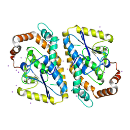
3NJB
DownloadVisualize
BU of 3njb by Molmil
Crystal structure of enoyl-coa hydratase from Mycobacterium smegmatis, iodide soak
Descriptor: Enoyl-CoA hydratase, IODIDE ION
Authors:Seattle Structural Genomics Center for Infectious Disease (SSGCID)
Deposit date:2010-06-17
Release date:2010-07-28
Last modified:2024-02-21
Method:X-RAY DIFFRACTION (2.2 Å)
Cite:SAD phasing using iodide ions in a high-throughput structural genomics environment.
J.Struct.Funct.Genom., 12, 2011
3OIB
DownloadVisualize
BU of 3oib by Molmil
Crystal structure of a putative ACYL-COA Dehydrogenase from mycobacterium smegmatis, Iodide soak
Descriptor: 1,2-ETHANEDIOL, Acyl-CoA dehydrogenase, DIHYDROFLAVINE-ADENINE DINUCLEOTIDE, ...
Authors:Seattle Structural Genomics Center for Infectious Disease (SSGCID)
Deposit date:2010-08-18
Release date:2010-09-01
Last modified:2024-05-22
Method:X-RAY DIFFRACTION (2.1 Å)
Cite:SAD phasing using iodide ions in a high-throughput structural genomics environment.
J Struct Funct Genomics, 12, 2011
4Q6U
DownloadVisualize
BU of 4q6u by Molmil
Crystal structure of a putative uncharacterized protein from Mycobacterium tuberculosis
Descriptor: PENTAETHYLENE GLYCOL, Uncharacterized protein
Authors:Seattle Structural Genomics Center for Infectious Disease (SSGCID)
Deposit date:2014-04-23
Release date:2014-06-18
Last modified:2018-03-07
Method:X-RAY DIFFRACTION (1.95 Å)
Cite:Mycobacterium tuberculosis Rv3651 is a triple sensor-domain protein.
Protein Sci., 27, 2018
3PFD
DownloadVisualize
BU of 3pfd by Molmil
Crystal structure of an Acyl-CoA dehydrogenase from Mycobacterium thermoresistibile bound to reduced flavin adenine dinucleotide solved by combined iodide ion SAD MR
Descriptor: Acyl-CoA dehydrogenase, DIHYDROFLAVINE-ADENINE DINUCLEOTIDE, IODIDE ION
Authors:Seattle Structural Genomics Center for Infectious Disease (SSGCID)
Deposit date:2010-10-28
Release date:2010-11-10
Last modified:2023-09-06
Method:X-RAY DIFFRACTION (2.1 Å)
Cite:SAD phasing using iodide ions in a high-throughput structural genomics environment.
J.STRUCT.FUNCT.GENOM., 12, 2011
4KYX
DownloadVisualize
BU of 4kyx by Molmil
Crystal structure of ADP-ribose pyrophosphatase MutT from Rickettsia felis
Descriptor: ADP-ribose pyrophosphatase MutT
Authors:Seattle Structural Genomics Center for Infectious Disease (SSGCID)
Deposit date:2013-05-29
Release date:2013-08-07
Last modified:2024-02-28
Method:X-RAY DIFFRACTION (1.8 Å)
Cite:Crystal structure of ADP-ribose pyrophosphatase MutT from Rickettsia felis
To be Published
4LW8
DownloadVisualize
BU of 4lw8 by Molmil
Crystal structure of a putative epimerase from Burkholderia cenocepacia J2315
Descriptor: 4-(2-HYDROXYETHYL)-1-PIPERAZINE ETHANESULFONIC ACID, Putative epimerase
Authors:Seattle Structural Genomics Center for Infectious Disease (SSGCID)
Deposit date:2013-07-26
Release date:2013-09-04
Last modified:2023-09-20
Method:X-RAY DIFFRACTION (2.1 Å)
Cite:Crystal structure of a putative epimerase from Burkholderia cenocepacia J2315
TO BE PUBLISHED
4O5F
DownloadVisualize
BU of 4o5f by Molmil
Crystal structure of Type III pantothenate kinase from Burkholderia thailandensis in complex with pantothenate and phosphate
Descriptor: ADENOSINE-5'-TRIPHOSPHATE, CHLORIDE ION, GLYCEROL, ...
Authors:Seattle Structural Genomics Center for Infectious Disease (SSGCID), Seattle Structural Genomics Center for Infectious Disease (SSGCID)
Deposit date:2013-12-19
Release date:2014-01-15
Last modified:2023-09-20
Method:X-RAY DIFFRACTION (1.55 Å)
Cite:Crystal structure of Type III pantothenate kinase from Burkholderia thailandensis in complex with pantothenate and phosphate
To be Published
7K74
DownloadVisualize
BU of 7k74 by Molmil
Crystal Structure of Fructose-1,6-bisphosphatase, type I, from Stenotrophomonas maltophilia K279a
Descriptor: Fructose-1,6-bisphosphatase class 1, PHOSPHATE ION
Authors:Seattle Structural Genomics Center for Infectious Disease (SSGCID)
Deposit date:2020-09-22
Release date:2020-10-21
Last modified:2023-10-18
Method:X-RAY DIFFRACTION (2.2 Å)
Cite:Crystal Structure of Fructose-1,6-bisphosphatase, type I, from Stenotrophomonas maltophilia K279a
to be published

220113

건을2024-05-22부터공개중

PDB statisticsPDBj update infoContact PDBjnumon