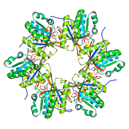
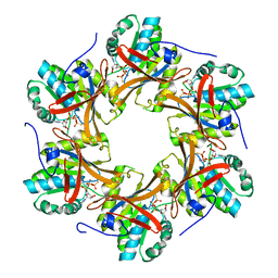
4TLE
 
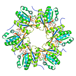 | | Crystal structure of N-terminal C1 domain of KaiC | | 分子名称: | CHLORIDE ION, Circadian clock protein kinase KaiC, MAGNESIUM ION, ... | | 著者 | Abe, J, Hiyama, T.B, Mukaiyama, A, Son, S, Akiyama, S. | | 登録日 | 2014-05-29 | | 公開日 | 2015-07-01 | | 最終更新日 | 2024-03-20 | | 実験手法 | X-RAY DIFFRACTION (1.936 Å) | | 主引用文献 | Atomic-scale origins of slowness in the cyanobacterial circadian clock
Science, 349, 2015
|
|
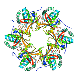
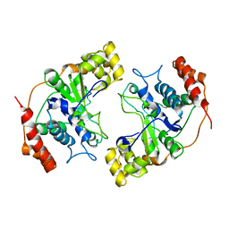
4TL7
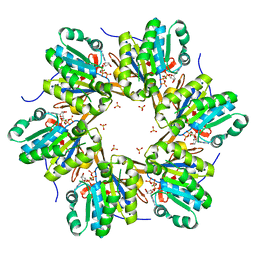 | | Crystal structure of N-terminal C1 domain of KaiC | | 分子名称: | ADENOSINE-5'-TRIPHOSPHATE, CHLORIDE ION, Circadian clock protein kinase KaiC, ... | | 著者 | Abe, J, Hiyama, T.B, Mukaiyama, A, Son, S, Akiyama, S. | | 登録日 | 2014-05-29 | | 公開日 | 2015-07-01 | | 最終更新日 | 2024-03-20 | | 実験手法 | X-RAY DIFFRACTION (1.936 Å) | | 主引用文献 | Circadian rhythms. Atomic-scale origins of slowness in the cyanobacterial circadian clock.
Science, 349, 2015
|
|
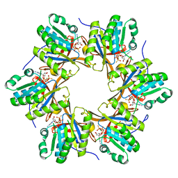
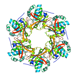
4TLC
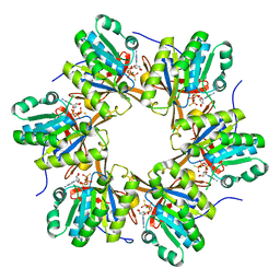 | | Crystal structure of N-terminal C1 domain of KaiC | | 分子名称: | CHLORIDE ION, Circadian clock protein kinase KaiC, MAGNESIUM ION, ... | | 著者 | Abe, J, Hiyama, T.B, Mukaiyama, A, Son, S, Akiyama, S. | | 登録日 | 2014-05-29 | | 公開日 | 2015-07-01 | | 最終更新日 | 2024-03-20 | | 実験手法 | X-RAY DIFFRACTION (2.09 Å) | | 主引用文献 | Atomic-scale origins of slowness in the cyanobacterial circadian clock
Science, 349, 2015
|
|
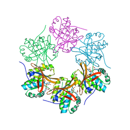
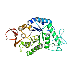
4TLA
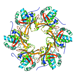 | | Crystal structure of N-terminal C1 domain of KaiC | | 分子名称: | ADENOSINE-5'-DIPHOSPHATE, CHLORIDE ION, Circadian clock protein kinase KaiC, ... | | 著者 | Abe, J, Hiyama, T.B, Mukaiyama, A, Son, S, Akiyama, S. | | 登録日 | 2014-05-29 | | 公開日 | 2015-07-01 | | 最終更新日 | 2024-03-20 | | 実験手法 | X-RAY DIFFRACTION (1.8 Å) | | 主引用文献 | Circadian rhythms. Atomic-scale origins of slowness in the cyanobacterial circadian clock.
Science, 349, 2015
|
|
4TLB
 | | Crystal structure of N-terminal C1 domain of KaiC | | 分子名称: | CHLORIDE ION, Circadian clock protein kinase KaiC, MAGNESIUM ION, ... | | 著者 | Abe, J, Hiyama, T.B, Mukaiyama, A, Son, S, Akiyama, S. | | 登録日 | 2014-05-29 | | 公開日 | 2015-07-01 | | 最終更新日 | 2024-03-20 | | 実験手法 | X-RAY DIFFRACTION (1.983 Å) | | 主引用文献 | Circadian rhythms. Atomic-scale origins of slowness in the cyanobacterial circadian clock.
Science, 349, 2015
|
|
4TL9
 | | Crystal structure of N-terminal C1 domain of KaiC | | 分子名称: | CHLORIDE ION, Circadian clock protein kinase KaiC, MAGNESIUM ION, ... | | 著者 | Abe, J, Hiyama, T.B, Mukaiyama, A, Son, S, Akiyama, S. | | 登録日 | 2014-05-29 | | 公開日 | 2015-07-01 | | 最終更新日 | 2024-03-20 | | 実験手法 | X-RAY DIFFRACTION (1.822 Å) | | 主引用文献 | Circadian rhythms. Atomic-scale origins of slowness in the cyanobacterial circadian clock.
Science, 349, 2015
|
|
4TL8
 | | Crystal structure of N-terminal C1 domain of KaiC | | 分子名称: | CHLORIDE ION, Circadian clock protein kinase KaiC, MAGNESIUM ION, ... | | 著者 | Abe, J, Hiyama, T.B, Mukaiyama, A, Son, S, Akiyama, S. | | 登録日 | 2014-05-29 | | 公開日 | 2015-07-01 | | 最終更新日 | 2023-11-08 | | 実験手法 | X-RAY DIFFRACTION (1.859 Å) | | 主引用文献 | Atomic-scale origins of slowness in the cyanobacterial circadian clock
Science, 349, 2015
|
|
4TL6
 | | Crystal structure of N-terminal domain of KaiC | | 分子名称: | CHLORIDE ION, Circadian clock protein kinase KaiC, MAGNESIUM ION, ... | | 著者 | Abe, J, Hiyama, T.B, Mukaiyama, A, Son, S, Akiyama, S. | | 登録日 | 2014-05-29 | | 公開日 | 2015-07-01 | | 最終更新日 | 2023-11-08 | | 実験手法 | X-RAY DIFFRACTION (1.763 Å) | | 主引用文献 | Circadian rhythms. Atomic-scale origins of slowness in the cyanobacterial circadian clock.
Science, 349, 2015
|
|
4TLD
 | | Crystal structure of N-terminal C1 domain of KaiC | | 分子名称: | CHLORIDE ION, Circadian clock protein kinase KaiC, MAGNESIUM ION, ... | | 著者 | Abe, J, Hiyama, T.B, Mukaiyama, A, Son, S, Akiyama, S. | | 登録日 | 2014-05-29 | | 公開日 | 2015-07-01 | | 最終更新日 | 2024-03-20 | | 実験手法 | X-RAY DIFFRACTION (1.949 Å) | | 主引用文献 | Atomic-scale origins of slowness in the cyanobacterial circadian clock
Science, 349, 2015
|
|
1MC8
 | | Crystal Structure of Flap Endonuclease-1 R42E mutant from Pyrococcus horikoshii | | 分子名称: | Flap Endonuclease-1 | | 著者 | Matsui, E, Musti, K.V, Abe, J, Yamazaki, K, Matsui, I, Harata, K. | | 登録日 | 2002-08-06 | | 公開日 | 2002-10-16 | | 最終更新日 | 2023-10-25 | | 実験手法 | X-RAY DIFFRACTION (3.1 Å) | | 主引用文献 | Molecular Structure and Novel DNA Binding Sites Located in Loops of Flap Endonuclease-1 from Pyrococcus horikoshii
J.BIOL.CHEM., 277, 2002
|
|
5YZ8
 | | Crystal Structure of N-terminal C1 domain of KaiC | | 分子名称: | CHLORIDE ION, Circadian Clock Protein Kinase KaiC, MAGNESIUM ION, ... | | 著者 | Mukaiyama, A, Furuike, Y, Abe, J, Akiyama, S. | | 登録日 | 2017-12-13 | | 公開日 | 2019-01-30 | | 最終更新日 | 2023-11-22 | | 実験手法 | X-RAY DIFFRACTION (2.81 Å) | | 主引用文献 | Conformational rearrangements of the C1 ring in KaiC measure the timing of assembly with KaiB.
Sci Rep, 8, 2018
|
|
1AMY
 | |
1AVA
 | | AMY2/BASI PROTEIN-PROTEIN COMPLEX FROM BARLEY SEED | | 分子名称: | BARLEY ALPHA-AMYLASE 2(CV MENUET), BARLEY ALPHA-AMYLASE/SUBTILISIN INHIBITOR, CALCIUM ION | | 著者 | Vallee, F, Kadziola, A, Bourne, Y, Juy, M, Svensson, B, Haser, R. | | 登録日 | 1997-09-15 | | 公開日 | 1999-03-16 | | 最終更新日 | 2024-04-03 | | 実験手法 | X-RAY DIFFRACTION (1.9 Å) | | 主引用文献 | Barley alpha-amylase bound to its endogenous protein inhibitor BASI: crystal structure of the complex at 1.9 A resolution.
Structure, 6, 1998
|
|
5B59
 | | Hen egg-white lysozyme modified with a keto-ABNO. | | 分子名称: | (2~{S})-2-azanyl-3-[(2~{R},3~{S})-2-oxidanyl-3-[[(1~{S},5~{R})-3-oxidanylidene-9-azabicyclo[3.3.1]nonan-9-yl]oxy]-1,2-dihydroindol-3-yl]propanal, Lysozyme C | | 著者 | Sasaki, D, Seki, Y, Sohma, Y, Oisaki, K, Kanai, M. | | 登録日 | 2016-04-28 | | 公開日 | 2016-09-14 | | 最終更新日 | 2023-11-08 | | 実験手法 | X-RAY DIFFRACTION (2 Å) | | 主引用文献 | Transition Metal-Free Tryptophan-Selective Bioconjugation of Proteins
J.Am.Chem.Soc., 138, 2016
|
|
1HT6
 | | CRYSTAL STRUCTURE AT 1.5A RESOLUTION OF THE BARLEY ALPHA-AMYLASE ISOZYME 1 | | 分子名称: | 1,2-ETHANEDIOL, ALPHA-AMYLASE ISOZYME 1, CALCIUM ION | | 著者 | Robert, X, Haser, R, Aghajari, N. | | 登録日 | 2000-12-29 | | 公開日 | 2003-07-08 | | 最終更新日 | 2024-04-03 | | 実験手法 | X-RAY DIFFRACTION (1.5 Å) | | 主引用文献 | The structure of barley alpha-amylase isozyme 1 reveals a novel role of domain C in substrate recognition and binding: a pair of sugar tongs
Structure, 11, 2003
|
|


