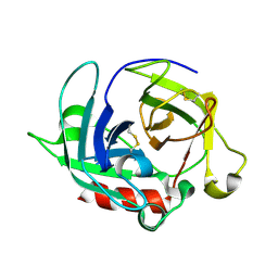
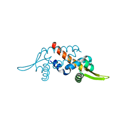
3VZP
 
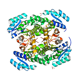 | | Crystal structure of PhaB from Ralstonia eutropha | | 分子名称: | 1,4-DIETHYLENE DIOXIDE, Acetoacetyl-CoA reductase, GLYCEROL, ... | | 著者 | Ikeda, K, Tanaka, Y, Tanaka, I, Yao, M. | | 登録日 | 2012-10-15 | | 公開日 | 2013-08-28 | | 最終更新日 | 2023-11-08 | | 実験手法 | X-RAY DIFFRACTION (1.792 Å) | | 主引用文献 | Directed evolution and structural analysis of NADPH-dependent Acetoacetyl Coenzyme A (Acetoacetyl-CoA) reductase from Ralstonia eutropha reveals two mutations responsible for enhanced kinetics
Appl.Environ.Microbiol., 79, 2013
|
|
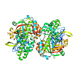
3VZS
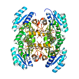 | | Crystal structure of PhaB from Ralstonia eutropha in complex with Acetoacetyl-CoA and NADP | | 分子名称: | ACETOACETYL-COENZYME A, Acetoacetyl-CoA reductase, NADP NICOTINAMIDE-ADENINE-DINUCLEOTIDE PHOSPHATE, ... | | 著者 | Ikeda, K, Tanaka, Y, Tanaka, I, Yao, M. | | 登録日 | 2012-10-15 | | 公開日 | 2013-08-28 | | 最終更新日 | 2023-11-08 | | 実験手法 | X-RAY DIFFRACTION (2.14 Å) | | 主引用文献 | Directed evolution and structural analysis of NADPH-dependent Acetoacetyl Coenzyme A (Acetoacetyl-CoA) reductase from Ralstonia eutropha reveals two mutations responsible for enhanced kinetics
Appl.Environ.Microbiol., 79, 2013
|
|
3W9V
 | | Crystal structure of refolded DING protein | | 分子名称: | GLYCEROL, PHOSPHATE ION, Phosphate-binding protein | | 著者 | Gai, Z.Q, Nakamura, A, Tanaka, Y, Hirano, N, Tanaka, I, Yao, M. | | 登録日 | 2013-04-17 | | 公開日 | 2013-10-30 | | 最終更新日 | 2023-11-08 | | 実験手法 | X-RAY DIFFRACTION (1.031 Å) | | 主引用文献 | Crystal structure analysis, overexpression and refolding behaviour of a DING protein with single mutation.
J.SYNCHROTRON RADIAT., 20, 2013
|
|
3W9W
 | | Crystal structure of DING protein | | 分子名称: | DING protein, GLYCEROL, PHOSPHATE ION | | 著者 | Gai, Z.Q, Nakamura, A, Tanaka, Y, Hirano, N, Tanaka, I, Yao, M. | | 登録日 | 2013-04-17 | | 公開日 | 2013-10-30 | | 最終更新日 | 2023-11-08 | | 実験手法 | X-RAY DIFFRACTION (1.35 Å) | | 主引用文献 | Crystal structure analysis, overexpression and refolding behaviour of a DING protein with single mutation.
J.SYNCHROTRON RADIAT., 20, 2013
|
|
3WY8
 | | Crystal Structure of Protease Anisep from Arthrobacter Nicotinovorans | | 分子名称: | Serine protease | | 著者 | Sone, T, Haraguchi, Y, Kuwahara, A, Ose, T, Takano, M, Abe, A, Tanaka, M, Tanaka, I, Asano, K. | | 登録日 | 2014-08-20 | | 公開日 | 2015-08-26 | | 最終更新日 | 2023-11-08 | | 実験手法 | X-RAY DIFFRACTION (1.7 Å) | | 主引用文献 | Structural characterization reveals the keratinolytic activity of an arthrobacter nicotinovorans protease.
Protein Pept.Lett., 22, 2015
|
|
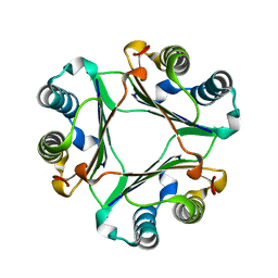
3A8E
 | | The structure of AxCesD octamer complexed with cellopentaose | | 分子名称: | Cellulose synthase operon protein D, beta-D-glucopyranose-(1-4)-beta-D-glucopyranose-(1-4)-beta-D-glucopyranose-(1-4)-beta-D-glucopyranose-(1-4)-beta-D-glucopyranose | | 著者 | Hu, S.Q, Tajima, K, Zhou, Y, Yao, M, Tanaka, I. | | 登録日 | 2009-10-05 | | 公開日 | 2010-09-22 | | 最終更新日 | 2023-11-01 | | 実験手法 | X-RAY DIFFRACTION (3 Å) | | 主引用文献 | Structure of bacterial cellulose synthase subunit D octamer with four inner passageways
Proc.Natl.Acad.Sci.USA, 107, 2010
|
|
3AJ2
 | | The structure of AxCeSD octamer (C-terminal HIS-tag) from Acetobacter xylinum | | 分子名称: | Cellulose synthase operon protein D | | 著者 | Hu, S.Q, Tajima, K, Zhou, Y, Tanaka, I, Yao, M. | | 登録日 | 2010-05-20 | | 公開日 | 2010-10-06 | | 最終更新日 | 2023-11-01 | | 実験手法 | X-RAY DIFFRACTION (2.7 Å) | | 主引用文献 | Structure of bacterial cellulose synthase subunit D octamer with four inner passageways
Proc.Natl.Acad.Sci.USA, 107, 2010
|
|
3AJ1
 | | The structure of AxCeSD octamer (N-terminal HIS-tag) from Acetobacter xylinum | | 分子名称: | Cellulose synthase operon protein D | | 著者 | Hu, S.Q, Tajima, K, Zhou, Y, Tanaka, I, Yao, M. | | 登録日 | 2010-05-20 | | 公開日 | 2010-10-06 | | 最終更新日 | 2011-07-13 | | 実験手法 | X-RAY DIFFRACTION (2.5 Å) | | 主引用文献 | Structure of bacterial cellulose synthase subunit D octamer with four inner passageways
Proc.Natl.Acad.Sci.USA, 107, 2010
|
|
3X2O
 | | Neutron and X-ray joint refined structure of PcCel45A apo form at 298K. | | 分子名称: | Endoglucanase V-like protein | | 著者 | Nakamura, A, Ishida, T, Kusaka, K, Yamada, T, Tanaka, I, Niimura, N, Samejima, M, Igarashi, K. | | 登録日 | 2014-12-22 | | 公開日 | 2015-10-07 | | 最終更新日 | 2019-12-18 | | 実験手法 | NEUTRON DIFFRACTION (1.5 Å), X-RAY DIFFRACTION | | 主引用文献 | "Newton's cradle" proton relay with amide-imidic acid tautomerization in inverting cellulase visualized by neutron crystallography.
Sci Adv, 1, 2015
|
|
3X2P
 | | Neutron and X-ray joint refined structure of PcCel45A with cellopentaose at 298K. | | 分子名称: | Endoglucanase V-like protein, beta-D-glucopyranose-(1-4)-beta-D-glucopyranose-(1-4)-beta-D-glucopyranose-(1-4)-beta-D-glucopyranose-(1-4)-beta-D-glucopyranose | | 著者 | Nakamura, A, Ishida, T, Kusaka, K, Yamada, T, Tanaka, I, Niimura, N, Samejima, M, Igarashi, K. | | 登録日 | 2014-12-22 | | 公開日 | 2015-10-14 | | 最終更新日 | 2020-07-29 | | 実験手法 | NEUTRON DIFFRACTION (1.518 Å), X-RAY DIFFRACTION | | 主引用文献 | "Newton's cradle" proton relay with amide-imidic acid tautomerization in inverting cellulase visualized by neutron crystallography.
Sci Adv, 1, 2015
|
|
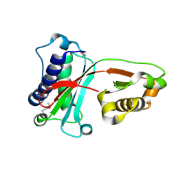
1UCA
 | | Crystal structure of the Ribonuclease MC1 from bitter gourd seeds complexed with 2'-UMP | | 分子名称: | PHOSPHORIC ACID MONO-[2-(2,4-DIOXO-3,4-DIHYDRO-2H-PYRIMIDIN-1-YL)-4-HYDROXY-5-HYDROXYMETHYL-TETRAHYDRO-FURAN-3-YL] ESTER, Ribonuclease MC | | 著者 | Suzuki, A, Yao, M, Tanaka, I, Numata, T, Kikukawa, S, Yamasaki, N, Kimura, M. | | 登録日 | 2003-04-10 | | 公開日 | 2003-04-29 | | 最終更新日 | 2023-10-25 | | 実験手法 | X-RAY DIFFRACTION (1.48 Å) | | 主引用文献 | Crystal structures of the ribonuclease MC1 from bitter gourd seeds, complexed with 2'-UMP or 3'-UMP, reveal structural basis for uridine specificity
Biochem.Biophys.Res.Commun., 275, 2000
|
|
1UC2
 | | Hypothetical Extein Protein of PH1602 from Pyrococcus horikoshii | | 分子名称: | SULFATE ION, beta-D-fructofuranose-(2-1)-alpha-D-glucopyranose, hypothetical protein PH1602 | | 著者 | Okada, C, Maegawa, Y, Yao, M, Tanaka, I. | | 登録日 | 2003-04-08 | | 公開日 | 2004-05-04 | | 最終更新日 | 2023-12-27 | | 実験手法 | X-RAY DIFFRACTION (2.15 Å) | | 主引用文献 | Crystal structure of an RtcB homolog protein (PH1602-extein protein) from Pyrococcus horikoshii reveals a novel fold
Proteins, 63, 2006
|
|
1UCD
 | | Crystal structure of Ribonuclease MC1 from bitter gourd seeds complexed with 5'-UMP | | 分子名称: | Ribonuclease MC, URACIL, URIDINE-5'-MONOPHOSPHATE | | 著者 | Suzuki, A, Numata, T, Yao, M, Kimura, M, Tanaka, I. | | 登録日 | 2003-04-10 | | 公開日 | 2004-05-18 | | 最終更新日 | 2023-10-25 | | 実験手法 | X-RAY DIFFRACTION (1.3 Å) | | 主引用文献 | Structure of RNase MC1 from bitter gourd seeds in complex with 5'UMP
To be published
|
|
1UC3
 | |
1UDD
 | | TenA homologue protein from P.horikoshii OT3 | | 分子名称: | transcriptional regulator | | 著者 | Itou, H, Yao, M, Watanabe, N, Tanaka, I. | | 登録日 | 2003-04-28 | | 公開日 | 2004-06-01 | | 最終更新日 | 2023-12-27 | | 実験手法 | X-RAY DIFFRACTION (2.15 Å) | | 主引用文献 | Structure analysis of PH1161 protein, a transcriptional activator TenA homologue from the hyperthermophilic archaeon Pyrococcus horikoshii.
Acta Crystallogr.,Sect.D, 60, 2004
|
|
1UB9
 | | Structure of the transcriptional regulator homologue protein from Pyrococcus horikoshii OT3 | | 分子名称: | Hypothetical protein PH1061 | | 著者 | Okada, U, Sakai, N, Tajika, Y, Yao, M, Watanabe, N, Tanaka, I. | | 登録日 | 2003-04-03 | | 公開日 | 2004-05-04 | | 最終更新日 | 2023-12-27 | | 実験手法 | X-RAY DIFFRACTION (2.05 Å) | | 主引用文献 | Structural analysis of the transcriptional regulator homolog protein from Pyrococcus horikoshii OT3.
Proteins, 63, 2006
|
|
1UCC
 | | Crystal structure of the Ribonuclease MC1 from bitter gourd seeds complexed with 3'-UMP. | | 分子名称: | 3'-URIDINEMONOPHOSPHATE, Ribonuclease MC | | 著者 | Suzuki, A, Yao, M, Tanaka, I, Numata, T, Kikukawa, S, Yamasaki, N, Kimura, M. | | 登録日 | 2003-04-10 | | 公開日 | 2003-04-29 | | 最終更新日 | 2023-10-25 | | 実験手法 | X-RAY DIFFRACTION (1.77 Å) | | 主引用文献 | Crystal structures of the ribonuclease MC1 from bitter gourd seeds, complexed with 2'-UMP or 3'-UMP, reveal structural basis for uridine specificity
Biochem.Biophys.Res.Commun., 275, 2000
|
|
1UIZ
 | | Crystal Structure Of Macrophage Migration Inhibitory Factor From Xenopus Laevis. | | 分子名称: | Macrophage Migration Inhibitory Factor | | 著者 | Suzuki, M, Takamura, Y, Maeno, M, Tochinai, S, Iyaguchi, D, Tanaka, I, Nishihira, J, Ishibashi, T. | | 登録日 | 2003-07-24 | | 公開日 | 2004-05-25 | | 最終更新日 | 2023-12-27 | | 実験手法 | X-RAY DIFFRACTION (2.5 Å) | | 主引用文献 | Xenopus laevis Macrophage Migration Inhibitory Factor Is Essential for Axis Formation and Neural Development.
J.Biol.Chem., 279, 2004
|
|
1VAJ
 | | Crystal Structure of Uncharacterized Protein PH0010 From Pyrococcus horikoshii | | 分子名称: | Hypothetical protein PH0010 | | 著者 | Tajika, Y, Sakai, N, Tamura, T, Yao, M, Watanabe, N, Tanaka, I. | | 登録日 | 2004-02-17 | | 公開日 | 2005-01-25 | | 最終更新日 | 2023-12-27 | | 実験手法 | X-RAY DIFFRACTION (1.82 Å) | | 主引用文献 | Crystal structure of PH0010 from Pyrococcus horikoshii, which is highly homologous to human AMMECR 1C-terminal region
Proteins, 58, 2005
|
|
1V9G
 | | Neutron Crystallographic analysis of the Z-DNA hexamer CGCGCG | | 分子名称: | 5'-D(*CP*GP*CP*GP*CP*G)-3', N,N'-BIS(3-AMMONIOPROPYL)BUTANE-1,4-DIAMINIUM | | 著者 | Chatake, T, Tanaka, I, Niimura, N. | | 登録日 | 2004-01-26 | | 公開日 | 2005-01-26 | | 最終更新日 | 2023-10-25 | | 実験手法 | NEUTRON DIFFRACTION (1.8 Å) | | 主引用文献 | The hydration structure of a Z-DNA hexameric duplex determined by a neutron diffraction technique.
Acta Crystallogr.,Sect.D, 61, 2005
|
|
1ULY
 | | Crystal structure analysis of the ArsR homologue DNA-binding protein from P. horikoshii OT3 | | 分子名称: | hypothetical protein PH1932 | | 著者 | Itou, H, Yao, M, Watanabe, N, Tanaka, I. | | 登録日 | 2003-09-17 | | 公開日 | 2004-10-19 | | 最終更新日 | 2023-12-27 | | 実験手法 | X-RAY DIFFRACTION (2.5 Å) | | 主引用文献 | Crystal structure of the PH1932 protein, a unique archaeal ArsR type winged-HTH transcription factor from Pyrococcus horikoshii OT3
Proteins, 70, 2008
|
|
1V7O
 | |
1V43
 | | Crystal Structure of ATPase subunit of ABC Sugar Transporter | | 分子名称: | sugar-binding transport ATP-binding protein | | 著者 | Ose, T, Fujie, T, Yao, M, Watanabe, N, Tanaka, I. | | 登録日 | 2003-11-08 | | 公開日 | 2004-11-16 | | 最終更新日 | 2023-10-25 | | 実験手法 | X-RAY DIFFRACTION (2.2 Å) | | 主引用文献 | Crystal structure of the ATP-binding cassette of multisugar transporter from Pyrococcus horikoshii OT3
Proteins, 57, 2004
|
|
1V4N
 | | Structure of 5'-deoxy-5'-methylthioadenosine phosphorylase homologue from Sulfolobus tokodaii | | 分子名称: | 271aa long hypothetical 5'-methylthioadenosine phosphorylase | | 著者 | Kitago, Y, Yasutake, Y, Sakai, N, Tsujimura, M, Yao, M, Watanabe, N, Kawarabayasi, Y, Tanaka, I. | | 登録日 | 2003-11-14 | | 公開日 | 2005-01-04 | | 最終更新日 | 2023-10-25 | | 実験手法 | X-RAY DIFFRACTION (2.45 Å) | | 主引用文献 | Crystal structure of Sulfolobus tokodaii MTAP
To be Published
|
|
1V30
 | | Crystal Structure Of Uncharacterized Protein PH0828 From Pyrococcus horikoshii | | 分子名称: | 2-[N-CYCLOHEXYLAMINO]ETHANE SULFONIC ACID, Hypothetical UPF0131 protein PH0828 | | 著者 | Tajika, Y, Sakai, N, Yao, M, Watanabe, N, Tanaka, I. | | 登録日 | 2003-10-21 | | 公開日 | 2004-11-09 | | 最終更新日 | 2023-12-27 | | 実験手法 | X-RAY DIFFRACTION (1.4 Å) | | 主引用文献 | Crystal structure of hypothetical protein PH0828 from Pyrococcus horikoshii.
Proteins, 57, 2004
|
|


