Loading
PDBj
MenuPDBj@FacebookPDBj@TwitterPDBj@YouTubewwPDB FoundationwwPDB
RCSB PDBPDBeBMRBAdv. SearchSearch help
Search by PDB author
1Y65
DownloadVisualize
BU of 1y65 by Molmil
Crystal structure of beta-hexosaminidase from Vibrio cholerae in complex with N-acetyl-D-glucosamine to a resolution of 1.85
Descriptor: 2-acetamido-2-deoxy-beta-D-glucopyranose, Beta-hexosaminidase
Authors:Gorman, J, Shapiro, L, Burley, S.K, New York SGX Research Center for Structural Genomics (NYSGXRC)
Deposit date:2004-12-03
Release date:2004-12-14
Last modified:2023-08-23
Method:X-RAY DIFFRACTION (1.85 Å)
Cite:Crystal structure of beta-hexosaminidase from Vibrio cholerae in complex with N-acetyl-D-glucosamine to a resolution of 1.85
To be Published
1Y7E
DownloadVisualize
BU of 1y7e by Molmil
The Crystal Structure of Aminopeptidase I from Borrelia burgdorferi B31
Descriptor: Probable M18-family aminopeptidase 1
Authors:Min, T, Gorman, J, Shapiro, L, Burley, S.K, New York SGX Research Center for Structural Genomics (NYSGXRC)
Deposit date:2004-12-08
Release date:2005-01-04
Last modified:2021-02-03
Method:X-RAY DIFFRACTION (3.2 Å)
Cite:The crystal structure of aminopeptidase I (yscI) from Borrelia burgdorferi
To be Published
1Y9E
DownloadVisualize
BU of 1y9e by Molmil
Crystal structure of Bacillus subtilis protein yhfP with NAD bound
Descriptor: NICOTINAMIDE-ADENINE-DINUCLEOTIDE, hypothetical protein yhfP
Authors:Min, T, Gorman, J, Shapiro, L, Burley, S.K, New York SGX Research Center for Structural Genomics (NYSGXRC)
Deposit date:2004-12-15
Release date:2004-12-21
Last modified:2021-02-03
Method:X-RAY DIFFRACTION (2.8 Å)
Cite:The crystal structure of hypothetical protein yhfP from Bacillus subtilis
To be Published
1YRH
DownloadVisualize
BU of 1yrh by Molmil
Crystal Structure Of Trp Repressor Binding Protein Wrba in complex with FMN
Descriptor: FLAVIN MONONUCLEOTIDE, trp repressor binding protein WrbA
Authors:Gorman, J, Shapiro, L, Burley, S.K, New York SGX Research Center for Structural Genomics (NYSGXRC)
Deposit date:2005-02-03
Release date:2005-02-15
Last modified:2021-02-03
Method:X-RAY DIFFRACTION (3.11 Å)
Cite:Crystal structures of the tryptophan repressor binding protein WrbA and complexes with flavin mononucleotide.
Protein Sci., 14, 2005
1ZVN
DownloadVisualize
BU of 1zvn by Molmil
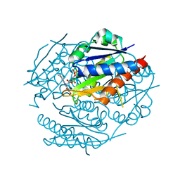
Crystal structure of chick MN-cadherin EC1
Descriptor: Cadherin 1
Authors:Patel, S.D, Ciatto, C, Chen, C.P, Bahna, F, Schieren, I, Rajebhosale, M, Jessell, T.M, Honig, B, Shapiro, L, Price, S.R.
Deposit date:2005-06-02
Release date:2006-04-25
Last modified:2024-02-14
Method:X-RAY DIFFRACTION (2.16 Å)
Cite:Type II cadherin ectodomain structures: implications for classical cadherin specificity.
Cell(Cambridge,Mass.), 124, 2006
1ZWL
DownloadVisualize
BU of 1zwl by Molmil
Structure of WrbA from Pseudomonas aeruginosa in complex with FMN
Descriptor: FLAVIN MONONUCLEOTIDE, Trp repressor binding protein WrbA
Authors:Gorman, J, Shapiro, L, Burley, S.K, New York SGX Research Center for Structural Genomics (NYSGXRC)
Deposit date:2005-06-03
Release date:2005-06-14
Last modified:2023-08-23
Method:X-RAY DIFFRACTION (2.8 Å)
Cite:Crystal structures of the tryptophan repressor binding protein WrbA and complexes with flavin mononucleotide.
Protein Sci., 14, 2005
2A4E
DownloadVisualize
BU of 2a4e by Molmil
Crystal structure of mouse cadherin-11 EC1-2
Descriptor: CALCIUM ION, Cadherin-11
Authors:Patel, S.D, Ciatto, C, Chen, C.P, Bahna, F, Arkus, N, Rajebhosale, M, Jessell, T.M, Honig, B, Price, S.R, Shapiro, L.
Deposit date:2005-06-28
Release date:2006-04-25
Last modified:2023-08-23
Method:X-RAY DIFFRACTION (3.2 Å)
Cite:Type II cadherin ectodomain structures: implications for classical cadherin specificity.
Cell(Cambridge,Mass.), 124, 2006
1ZWK
DownloadVisualize
BU of 1zwk by Molmil
Structure of WrbA from Pseudomonas aeruginosa
Descriptor: PHOSPHATE ION, Trp repressor binding protein WrbA
Authors:Gorman, J, Shapiro, L, Burley, S.K, New York SGX Research Center for Structural Genomics (NYSGXRC)
Deposit date:2005-06-03
Release date:2005-06-14
Last modified:2023-08-23
Method:X-RAY DIFFRACTION (2.6 Å)
Cite:Crystal structures of the tryptophan repressor binding protein WrbA and complexes with flavin mononucleotide.
Protein Sci., 14, 2005
1ZXK
DownloadVisualize
BU of 1zxk by Molmil
Crystal Structure of Cadherin8 EC1 domain
Descriptor: Cadherin-8
Authors:Patel, S.D, Ciatto, C, Chen, C.P, Bahna, F, Arkus, N, Schieren, I, Rajebhosale, M, Jessell, T.M, Honig, B, Price, S.R, Shapiro, L.
Deposit date:2005-06-08
Release date:2006-04-25
Last modified:2023-08-23
Method:X-RAY DIFFRACTION (2 Å)
Cite:Type II cadherin ectodomain structures: implications for classical cadherin specificity.
Cell(Cambridge,Mass.), 124, 2006
2A62
DownloadVisualize
BU of 2a62 by Molmil
Crystal structure of mouse cadherin-8 EC1-3
Descriptor: CALCIUM ION, Cadherin-8
Authors:Patel, S.D, Ciatto, C, Chen, C.P, Bahna, F, Arkus, N, Schieren, I, Jessell, T.M, Honig, B, Price, S.R, Shapiro, L.
Deposit date:2005-07-01
Release date:2006-04-25
Last modified:2024-02-14
Method:X-RAY DIFFRACTION (4.5 Å)
Cite:Type II cadherin ectodomain structures: implications for classical cadherin specificity.
Cell(Cambridge,Mass.), 124, 2006
2A1F
DownloadVisualize
BU of 2a1f by Molmil
Crystal Structure of Uridylate kinase
Descriptor: Uridylate kinase
Authors:Gorman, J, Shapiro, L, Burley, S.K, New York SGX Research Center for Structural Genomics (NYSGXRC)
Deposit date:2005-06-20
Release date:2005-06-28
Last modified:2023-08-23
Method:X-RAY DIFFRACTION (2.1 Å)
Cite:Crystal Structure of Uridylate kinase
To be Published
2A4C
DownloadVisualize
BU of 2a4c by Molmil
Crystal structure of mouse cadherin-11 EC1
Descriptor: Cadherin-11
Authors:Patel, S.D, Ciatto, C, Chen, C.P, Bahna, F, Schieren, I, Rajebhosale, M, Jessell, T.M, Honig, B, Price, S.R, Shapiro, L.
Deposit date:2005-06-28
Release date:2006-04-25
Last modified:2023-08-23
Method:X-RAY DIFFRACTION (2.9 Å)
Cite:Type II cadherin ectodomain structures: implications for classical cadherin specificity.
Cell(Cambridge,Mass.), 124, 2006
2AKO
DownloadVisualize
BU of 2ako by Molmil
Crystal structure of Glutamate 5-kinase from Campylobacter jejuni
Descriptor: ADENOSINE-5'-DIPHOSPHATE, Glutamate 5-kinase
Authors:Gorman, J, Shapiro, L, Burley, S.K, New York SGX Research Center for Structural Genomics (NYSGXRC)
Deposit date:2005-08-03
Release date:2005-08-09
Last modified:2021-02-03
Method:X-RAY DIFFRACTION (2.2 Å)
Cite:Crystal structure of Glutamate 5-kinase from Campylobacter jejuni
To be Published
2AWD
DownloadVisualize
BU of 2awd by Molmil
Crystal structure of LacC from Enterococcus faecalis
Descriptor: BROMIDE ION, tagatose-6-phosphate kinase
Authors:Gorman, J, Shapiro, L, Burley, S.K, New York SGX Research Center for Structural Genomics (NYSGXRC)
Deposit date:2005-08-31
Release date:2005-09-13
Last modified:2021-02-03
Method:X-RAY DIFFRACTION (2 Å)
Cite:Crystal structure of LacC from Enterococcus faecalis
To be Published
6CG7
DownloadVisualize
BU of 6cg7 by Molmil
mouse cadherin-22 EC1-2 adhesive fragment
Descriptor: CALCIUM ION, Cadherin-22
Authors:Brasch, J, Harrison, O.J, Shapiro, L.
Deposit date:2018-02-19
Release date:2018-05-09
Last modified:2023-10-04
Method:X-RAY DIFFRACTION (2.705 Å)
Cite:Homophilic and Heterophilic Interactions of Type II Cadherins Identify Specificity Groups Underlying Cell-Adhesive Behavior.
Cell Rep, 23, 2018
6CGS
DownloadVisualize
BU of 6cgs by Molmil
mouse cadherin-7 EC1-2 adhesive fragment
Descriptor: CALCIUM ION, Cadherin-7, GLYCEROL
Authors:Brasch, J, Harrison, O.J, Kaczynska, A, Shapiro, L.
Deposit date:2018-02-20
Release date:2018-05-09
Last modified:2023-10-04
Method:X-RAY DIFFRACTION (1.72 Å)
Cite:Homophilic and Heterophilic Interactions of Type II Cadherins Identify Specificity Groups Underlying Cell-Adhesive Behavior.
Cell Rep, 23, 2018
6CV7
DownloadVisualize
BU of 6cv7 by Molmil
Mouse Protocadherin-15 Extracellular Cadherin Domains 1 through 3
Descriptor: 1,2-ETHANEDIOL, 2-acetamido-2-deoxy-beta-D-glucopyranose, CALCIUM ION, ...
Authors:Dionne, G, Shapiro, L.
Deposit date:2018-03-27
Release date:2018-08-01
Last modified:2023-10-04
Method:X-RAY DIFFRACTION (1.692 Å)
Cite:Mechanotransduction by PCDH15 Relies on a Novel cis-Dimeric Architecture.
Neuron, 99, 2018
6CG6
DownloadVisualize
BU of 6cg6 by Molmil
mouse cadherin-10 EC1-2 adhesive fragment
Descriptor: 1,2-ETHANEDIOL, CALCIUM ION, Cadherin-10, ...
Authors:Brasch, J, Harrison, O.J, Shapiro, L.
Deposit date:2018-02-19
Release date:2018-05-09
Last modified:2023-10-04
Method:X-RAY DIFFRACTION (2.707 Å)
Cite:Homophilic and Heterophilic Interactions of Type II Cadherins Identify Specificity Groups Underlying Cell-Adhesive Behavior.
Cell Rep, 23, 2018
6CGU
DownloadVisualize
BU of 6cgu by Molmil
mouse cadherin-6 EC1-2 adhesive fragment
Descriptor: CALCIUM ION, Cadherin-6
Authors:Brasch, J, Harrison, O.J, Shapiro, L.
Deposit date:2018-02-20
Release date:2018-05-09
Last modified:2023-10-04
Method:X-RAY DIFFRACTION (1.9 Å)
Cite:Homophilic and Heterophilic Interactions of Type II Cadherins Identify Specificity Groups Underlying Cell-Adhesive Behavior.
Cell Rep, 23, 2018
6CGB
DownloadVisualize
BU of 6cgb by Molmil
chimera of mouse cadherin-11 EC1 and mouse cadherin-6 EC2
Descriptor: ACETATE ION, CALCIUM ION, Cadherin-11, ...
Authors:Brasch, J, Harrison, O.J, Shapiro, L, Kaeser, B.
Deposit date:2018-02-19
Release date:2018-05-09
Last modified:2023-10-04
Method:X-RAY DIFFRACTION (2.994 Å)
Cite:Homophilic and Heterophilic Interactions of Type II Cadherins Identify Specificity Groups Underlying Cell-Adhesive Behavior.
Cell Rep, 23, 2018
6E6B
DownloadVisualize
BU of 6e6b by Molmil
Crystal structure of the Protocadherin GammaB4 extracellular domain
Descriptor: 2-acetamido-2-deoxy-beta-D-glucopyranose, 2-acetamido-2-deoxy-beta-D-glucopyranose-(1-4)-2-acetamido-2-deoxy-beta-D-glucopyranose, 2-acetamido-2-deoxy-beta-D-glucopyranose-(1-4)-[alpha-L-fucopyranose-(1-6)]2-acetamido-2-deoxy-beta-D-glucopyranose, ...
Authors:Goodman, K.M, Mannepalli, S, Bahna, F, Honig, B, Shapiro, L.
Deposit date:2018-07-24
Release date:2019-04-10
Last modified:2023-10-11
Method:X-RAY DIFFRACTION (4.52 Å)
Cite:Visualization of clustered protocadherin neuronal self-recognition complexes.
Nature, 569, 2019
3LNE
DownloadVisualize
BU of 3lne by Molmil
Crystal structure of E-cadherin EC12 K14E
Descriptor: CALCIUM ION, Cadherin-1, GLYCEROL
Authors:Jin, X, Harrison, O, Shapiro, L.
Deposit date:2010-02-02
Release date:2010-03-02
Last modified:2023-09-06
Method:X-RAY DIFFRACTION (2 Å)
Cite:Two-step adhesive binding by classical cadherins.
Nat.Struct.Mol.Biol., 17, 2010
3LNF
DownloadVisualize
BU of 3lnf by Molmil
Crystal structure of E-cadherin EC12 K14EW2A
Descriptor: CALCIUM ION, Cadherin-1
Authors:Jin, X, Harrison, O, Shapiro, L.
Deposit date:2010-02-02
Release date:2010-03-02
Last modified:2023-09-06
Method:X-RAY DIFFRACTION (2.5 Å)
Cite:Two-step adhesive binding by classical cadherins.
Nat.Struct.Mol.Biol., 17, 2010
3LNI
DownloadVisualize
BU of 3lni by Molmil
Crystal structure of E-cadherin EC12 E89A
Descriptor: CALCIUM ION, Cadherin-1
Authors:Harrison, O, Jin, X, Shapiro, L.
Deposit date:2010-02-02
Release date:2010-03-02
Last modified:2023-09-06
Method:X-RAY DIFFRACTION (2.3 Å)
Cite:Two-step adhesive binding by classical cadherins.
Nat.Struct.Mol.Biol., 17, 2010
3LNH
DownloadVisualize
BU of 3lnh by Molmil
Crystal structure of E-cadherin EC12 W2A
Descriptor: CALCIUM ION, Cadherin-1
Authors:Harrison, O, Jin, X, Shapiro, L.
Deposit date:2010-02-02
Release date:2010-03-02
Last modified:2023-09-06
Method:X-RAY DIFFRACTION (2.6 Å)
Cite:Two-step adhesive binding by classical cadherins.
Nat.Struct.Mol.Biol., 17, 2010

219869

건을2024-05-15부터공개중

PDB statisticsPDBj update infoContact PDBjnumon