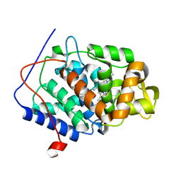
1L2K
 
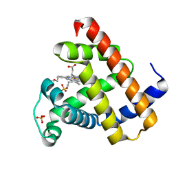 | | Neutron Structure Determination of Sperm Whale Met-Myoglobin at 1.5A Resolution. | | 分子名称: | AMMONIUM CATION WITH D, MYOGLOBIN, PROTOPORPHYRIN IX CONTAINING FE, ... | | 著者 | Ostermann, A, Tanaka, I, Engler, N, Niimura, N, Parak, F.G. | | 登録日 | 2002-02-21 | | 公開日 | 2002-08-21 | | 最終更新日 | 2024-04-03 | | 実験手法 | NEUTRON DIFFRACTION (1.5 Å) | | 主引用文献 | Hydrogen and deuterium in myoglobin as seen by a neutron structure determination at 1.5 A resolution.
Biophys.Chem., 95, 2002
|
|
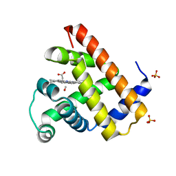
1DO3
 | | CARBONMONOXY-MYOGLOBIN (MUTANT L29W) AFTER PHOTOLYSIS AT T>180K | | 分子名称: | CARBON MONOXIDE, MYOGLOBIN, PROTOPORPHYRIN IX CONTAINING FE, ... | | 著者 | Ostermann, A, Waschipky, R, Parak, F.G, Nienhaus, G.U. | | 登録日 | 1999-12-18 | | 公開日 | 2000-01-05 | | 最終更新日 | 2023-09-20 | | 実験手法 | X-RAY DIFFRACTION (1.55 Å) | | 主引用文献 | Ligand binding and conformational motions in myoglobin.
Nature, 404, 2000
|
|
1DO7
 | | CARBONMONOXY-MYOGLOBIN (MUTANT L29W) REBINDING STRUCTURE AFTER PHOTOLYSIS AT T< 180K | | 分子名称: | CARBON MONOXIDE, MYOGLOBIN, PROTOPORPHYRIN IX CONTAINING FE, ... | | 著者 | Ostermann, A, Waschipky, R, Parak, F.G, Nienhaus, G.U. | | 登録日 | 1999-12-19 | | 公開日 | 2000-01-05 | | 最終更新日 | 2023-09-20 | | 実験手法 | X-RAY DIFFRACTION (1.85 Å) | | 主引用文献 | Ligand binding and conformational motions in myoglobin.
Nature, 404, 2000
|
|
1DO4
 | | CARBONMONOXY-MYOGLOBIN (MUTANT L29W) AFTER PHOTOLYSIS AT T<180K | | 分子名称: | CARBON MONOXIDE, MYOGLOBIN, PROTOPORPHYRIN IX CONTAINING FE, ... | | 著者 | Ostermann, A, Waschipky, R, Parak, F.G, Nienhaus, G.U. | | 登録日 | 1999-12-18 | | 公開日 | 2000-01-05 | | 最終更新日 | 2023-09-20 | | 実験手法 | X-RAY DIFFRACTION (1.7 Å) | | 主引用文献 | Ligand binding and conformational motions in myoglobin.
Nature, 404, 2000
|
|
1DO1
 | | CARBONMONOXY-MYOGLOBIN MUTANT L29W AT 105K | | 分子名称: | CARBON MONOXIDE, MYOGLOBIN, PROTOPORPHYRIN IX CONTAINING FE, ... | | 著者 | Ostermann, A, Waschipky, R, Parak, F.G, Nienhaus, G.U. | | 登録日 | 1999-12-18 | | 公開日 | 2000-01-05 | | 最終更新日 | 2023-09-20 | | 実験手法 | X-RAY DIFFRACTION (1.5 Å) | | 主引用文献 | Ligand binding and conformational motions in myoglobin.
Nature, 404, 2000
|
|
2ZWB
 | | Neutron crystal structure of wild type human lysozyme in D2O | | 分子名称: | Lysozyme C | | 著者 | Chiba-Kamoshida, K, Matsui, T, Chatake, T, Ohhara, T, Ostermann, A, Tanaka, I, Yutani, K, Niimura, N. | | 登録日 | 2008-12-02 | | 公開日 | 2009-12-08 | | 最終更新日 | 2023-11-01 | | 実験手法 | NEUTRON DIFFRACTION (1.8 Å) | | 主引用文献 | Site-specific softening of peptide bonds by localized deuterium observed by neutron crystallography of human lysozyme
To be Published
|
|
6TAE
 | | Neutron structure of ferric ascorbate peroxidase | | 分子名称: | Ascorbate peroxidase, PROTOPORPHYRIN IX CONTAINING FE, SULFATE ION | | 著者 | Kwon, H, Basran, J, Devos, J.M, Schrader, T.E, Ostermann, A, Blakeley, M.P, Raven, E.L, Moody, P.C.E. | | 登録日 | 2019-10-29 | | 公開日 | 2020-03-18 | | 最終更新日 | 2020-04-08 | | 実験手法 | NEUTRON DIFFRACTION (1.9 Å), X-RAY DIFFRACTION | | 主引用文献 | Visualizing the protons in a metalloenzyme electron proton transfer pathway.
Proc.Natl.Acad.Sci.USA, 117, 2020
|
|
3FE0
 | | X-ray crystal structure of wild type human lysozyme in D2O | | 分子名称: | Lysozyme C | | 著者 | Chiba-Kamoshida, K, Matsui, T, Chatake, T, Ohhara, T, Ostermann, A, Tanaka, I, Yutani, K, Niimura, N. | | 登録日 | 2008-11-27 | | 公開日 | 2009-12-08 | | 最終更新日 | 2023-11-01 | | 実験手法 | X-RAY DIFFRACTION (1.5 Å) | | 主引用文献 | Site-specific softening of peptide bonds by localized deuterium observed by neutron crystallography of human lysozyme hydrogen
To be Published
|
|
6L27
 | | X-ray crystal structure of the mutant green fluorescent protein | | 分子名称: | Green fluorescent protein | | 著者 | Adachi, M, Shimizu, R, Shibazaki, C, Kagotani, Y, Ostermann, A, Schrader, T.E. | | 登録日 | 2019-10-02 | | 公開日 | 2020-04-01 | | 最終更新日 | 2023-11-15 | | 実験手法 | X-RAY DIFFRACTION (0.77 Å) | | 主引用文献 | Direct Observation of the Protonation States in the Mutant Green Fluorescent Protein.
J Phys Chem Lett, 11, 2020
|
|
6L26
 | | Neutron crystal structure of the mutant green fluorescent protein (EGFP) | | 分子名称: | Green fluorescent protein, trideuteriooxidanium | | 著者 | Adachi, M, Shimizu, R, Shibazaki, C, Kagotani, Y, Ostermann, A, Schrader, T.E. | | 登録日 | 2019-10-02 | | 公開日 | 2020-04-08 | | 最終更新日 | 2023-11-22 | | 実験手法 | NEUTRON DIFFRACTION (1.444 Å) | | 主引用文献 | Direct Observation of the Protonation States in the Mutant Green Fluorescent Protein.
J Phys Chem Lett, 11, 2020
|
|
6XV4
 | | Neutron structure of ferric ascorbate peroxidase-ascorbate complex | | 分子名称: | ASCORBIC ACID, Ascorbate peroxidase, POTASSIUM ION, ... | | 著者 | Kwon, H, Basran, J, Devos, J.M, Schrader, T.E, Ostermann, A, Blakeley, M.P, Raven, E.L, Moody, P.C.E. | | 登録日 | 2020-01-21 | | 公開日 | 2020-03-18 | | 最終更新日 | 2020-07-29 | | 実験手法 | NEUTRON DIFFRACTION (1.9 Å), X-RAY DIFFRACTION | | 主引用文献 | Visualizing the protons in a metalloenzyme electron proton transfer pathway.
Proc.Natl.Acad.Sci.USA, 117, 2020
|
|
4XQD
 | | X-ray structure analysis of xylanase-WT at pH4.0 | | 分子名称: | 2-AMINO-2-HYDROXYMETHYL-PROPANE-1,3-DIOL, Endo-1,4-beta-xylanase 2, IODIDE ION | | 著者 | Wan, Q, Park, J.M, Riccardi, D.M, Hanson, L.B, Fisher, Z, Smith, J.C, Ostermann, A, Schrader, T, Graham, D.E, Coates, L, Langan, P, Kovalevsky, A.Y. | | 登録日 | 2015-01-19 | | 公開日 | 2015-09-23 | | 最終更新日 | 2023-09-27 | | 実験手法 | X-RAY DIFFRACTION (1.5 Å) | | 主引用文献 | Direct determination of protonation states and visualization of hydrogen bonding in a glycoside hydrolase with neutron crystallography.
Proc.Natl.Acad.Sci.USA, 112, 2015
|
|
7F4X
 | | Joint neutron and X-ray crystal structure of the nucleotide-binding domain of Hsp72 in complex with ADP | | 分子名称: | ADENOSINE-5'-DIPHOSPHATE, Heat shock 70 kDa protein 1B, MAGNESIUM ION, ... | | 著者 | Yokoyama, T, Ostermann, A, Schrader, T.E. | | 登録日 | 2021-06-21 | | 公開日 | 2022-06-29 | | 最終更新日 | 2024-04-03 | | 実験手法 | NEUTRON DIFFRACTION (1.6 Å), X-RAY DIFFRACTION | | 主引用文献 | Neutron crystallographic analysis of the nucleotide-binding domain of Hsp72 in complex with ADP.
Iucrj, 9, 2022
|
|
4XQW
 | | X-ray structure analysis of xylanase-N44E with MES at pH6.0 | | 分子名称: | 2-(N-MORPHOLINO)-ETHANESULFONIC ACID, Endo-1,4-beta-xylanase 2, IODIDE ION | | 著者 | Wan, Q, Park, J.M, Riccardi, D.M, Hanson, L.B, Fisher, Z, Smith, J.C, Ostermann, A, Schrader, T, Graham, D.E, Coates, L, Langan, P, Kovalevsky, A.Y. | | 登録日 | 2015-01-20 | | 公開日 | 2015-09-23 | | 最終更新日 | 2023-09-27 | | 実験手法 | X-RAY DIFFRACTION (1.5 Å) | | 主引用文献 | Direct determination of protonation states and visualization of hydrogen bonding in a glycoside hydrolase with neutron crystallography.
Proc.Natl.Acad.Sci.USA, 112, 2015
|
|
4XPV
 | | Neutron and X-ray structure analysis of xylanase: N44D at pH6 | | 分子名称: | Endo-1,4-beta-xylanase 2, IODIDE ION | | 著者 | Wan, Q, Park, J.M, Riccardi, D.M, Hanson, L.B, Fisher, Z, Smith, J.C, Ostermann, A, Schrader, T, Graham, D.E, Coates, L, Langan, P, Kovalevsky, A.Y. | | 登録日 | 2015-01-18 | | 公開日 | 2015-09-30 | | 最終更新日 | 2023-09-27 | | 実験手法 | NEUTRON DIFFRACTION (1.7 Å), X-RAY DIFFRACTION | | 主引用文献 | Direct determination of protonation states and visualization of hydrogen bonding in a glycoside hydrolase with neutron crystallography.
Proc.Natl.Acad.Sci.USA, 112, 2015
|
|
4XQ4
 | | X-ray structure analysis of xylanase - N44D | | 分子名称: | Endo-1,4-beta-xylanase 2, IODIDE ION | | 著者 | Wan, Q, Park, J.M, Riccardi, D.M, Hanson, L.B, Fisher, Z, Smith, J.C, Ostermann, A, Schrader, T, Graham, D.E, Coates, L, Langan, P, Kovalevsky, A.Y. | | 登録日 | 2015-01-19 | | 公開日 | 2015-09-23 | | 最終更新日 | 2023-09-27 | | 実験手法 | X-RAY DIFFRACTION (1.25 Å) | | 主引用文献 | Direct determination of protonation states and visualization of hydrogen bonding in a glycoside hydrolase with neutron crystallography.
Proc.Natl.Acad.Sci.USA, 112, 2015
|
|
4CVJ
 | | Neutron Structure of Compound I intermediate of Cytochrome c Peroxidase - Deuterium exchanged 100 K | | 分子名称: | CYTOCHROME C PEROXIDASE, MITOCHONDRIAL, PROTOPORPHYRIN IX CONTAINING FE | | 著者 | Casadei, C.M, Gumiero, A, Blakeley, M.P, Ostermann, A, Raven, E.L, Moody, P.C.E. | | 登録日 | 2014-03-27 | | 公開日 | 2014-07-16 | | 最終更新日 | 2018-11-14 | | 実験手法 | NEUTRON DIFFRACTION (2.182 Å), X-RAY DIFFRACTION | | 主引用文献 | Neutron Cryo-Crystallography Captures the Protonation State of Ferryl Heme in a Peroxidase
Science, 345, 2014
|
|
4CVI
 | | Neutron Structure of Ferric Cytochrome c Peroxidase - Deuterium exchanged at room temperature | | 分子名称: | CYTOCHROME C PEROXIDASE, MITOCHONDRIAL, PROTOPORPHYRIN IX CONTAINING FE | | 著者 | Casadei, C.M, Gumiero, A, Blakeley, M.P, Ostermann, A, Raven, E.L, Moody, P.C.E. | | 登録日 | 2014-03-27 | | 公開日 | 2014-07-16 | | 最終更新日 | 2018-06-06 | | 実験手法 | NEUTRON DIFFRACTION (2.1 Å), X-RAY DIFFRACTION | | 主引用文献 | Neutron Cryo-Crystallography Captures the Protonation State of Ferryl Heme in a Peroxidase
Science, 345, 2014
|
|
4BD0
 | | X-ray structure of a perdeuterated Toho-1 R274N R276N double mutant Beta-lactamase in complex with a fully deuterated boronic acid (BZB) | | 分子名称: | BENZO[B]THIOPHENE-2-BORONIC ACID, BETA-LACTAMASE TOHO-1, SULFATE ION | | 著者 | Tomanicek, S.J, Weiss, K.L, Standaert, R.F, Ostermann, A, Schrader, T.E, Ng, J.D, Coates, L. | | 登録日 | 2012-10-04 | | 公開日 | 2013-01-09 | | 最終更新日 | 2023-12-20 | | 実験手法 | X-RAY DIFFRACTION (1.207 Å) | | 主引用文献 | Neutron and X-Ray Crystal Structures of a Perdeuterated Enzyme Inhibitor Complex Reveal the Catalytic Proton Network of the Toho-1 Beta-Lactamase for the Acylation Reaction.
J.Biol.Chem., 288, 2013
|
|
4BD1
 | | Neutron structure of a perdeuterated Toho-1 R274N R276N double mutant Beta-lactamase in complex with a fully deuterated boronic acid (BZB) | | 分子名称: | BENZO[B]THIOPHENE-2-BORONIC ACID, TOHO-1 BETA-LACTAMASE | | 著者 | Tomanicek, S.J, Weiss, K.L, Standaert, R.F, Ostermann, A, Schrader, T.E, Ng, J.D, Coates, L. | | 登録日 | 2012-10-04 | | 公開日 | 2013-01-16 | | 最終更新日 | 2017-03-22 | | 実験手法 | NEUTRON DIFFRACTION (2.002 Å) | | 主引用文献 | Neutron and X-Ray Crystal Structures of a Perdeuterated Enzyme Inhibitor Complex Reveal the Catalytic Proton Network of the Toho-1 Beta-Lactamase for the Acylation Reaction.
J.Biol.Chem., 288, 2013
|
|
4C3Q
 | | Neutron structure of a perdeuterated Toho-1 R274N R276N double mutant Beta-lactamase in complex with a fully deuterated boronic acid (BZB) at 100K | | 分子名称: | BENZO[B]THIOPHENE-2-BORONIC ACID, BETA-LACTAMASE TOHO-1 | | 著者 | Coates, L, Tomanicek, S.J, Schrader, T, Weiss, K.L, Ng, J.D, Ostermann, A. | | 登録日 | 2013-08-26 | | 公開日 | 2014-07-09 | | 最終更新日 | 2017-03-22 | | 実験手法 | NEUTRON DIFFRACTION (2.2 Å) | | 主引用文献 | Cryogenic Neutron Protein Crystallography: Routine Methods and Potential Benefits
J.Appl.Crystallogr., 47, 2014
|
|
5A92
 | | 15K X-ray structure with Cefotaxime: Exploring the Mechanism of beta- Lactam Ring Protonation in the Class A beta-lactamase Acylation Mechanism Using Neutron and X-ray Crystallography | | 分子名称: | BETA-LACTAMASE CTX-M-97, CEFOTAXIME, C3' cleaved, ... | | 著者 | Vandavasi, V.G, Weiss, K.L, Cooper, J.B, Erskine, P.T, Tomanicek, S.J, Ostermann, A, Schrader, T.E, Ginell, S.L, Coates, L. | | 登録日 | 2015-07-17 | | 公開日 | 2015-12-16 | | 最終更新日 | 2018-10-03 | | 実験手法 | X-RAY DIFFRACTION (1.05 Å) | | 主引用文献 | Exploring the Mechanism of Beta-Lactam Ring Protonation in the Class a Beta-Lactamase Acylation Mechanism Using Neutron and X-Ray Crystallography.
J.Med.Chem., 59, 2016
|
|
5A90
 | | 100K Neutron Ligand Free: Exploring the Mechanism of beta-Lactam Ring Protonation in the Class A beta-lactamase Acylation Mechanism Using Neutron and X-ray Crystallography | | 分子名称: | BETA-LACTAMASE CTX-M-97 | | 著者 | Vandavasi, V.G, Weiss, K.L, Cooper, J.B, Erskine, P.T, Tomanicek, S.J, Ostermann, A, Schrader, T.E, Ginell, S.L, Coates, L. | | 登録日 | 2015-07-17 | | 公開日 | 2015-12-16 | | 最終更新日 | 2017-03-22 | | 実験手法 | NEUTRON DIFFRACTION (1.7 Å) | | 主引用文献 | Exploring the Mechanism of Beta-Lactam Ring Protonation in the Class a Beta-Lactamase Acylation Mechanism Using Neutron and X-Ray Crystallography.
J.Med.Chem., 59, 2016
|
|
5A91
 | | 15K X-ray ligand free: Exploring the Mechanism of beta-Lactam Ring Protonation in the Class A beta-lactamase Acylation Mechanism Using Neutron and X-ray Crystallography | | 分子名称: | SULFATE ION | | 著者 | Vandavasi, V.G, Weiss, K.L, Cooper, J.B, Erskine, P.T, Tomanicek, S.J, Ostermann, A, Schrader, T.E, Ginell, S.L, Coates, L. | | 登録日 | 2015-07-17 | | 公開日 | 2015-12-16 | | 最終更新日 | 2019-10-09 | | 実験手法 | X-RAY DIFFRACTION (1.2 Å) | | 主引用文献 | Exploring the Mechanism of Beta-Lactam Ring Protonation in the Class a Beta-Lactamase Acylation Mechanism Using Neutron and X-Ray Crystallography.
J.Med.Chem., 59, 2016
|
|
5A93
 | | 293K Joint X-ray Neutron with Cefotaxime: EXPLORING THE MECHANISM OF BETA-LACTAM RING PROTONATION IN THE CLASS A BETA-LACTAMASE ACYLATION MECHANISM USING NEUTRON AND X-RAY CRYSTALLOGRAPHY | | 分子名称: | BETA-LACTAMASE CTX-M-97, CEFOTAXIME, C3' cleaved, ... | | 著者 | Vandavasi, V.G, Weiss, K.L, Cooper, J.B, Erskine, P.T, Tomanicek, S.J, Ostermann, A, Schrader, T.E, Ginell, S.L, Coates, L. | | 登録日 | 2015-07-17 | | 公開日 | 2015-12-16 | | 最終更新日 | 2024-01-10 | | 実験手法 | NEUTRON DIFFRACTION (1.598 Å), X-RAY DIFFRACTION | | 主引用文献 | Exploring the Mechanism of Beta-Lactam Ring Protonation in the Class a Beta-Lactamase Acylation Mechanism Using Neutron and X-Ray Crystallography.
J.Med.Chem., 59, 2016
|
|


