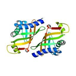
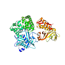
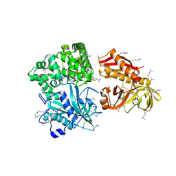
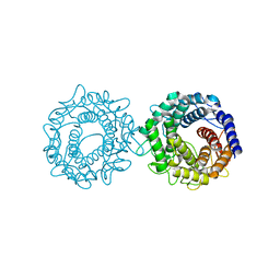
2D31
 
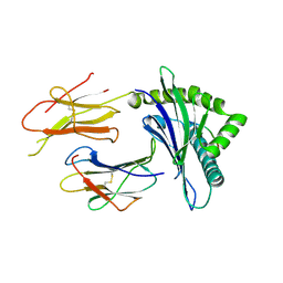 | | Crystal structure of disulfide-linked HLA-G dimer | | 分子名称: | 9-mer peptide from Histone H2A, Beta-2-microglobulin, HLA class I histocompatibility antigen, ... | | 著者 | Shiroishi, M, Kuroki, K, Ose, T, Rasubala, L, Shiratori, I, Arase, H, Tsumoto, K, Kumagai, I, Kohda, D, Maenaka, K. | | 登録日 | 2005-09-23 | | 公開日 | 2006-03-14 | | 最終更新日 | 2023-10-25 | | 実験手法 | X-RAY DIFFRACTION (3.2 Å) | | 主引用文献 | Efficient Leukocyte Ig-like Receptor Signaling and Crystal Structure of Disulfide-linked HLA-G Dimer
J.Biol.Chem., 281, 2006
|
|
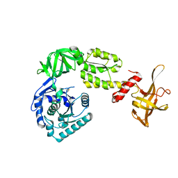
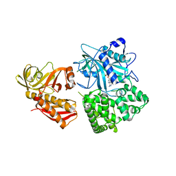
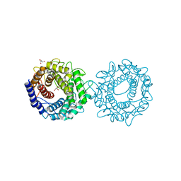
2D3V
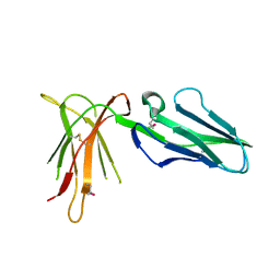 | | Crystal Structure of Leukocyte Ig-like Receptor A5 (LILRA5/LIR9/ILT11) | | 分子名称: | leukocyte immunoglobulin-like receptor subfamily A member 5 isoform 1 | | 著者 | Shiroishi, M, Kajikawa, M, Kuroki, K, Ose, T, Kohda, D, Maenaka, K. | | 登録日 | 2005-10-03 | | 公開日 | 2006-06-06 | | 最終更新日 | 2011-07-13 | | 実験手法 | X-RAY DIFFRACTION (1.85 Å) | | 主引用文献 | Crystal structure of the human monocyte-activating receptor,
J.Biol.Chem., 281, 2006
|
|
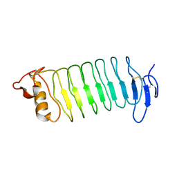
3VI6
 | | Crystal Structure of human ribosomal protein L30e | | 分子名称: | 60S ribosomal protein L30, FORMIC ACID | | 著者 | Kawaguchi, A, Ose, T, Yao, M, Tanaka, I. | | 登録日 | 2011-09-21 | | 公開日 | 2011-12-21 | | 最終更新日 | 2023-11-08 | | 実験手法 | X-RAY DIFFRACTION (1.59 Å) | | 主引用文献 | Crystallization and preliminary X-ray structure analysis of human ribosomal protein L30e
Acta Crystallogr.,Sect.F, 67, 2011
|
|
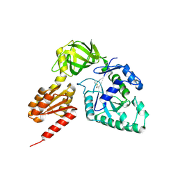
3WBK
 | | crystal structure analysis of eukaryotic translation initiation factor 5B and 1A complex | | 分子名称: | Eukaryotic translation initiation factor 1A, Eukaryotic translation initiation factor 5B | | 著者 | Zheng, A, Yamamoto, R, Ose, T, Yu, J, Tanaka, I, Yao, M. | | 登録日 | 2013-05-20 | | 公開日 | 2014-11-19 | | 最終更新日 | 2023-11-08 | | 実験手法 | X-RAY DIFFRACTION (3.3 Å) | | 主引用文献 | X-ray structures of eIF5B and the eIF5B-eIF1A complex: the conformational flexibility of eIF5B is restricted on the ribosome by interaction with eIF1A
Acta Crystallogr.,Sect.D, 70, 2014
|
|
3WH2
 | | Human Mincle in complex with citrate | | 分子名称: | C-type lectin domain family 4 member E, CALCIUM ION, CITRATE ANION | | 著者 | Furukawa, A, Kamishikiryo, J, Mori, D, Toyonaga, K, Okabe, Y, Toji, A, Kanda, R, Miyake, Y, Ose, T, Yamasaki, S, Maenaka, K. | | 登録日 | 2013-08-21 | | 公開日 | 2013-10-23 | | 最終更新日 | 2023-11-08 | | 実験手法 | X-RAY DIFFRACTION (1.3 Å) | | 主引用文献 | Structural analysis for glycolipid recognition by the C-type lectins Mincle and MCL
Proc.Natl.Acad.Sci.USA, 110, 2013
|
|
3WHD
 | | C-type lectin, human MCL | | 分子名称: | C-type lectin domain family 4 member D, CALCIUM ION | | 著者 | Furukawa, A, Kamishikiryo, J, Mori, D, Toyonaga, K, Okabe, Y, Toji, A, Kanda, R, Miyake, Y, Ose, T, Yamasaki, S, Maenaka, K. | | 登録日 | 2013-08-24 | | 公開日 | 2013-10-23 | | 最終更新日 | 2013-12-11 | | 実験手法 | X-RAY DIFFRACTION (2.29 Å) | | 主引用文献 | Structural analysis for glycolipid recognition by the C-type lectins Mincle and MCL
Proc.Natl.Acad.Sci.USA, 110, 2013
|
|
3WMD
 | | Crystal structure of epoxide hydrolase MonBI | | 分子名称: | Probable monensin biosynthesis isomerase | | 著者 | Minami, A, Ose, T, Sato, K, Oikawa, A, Kuroki, K, Maenaka, K, Oguri, H, Oikawa, H. | | 登録日 | 2013-11-16 | | 公開日 | 2014-01-15 | | 最終更新日 | 2024-03-20 | | 実験手法 | X-RAY DIFFRACTION (1.999 Å) | | 主引用文献 | Allosteric regulation of epoxide opening cascades by a pair of epoxide hydrolases in monensin biosynthesis
Acs Chem.Biol., 9, 2014
|
|
3WBI
 | | Crystal structure analysis of eukaryotic translation initiation factor 5B structure I | | 分子名称: | Eukaryotic translation initiation factor 5B | | 著者 | Zheng, A, Yamamoto, R, Ose, T, Yu, J, Tanaka, I, Yao, M. | | 登録日 | 2013-05-20 | | 公開日 | 2014-11-19 | | 最終更新日 | 2017-11-22 | | 実験手法 | X-RAY DIFFRACTION (2.35 Å) | | 主引用文献 | X-ray structures of eIF5B and the eIF5B-eIF1A complex: the conformational flexibility of eIF5B is restricted on the ribosome by interaction with eIF1A
Acta Crystallogr.,Sect.D, 70, 2014
|
|
3WH3
 | | human Mincle, ligand free form | | 分子名称: | C-type lectin domain family 4 member E, CALCIUM ION | | 著者 | Furukawa, A, Kamishikiryo, J, Mori, D, Toyonaga, K, Okabe, Y, Toji, A, Kanda, R, Miyake, Y, Ose, T, Yamasaki, S, Maenaka, K. | | 登録日 | 2013-08-21 | | 公開日 | 2013-10-23 | | 最終更新日 | 2023-11-08 | | 実験手法 | X-RAY DIFFRACTION (1.32 Å) | | 主引用文献 | Structural analysis for glycolipid recognition by the C-type lectins Mincle and MCL
Proc.Natl.Acad.Sci.USA, 110, 2013
|
|
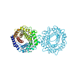
3WO9
 | | Crystal structure of the lamprey variable lymphocyte receptor C | | 分子名称: | Variable lymphocyte receptor C | | 著者 | Kanda, R, Sutoh, Y, Kasamatsu, J, Maenaka, K, Kasahara, M, Ose, T. | | 登録日 | 2013-12-20 | | 公開日 | 2014-03-05 | | 最終更新日 | 2023-11-08 | | 実験手法 | X-RAY DIFFRACTION (2.3 Å) | | 主引用文献 | Crystal structure of the lamprey variable lymphocyte receptor C reveals an unusual feature in its N-terminal capping module.
Plos One, 9, 2014
|
|
3WBJ
 | | Crystal structure analysis of eukaryotic translation initiation factor 5B structure II | | 分子名称: | Eukaryotic translation initiation factor 5B | | 著者 | Zheng, A, Yamamoto, R, Ose, T, Yu, J, Tanaka, I, Yao, M. | | 登録日 | 2013-05-20 | | 公開日 | 2014-11-19 | | 最終更新日 | 2023-11-08 | | 実験手法 | X-RAY DIFFRACTION (2.495 Å) | | 主引用文献 | X-ray structures of eIF5B and the eIF5B-eIF1A complex: the conformational flexibility of eIF5B is restricted on the ribosome by interaction with eIF1A
Acta Crystallogr.,Sect.D, 70, 2014
|
|
2ZZF
 | | Crystal structure of alanyl-tRNA synthetase without oligomerization domain | | 分子名称: | Alanyl-tRNA synthetase, ZINC ION | | 著者 | Sokabe, M, Ose, T, Tokunaga, K, Nakamura, A, Nureki, O, Yao, M, Tanaka, I. | | 登録日 | 2009-02-10 | | 公開日 | 2009-07-21 | | 最終更新日 | 2023-11-01 | | 実験手法 | X-RAY DIFFRACTION (2.7 Å) | | 主引用文献 | The structure of alanyl-tRNA synthetase with editing domain.
Proc.Natl.Acad.Sci.USA, 106, 2009
|
|
2ZZG
 | | Crystal structure of alanyl-tRNA synthetase in complex with 5''-O-(N-(L-alanyl)-sulfamyoxyl) adenine without oligomerization domain | | 分子名称: | '5'-O-(N-(L-ALANYL)-SULFAMOYL)ADENOSINE, Alanyl-tRNA synthetase, ZINC ION | | 著者 | Sokabe, M, Ose, T, Tokunaga, K, Nakamura, A, Nureki, O, Yao, M, Tanaka, I. | | 登録日 | 2009-02-10 | | 公開日 | 2009-07-21 | | 最終更新日 | 2023-11-01 | | 実験手法 | X-RAY DIFFRACTION (3.1 Å) | | 主引用文献 | The structure of alanyl-tRNA synthetase with editing domain.
Proc.Natl.Acad.Sci.USA, 106, 2009
|
|
2ZZE
 | | Crystal structure of alanyl-tRNA synthetase without oligomerization domain in lysine-methylated form | | 分子名称: | Alanyl-tRNA synthetase, ZINC ION | | 著者 | Sokabe, M, Ose, T, Tokunaga, K, Nakamura, A, Nureki, O, Yao, M, Tanaka, I. | | 登録日 | 2009-02-10 | | 公開日 | 2009-07-21 | | 最終更新日 | 2023-11-15 | | 実験手法 | X-RAY DIFFRACTION (2.16 Å) | | 主引用文献 | The structure of alanyl-tRNA synthetase with editing domain.
Proc.Natl.Acad.Sci.USA, 106, 2009
|
|
5H7K
 | | Crystal structure of Elongation factor 2 GDP-form | | 分子名称: | Elongation factor 2, GUANOSINE-5'-DIPHOSPHATE | | 著者 | Tanzawa, T, Kato, K, Uchiumi, T, Yao, M. | | 登録日 | 2016-11-18 | | 公開日 | 2018-02-21 | | 最終更新日 | 2024-03-20 | | 実験手法 | X-RAY DIFFRACTION (1.599 Å) | | 主引用文献 | The C-terminal helix of ribosomal P stalk recognizes a hydrophobic groove of elongation factor 2 in a novel fashion
Nucleic Acids Res., 46, 2018
|
|
5H7L
 | | Complex of Elongation factor 2-50S ribosomal protein L12 | | 分子名称: | 50S ribosomal protein L12, Elongation factor 2, PHOSPHOMETHYLPHOSPHONIC ACID GUANYLATE ESTER | | 著者 | Tanzawa, T, Kato, K, Uchiumi, T, Yao, M. | | 登録日 | 2016-11-18 | | 公開日 | 2018-02-21 | | 最終更新日 | 2018-05-02 | | 実験手法 | X-RAY DIFFRACTION (3.1 Å) | | 主引用文献 | The C-terminal helix of ribosomal P stalk recognizes a hydrophobic groove of elongation factor 2 in a novel fashion
Nucleic Acids Res., 46, 2018
|
|
5H7J
 | | Crystal structure of Elongation factor 2 | | 分子名称: | Elongation factor 2, PHOSPHOMETHYLPHOSPHONIC ACID GUANYLATE ESTER | | 著者 | Tanzawa, T, Kato, K, Uchiumi, T, Yao, M. | | 登録日 | 2016-11-18 | | 公開日 | 2018-02-21 | | 最終更新日 | 2018-05-02 | | 実験手法 | X-RAY DIFFRACTION (2.3 Å) | | 主引用文献 | The C-terminal helix of ribosomal P stalk recognizes a hydrophobic groove of elongation factor 2 in a novel fashion
Nucleic Acids Res., 46, 2018
|
|
8H1M
 | | Crystal structure of glucose-2-epimerase mutant_D254A from Runella slithyformis Runsl_4512 | | 分子名称: | FORMIC ACID, N-acylglucosamine 2-epimerase | | 著者 | Wang, H, Sun, X.M, Saburi, W, Yu, J, Yao, M. | | 登録日 | 2022-10-03 | | 公開日 | 2023-07-12 | | 実験手法 | X-RAY DIFFRACTION (1.6 Å) | | 主引用文献 | Structural insights into the substrate specificity and activity of a novel mannose 2-epimerase from Runella slithyformis.
Acta Crystallogr D Struct Biol, 79, 2023
|
|
8H1N
 | | Crystal structure of glucose-2-epimerase mutant_D254A in complex with D-Glucitol from Runella slithyformis Runsl_4512 | | 分子名称: | FORMIC ACID, N-acylglucosamine 2-epimerase, sorbitol | | 著者 | Wang, H, Sun, X.M, Saburi, W, Yu, J, Yao, M. | | 登録日 | 2022-10-03 | | 公開日 | 2023-07-12 | | 最終更新日 | 2023-11-29 | | 実験手法 | X-RAY DIFFRACTION (2.67 Å) | | 主引用文献 | Structural insights into the substrate specificity and activity of a novel mannose 2-epimerase from Runella slithyformis.
Acta Crystallogr D Struct Biol, 79, 2023
|
|
8H1K
 | | Crystal structure of glucose-2-epimerase from Runella slithyformis Runsl_4512 | | 分子名称: | FORMIC ACID, GLYCEROL, N-acylglucosamine 2-epimerase | | 著者 | Wang, H, Sun, X.M, Saburi, W, Yu, J, Yao, M. | | 登録日 | 2022-10-03 | | 公開日 | 2023-07-12 | | 最終更新日 | 2023-11-29 | | 実験手法 | X-RAY DIFFRACTION (1.6 Å) | | 主引用文献 | Structural insights into the substrate specificity and activity of a novel mannose 2-epimerase from Runella slithyformis.
Acta Crystallogr D Struct Biol, 79, 2023
|
|
8H1L
 | | Crystal structure of glucose-2-epimerase in complex with D-Glucitol from Runella slithyformis Runsl_4512 | | 分子名称: | N-acylglucosamine 2-epimerase, sorbitol | | 著者 | Wang, H, Sun, X.M, Saburi, W, Yu, J, Yao, M. | | 登録日 | 2022-10-03 | | 公開日 | 2023-07-12 | | 最終更新日 | 2023-11-29 | | 実験手法 | X-RAY DIFFRACTION (2.33 Å) | | 主引用文献 | Structural insights into the substrate specificity and activity of a novel mannose 2-epimerase from Runella slithyformis.
Acta Crystallogr D Struct Biol, 79, 2023
|
|
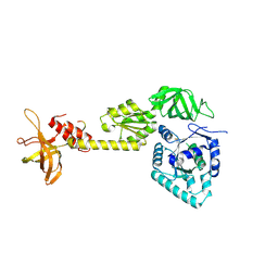
3AX3
 | |
3AX2
 | | Crystal structure of rat TOM20-ALDH presequence complex: a disulfide-tethered complex with a non-optimized, long linker | | 分子名称: | Aldehyde dehydrogenase, mitochondrial, Mitochondrial import receptor subunit TOM20 homolog, ... | | 著者 | Saitoh, T, Maita, Y, Kohda, D. | | 登録日 | 2011-03-28 | | 公開日 | 2011-07-06 | | 最終更新日 | 2023-11-01 | | 実験手法 | X-RAY DIFFRACTION (1.9 Å) | | 主引用文献 | Crystallographic snapshots of tom20-mitochondrial presequence interactions with disulfide-stabilized peptides.
Biochemistry, 50, 2011
|
|
3AX5
 | | Crystal structure of rat TOM20-ALDH presequence complex: A complex (form1) between Tom20 and a disulfide-bridged presequence peptide containing D-Cys and L-Cys at the i and i+3 positions. | | 分子名称: | Aldehyde dehydrogenase, mitochondrial, Mitochondrial import receptor subunit TOM20 homolog, ... | | 著者 | Saitoh, T, Maita, Y, Kohda, D. | | 登録日 | 2011-03-29 | | 公開日 | 2011-07-06 | | 最終更新日 | 2023-11-01 | | 実験手法 | X-RAY DIFFRACTION (2.2 Å) | | 主引用文献 | Crystallographic snapshots of tom20-mitochondrial presequence interactions with disulfide-stabilized peptides.
Biochemistry, 50, 2011
|
|


