4JIV
 
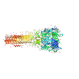
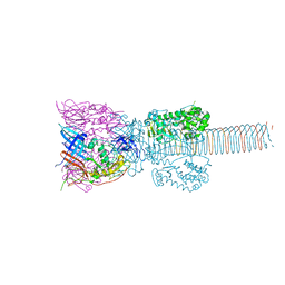
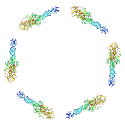
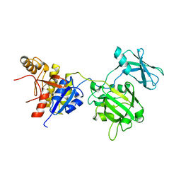
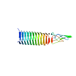 | | VCA0105 PAAR-repeat protein from Vibrio cholerae in complex with a VgrG-like beta-helix that is based on a fragment of T4 gp5 | | 分子名称: | 9-OCTADECENOIC ACID, MAGNESIUM ION, PALMITIC ACID, ... | | 著者 | Buth, S.A, Leiman, P.G, Shneider, M.M. | | 登録日 | 2013-03-07 | | 公開日 | 2013-08-14 | | 最終更新日 | 2024-03-20 | | 実験手法 | X-RAY DIFFRACTION (1.903 Å) | | 主引用文献 | PAAR-repeat proteins sharpen and diversify the type VI secretion system spike.
Nature, 500, 2013
|
|
4JK5
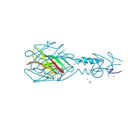
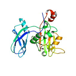
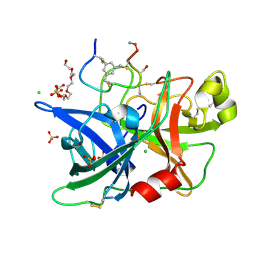 | | Human urokinase-type Plasminogen Activator (uPA) in complex with a bicyclic peptide inhibitor (UK18-D-Ser) | | 分子名称: | 1,3,5-tris(bromomethyl)benzene, CHLORIDE ION, HEXAETHYLENE GLYCOL, ... | | 著者 | Buth, S.A, Leiman, P.G, Chen, S, Heinis, C. | | 登録日 | 2013-03-09 | | 公開日 | 2013-07-17 | | 最終更新日 | 2023-09-20 | | 実験手法 | X-RAY DIFFRACTION (1.55 Å) | | 主引用文献 | Improving binding affinity and stability of Peptide ligands by substituting glycines with d-amino acids.
Chembiochem, 14, 2013
|
|
4JK6
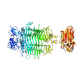
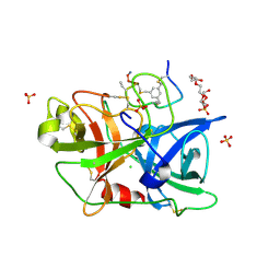 | | Human urokinase-type Plasminogen Activator (uPA) in complex with a bicyclic peptide inhibitor (UK18-D-Aba) | | 分子名称: | 1,3,5-tris(bromomethyl)benzene, CHLORIDE ION, HEXAETHYLENE GLYCOL, ... | | 著者 | Buth, S.A, Leiman, P.G, Chen, S, Heinis, C. | | 登録日 | 2013-03-09 | | 公開日 | 2013-07-17 | | 最終更新日 | 2023-12-06 | | 実験手法 | X-RAY DIFFRACTION (2.2 Å) | | 主引用文献 | Improving binding affinity and stability of Peptide ligands by substituting glycines with d-amino acids.
Chembiochem, 14, 2013
|
|
4KU0
 | |
4MTK
 | |
4MTM
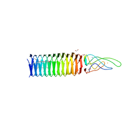
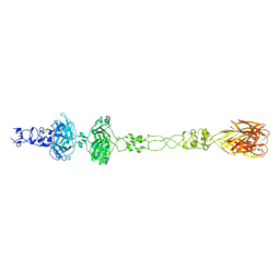
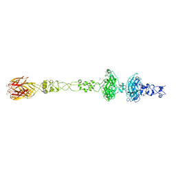
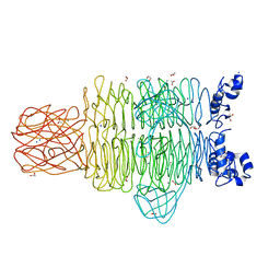
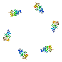
 | | Crystal structure of the tail fiber gp53 from Acinetobacter baumannii bacteriophage AP22 | | 分子名称: | 1,2-ETHANEDIOL, BROMIDE ION, GLYCEROL, ... | | 著者 | Sycheva, L.V, Shneider, M.M, Leiman, P.G. | | 登録日 | 2013-09-19 | | 公開日 | 2014-10-01 | | 最終更新日 | 2024-02-28 | | 実験手法 | X-RAY DIFFRACTION (1.368 Å) | | 主引用文献 | Crystal Structure of the putative tail fiber protein gp53 from the Acinetobacter baumannii bacteriophage AP22
Biorxiv, 2019
|
|
6C72
 | | Crystal structure of a tailspike protein gp49 from Acinetobacter baumannii bacteriophage Fri1, a capsular polysaccharide depolymerase | | 分子名称: | Particle-associated glycoside hydrolase, ZINC ION | | 著者 | Buth, S.A, Garcon, A, Popova, A.V, Shneider, M.M, Leiman, P.G. | | 登録日 | 2018-01-19 | | 公開日 | 2019-01-23 | | 最終更新日 | 2024-04-03 | | 実験手法 | X-RAY DIFFRACTION (2.396 Å) | | 主引用文献 | Exopolysaccharide degrading enzymes - specificity determinants of Acinetobacter baumannii phages
To Be Published
|
|
6CL5
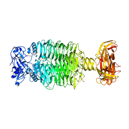
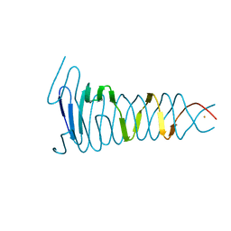
 | | Structure of P. aeruginosa R1 pyocin fiber PALES_06171 comprising C-terminal residues 323-701 | | 分子名称: | FE (III) ION, MAGNESIUM ION, SODIUM ION, ... | | 著者 | Buth, S.A, Shneider, M.M, Leiman, P.G. | | 登録日 | 2018-03-01 | | 公開日 | 2018-06-27 | | 最終更新日 | 2023-10-04 | | 実験手法 | X-RAY DIFFRACTION (2.324 Å) | | 主引用文献 | Structure and Analysis of R1 and R2 Pyocin Receptor-Binding Fibers.
Viruses, 10, 2018
|
|
1K28
 | | The Structure of the Bacteriophage T4 Cell-Puncturing Device | | 分子名称: | BASEPLATE STRUCTURAL PROTEIN GP27, PHOSPHATE ION, POTASSIUM ION, ... | | 著者 | Kanamaru, S, Leiman, P.G, Kostyuchenko, V.A, Chipman, P.R, Mesyanzhinov, V.V, Arisaka, F, Rossmann, M.G. | | 登録日 | 2001-09-26 | | 公開日 | 2002-02-06 | | 最終更新日 | 2011-07-13 | | 実験手法 | X-RAY DIFFRACTION (2.9 Å) | | 主引用文献 | Structure of the cell-puncturing device of bacteriophage T4.
Nature, 415, 2002
|
|
6CL6
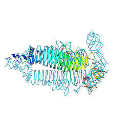
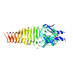
 | | Structure of P. aeruginosa R2 pyocin fiber PA0620 comprising C-terminal residues 323-691 | | 分子名称: | 1,2-ETHANEDIOL, CALCIUM ION, FE (III) ION, ... | | 著者 | Buth, S.A, Shneider, M.M, Leiman, P.G. | | 登録日 | 2018-03-01 | | 公開日 | 2018-06-27 | | 最終更新日 | 2024-04-03 | | 実験手法 | X-RAY DIFFRACTION (1.898 Å) | | 主引用文献 | Structure and Analysis of R1 and R2 Pyocin Receptor-Binding Fibers.
Viruses, 10, 2018
|
|
6E1R
 | | Crystal structure of the Acinetobacter phage vB_ApiP_P1 tailspike protein | | 分子名称: | CHLORIDE ION, SODIUM ION, Tailspike protein | | 著者 | Plattner, M, Shneider, M.M, Oliveira, H, Azeredo, J, Leiman, P.G. | | 登録日 | 2018-07-10 | | 公開日 | 2019-07-17 | | 実験手法 | X-RAY DIFFRACTION (2.693 Å) | | 主引用文献 | Crystal structure of the Acinetobacter phage vB_ApiP_P1 tailspike protein
To Be Published
|
|
6E0W
 | | Crystal structure of the colanidase tailspike protein gp150 of Phage Phi92 complexed with one repeating unit of colanic acid | | 分子名称: | 1,2-ETHANEDIOL, 1,5-anhydro-4,6-O-[(1R)-1-carboxyethylidene]-D-galactitol, Bacteriophage Phi92 gp150, ... | | 著者 | Plattner, M, Browning, C, Gerardy-Schahn, R, Shneider, M.M, Leiman, P.G, Schwarzer, D. | | 登録日 | 2018-07-07 | | 公開日 | 2019-07-17 | | 最終更新日 | 2023-10-11 | | 実験手法 | X-RAY DIFFRACTION (1.803 Å) | | 主引用文献 | Crystal structure of the colanidase tailspike protein gp150 of Phage Phi92 complexed with one repeating unit of colanic acid
To Be Published
|
|
6EU4
 | |
4OSD
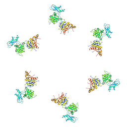
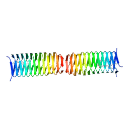 | | Dimer of a C-terminal fragment of phage T4 gp5 beta-helix | | 分子名称: | 9-OCTADECENOIC ACID, MAGNESIUM ION, PALMITIC ACID, ... | | 著者 | Buth, S.A, Leiman, P.G, Shneider, M.M. | | 登録日 | 2014-02-12 | | 公開日 | 2015-02-18 | | 最終更新日 | 2023-11-08 | | 実験手法 | X-RAY DIFFRACTION (1.96 Å) | | 主引用文献 | Structure and Biophysical Properties of a Triple-Stranded Beta-Helix Comprising the Central Spike of Bacteriophage T4.
Viruses, 7, 2015
|
|
4QNL
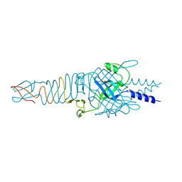
 | | Crystal structure of tail fiber protein gp63.1 from E. coli phage G7C | | 分子名称: | 1,2-ETHANEDIOL, CHLORIDE ION, DI(HYDROXYETHYL)ETHER, ... | | 著者 | Riccio, C, Browning, C, Prokhorov, N, Letarov, A, Leiman, P.G. | | 登録日 | 2014-06-18 | | 公開日 | 2015-06-24 | | 最終更新日 | 2017-12-20 | | 実験手法 | X-RAY DIFFRACTION (2.411 Å) | | 主引用文献 | Function of bacteriophage G7C esterase tailspike in host cell adsorption.
Mol. Microbiol., 105, 2017
|
|
4RU4
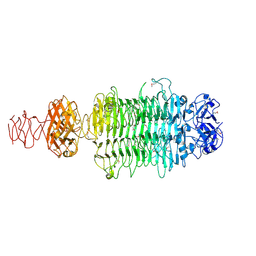
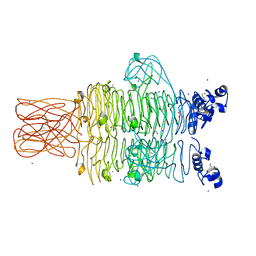 | | Crystal structure of the tailspike protein gp49 from Pseudomonas phage LKA1 | | 分子名称: | 1,2-ETHANEDIOL, CALCIUM ION, SODIUM ION, ... | | 著者 | Browning, C, Shneider, M.M, Leiman, P.G. | | 登録日 | 2014-11-18 | | 公開日 | 2015-11-18 | | 最終更新日 | 2024-02-28 | | 実験手法 | X-RAY DIFFRACTION (1.903 Å) | | 主引用文献 | The O-specific polysaccharide lyase from the phage LKA1 tailspike reduces Pseudomonas virulence.
Sci Rep, 7, 2017
|
|
4RU5
 | | Crystal Structure of the Pseudomonas phage phi297 tailspike gp61 | | 分子名称: | 1,2-ETHANEDIOL, ACETATE ION, CALCIUM ION, ... | | 著者 | Browning, C, Sycheva, L.V, Shneider, M.M, Leiman, P.G. | | 登録日 | 2014-11-18 | | 公開日 | 2015-11-18 | | 最終更新日 | 2024-02-28 | | 実験手法 | X-RAY DIFFRACTION (1.52 Å) | | 主引用文献 | The O-specific polysaccharide lyase from the phage LKA1 tailspike reduces Pseudomonas virulence.
Sci Rep, 7, 2017
|
|
4S36
 | |
4RU3
 | |
4S37
 | |
1PDI
 | | Fitting of the C-terminal part of the short tail fibers into the cryo-EM reconstruction of T4 baseplate | | 分子名称: | Short tail fiber protein | | 著者 | Kostyuchenko, V.A, Leiman, P.G, Chipman, P.R, Kanamaru, S, van Raaij, M.J, Arisaka, F, Mesyanzhinov, V.V, Rossmann, M.G. | | 登録日 | 2003-05-19 | | 公開日 | 2003-09-09 | | 最終更新日 | 2024-02-14 | | 実験手法 | ELECTRON MICROSCOPY (12 Å) | | 主引用文献 | Three-dimensional structure of bacteriophage T4 baseplate
Nat.Struct.Biol., 10, 2003
|
|
1PDP
 | | Fitting of gp9 structure into the bacteriophage T4 baseplate cryoEM reconstruction | | 分子名称: | Baseplate structural protein Gp9 | | 著者 | Kostyuchenko, V.A, Leiman, P.G, Chipman, P.R, Kanamaru, S, van Raaij, M.J, Arisaka, F, Mesyanzhinov, V.V, Rossmann, M.G. | | 登録日 | 2003-05-19 | | 公開日 | 2003-09-09 | | 最終更新日 | 2024-02-14 | | 実験手法 | ELECTRON MICROSCOPY (12 Å) | | 主引用文献 | Three-dimensional structure of the bacteriophage T4 baseplate
Nat.Struct.Biol., 10, 2003
|
|
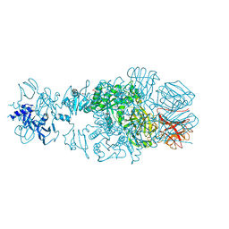
3FO8
 | | Crystal structure of the bacteriophage T4 tail sheath protein, protease resistant fragment gp18PR | | 分子名称: | ACETATE ION, Tail sheath protein Gp18 | | 著者 | Aksyuk, A.A, Leiman, P.G, Kurochkina, L.P, Shneider, M.M, Kostyuchenko, V.A, Mesyanzhinov, V.V, Rossmann, M.G. | | 登録日 | 2008-12-29 | | 公開日 | 2009-03-10 | | 最終更新日 | 2024-02-21 | | 実験手法 | X-RAY DIFFRACTION (1.8 Å) | | 主引用文献 | The tail sheath structure of bacteriophage T4: a molecular machine for infecting bacteria.
Embo J., 28, 2009
|
|
3FOI
 | | Fitting of gp18M crystal structure into 3D cryo-EM reconstruction of bacteriophage T4 contracted tail | | 分子名称: | Tail sheath protein Gp18 | | 著者 | Aksyuk, A.A, Leiman, P.G, Kurochkina, L.P, Shneider, M.M, Kostyuchenko, V.A, Mesyanzhinov, V.V, Rossmann, M.G. | | 登録日 | 2008-12-30 | | 公開日 | 2009-03-10 | | 最終更新日 | 2024-02-21 | | 実験手法 | ELECTRON MICROSCOPY (16 Å) | | 主引用文献 | The tail sheath structure of bacteriophage T4: a molecular machine for infecting bacteria.
Embo J., 28, 2009
|
|
3FOA
 | | Crystal structure of the bacteriophage T4 tail sheath protein, deletion mutant gp18M | | 分子名称: | Tail sheath protein Gp18 | | 著者 | Aksyuk, A.A, Leiman, P.G, Kurochkina, L.P, Shneider, M.M, Kostyuchenko, V.A, Mesyanzhinov, V.V, Rossmann, M.G. | | 登録日 | 2008-12-29 | | 公開日 | 2009-03-10 | | 最終更新日 | 2023-09-06 | | 実験手法 | X-RAY DIFFRACTION (3.5 Å) | | 主引用文献 | The tail sheath structure of bacteriophage T4: a molecular machine for infecting bacteria.
Embo J., 28, 2009
|
|


