3DQH
 
 | |
3DQ9
 | |
3DQM
 | |
3DQ2
 | |
3DQC
 | |
3DQ6
 | |
3DQL
 | |
3DQ3
 | |
3DQD
 | |
3DQN
 | |
3DPW
 | |
3DQO
 | |
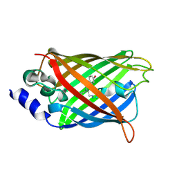
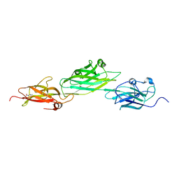
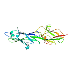
6JLA
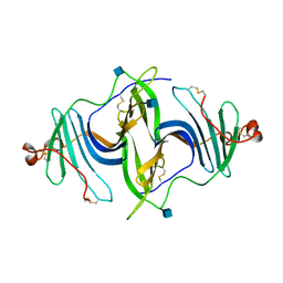 | | Crystal structure of a mouse ependymin related protein | | 分子名称: | 2-acetamido-2-deoxy-beta-D-glucopyranose, 2-acetamido-2-deoxy-beta-D-glucopyranose-(1-4)-[alpha-L-fucopyranose-(1-6)]2-acetamido-2-deoxy-beta-D-glucopyranose, Mammalian ependymin-related protein 1 | | 著者 | Park, S. | | 登録日 | 2019-03-04 | | 公開日 | 2020-03-04 | | 最終更新日 | 2020-09-16 | | 実験手法 | X-RAY DIFFRACTION (2.4 Å) | | 主引用文献 | Structures of three ependymin-related proteins suggest their function as a hydrophobic molecule binder.
Iucrj, 6, 2019
|
|
6JLD
 | |
6JL9
 | |
8SD7
 | |
8SF1
 | |
8SD9
 | |
8SD8
 | |
8SD6
 | |
8SD1
 | |
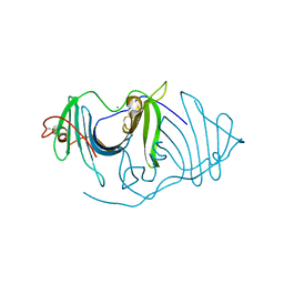
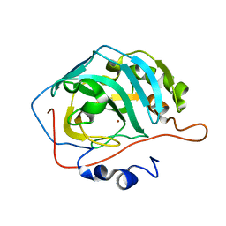
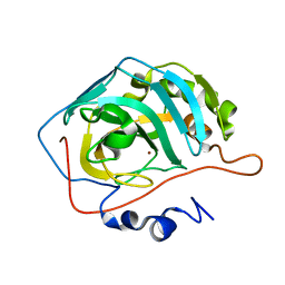
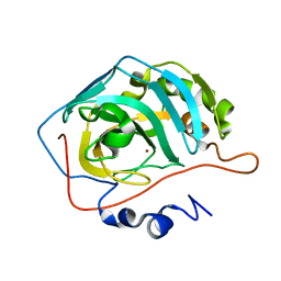
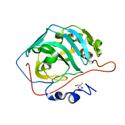
6PDV
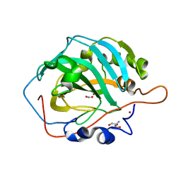 | | Cu-Carbonic Anhydrase II, A Nitrite Reductase | | 分子名称: | 2-AMINO-2-HYDROXYMETHYL-PROPANE-1,3-DIOL, COPPER (II) ION, Carbonic anhydrase 2, ... | | 著者 | Andring, J.T, McKenna, R. | | 登録日 | 2019-06-19 | | 公開日 | 2020-03-11 | | 最終更新日 | 2023-10-11 | | 実験手法 | X-RAY DIFFRACTION (1.23 Å) | | 主引用文献 | Structure and mechanism of copper-carbonic anhydrase II: a nitrite reductase.
Iucrj, 7, 2020
|
|
6PEA
 | |
4HSS
 | |
4HSQ
 | |


