4M7M
 
 | |
5F58
 | |
5F6B
 | |
5FAZ
 | |
5FEN
 | |
5FFH
 | |
5F7G
 | |
2DPQ
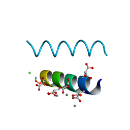 | | The crystal structures of the calcium-bound con-G and con-T(K7gamma) dimeric peptides demonstrate a novel metal-dependent helix-forming motif | | 分子名称: | CALCIUM ION, CHLORIDE ION, Conantokin-G | | 著者 | Cnudde, S.E, Prorok, M, Dai, Q, Castellino, F.J, Geiger, J.H. | | 登録日 | 2006-05-13 | | 公開日 | 2007-04-24 | | 最終更新日 | 2024-04-03 | | 実験手法 | X-RAY DIFFRACTION (1.25 Å) | | 主引用文献 | The crystal structures of the calcium-bound con-G and con-T[K7gamma] dimeric peptides demonstrate a metal-dependent helix-forming motif
J.Am.Chem.Soc., 129, 2007
|
|
2DOI
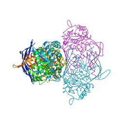
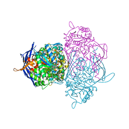
 | | The X-ray crystallographic structure of the angiogenesis inhibitor, angiostatin, bound to a peptide from the group A streptococcus protein PAM | | 分子名称: | Angiostatin, Plasminogen-binding group A streptococcal M-like protein PAM | | 著者 | Cnudde, S.E, Prorok, M, Castellino, F.J, Geiger, J.H. | | 登録日 | 2006-04-29 | | 公開日 | 2006-12-05 | | 最終更新日 | 2023-10-25 | | 実験手法 | X-RAY DIFFRACTION (3.1 Å) | | 主引用文献 | X-ray crystallographic structure of the angiogenesis inhibitor, angiostatin, bound to a peptide from the group A streptococcal surface protein PAM
Biochemistry, 45, 2006
|
|
2DOH
 | | The X-ray crystallographic structure of the angiogenesis inhibitor, angiostatin, bound a to a peptide from the group A streptococcal surface protein PAM | | 分子名称: | 1,4-DIETHYLENE DIOXIDE, Angiostatin, Plasminogen-binding group A streptococcal M-like protein PAM | | 著者 | Cnudde, S.E, Prorok, M, Castellino, F.J, Geiger, J.H. | | 登録日 | 2006-04-29 | | 公開日 | 2006-12-05 | | 最終更新日 | 2023-10-25 | | 実験手法 | X-RAY DIFFRACTION (2.3 Å) | | 主引用文献 | X-ray crystallographic structure of the angiogenesis inhibitor, angiostatin, bound to a peptide from the group A streptococcal surface protein PAM
Biochemistry, 45, 2006
|
|
2DPR
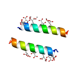 | | The crystal structures of the calcium-bound con-G and con-T(K7Glu) dimeric peptides demonstrate a novel metal-dependent helix-forming motif | | 分子名称: | CALCIUM ION, Conantokin-T | | 著者 | Cnudde, S.E, Prorok, M, Dai, Q, Castellino, F.J, Geiger, J.H. | | 登録日 | 2006-05-13 | | 公開日 | 2007-04-24 | | 最終更新日 | 2024-04-03 | | 実験手法 | X-RAY DIFFRACTION (1.7 Å) | | 主引用文献 | The crystal structures of the calcium-bound con-G and con-T[K7gamma] dimeric peptides demonstrate a metal-dependent helix-forming motif
J.Am.Chem.Soc., 129, 2007
|
|
2FRS
 | |
2FS6
 | |
2FR3
 | |
2FS7
 | |
2G78
 | |
2G7B
 | |
2G79
 | |
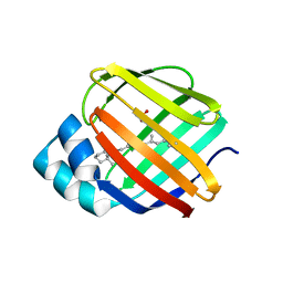
1KI0
 | | The X-ray Structure of Human Angiostatin | | 分子名称: | ANGIOSTATIN, BICINE | | 著者 | Abad, M.C, Arni, R.K, Grella, D.K, Castellino, F.J, Tulinsky, A, Geiger, J.H. | | 登録日 | 2001-12-02 | | 公開日 | 2002-05-29 | | 最終更新日 | 2021-10-27 | | 実験手法 | X-RAY DIFFRACTION (1.75 Å) | | 主引用文献 | The X-ray crystallographic structure of the angiogenesis inhibitor angiostatin.
J.Mol.Biol., 318, 2002
|
|
1M7X
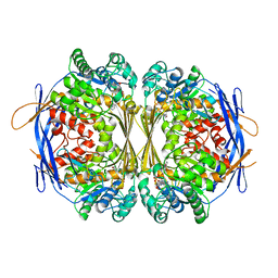
 | | The X-ray Crystallographic Structure of Branching Enzyme | | 分子名称: | 1,4-alpha-glucan Branching Enzyme | | 著者 | Abad, M.C, Binderup, K, Rios-Steiner, J, Arni, R.K, Preiss, J, Geiger, J.H. | | 登録日 | 2002-07-23 | | 公開日 | 2002-09-18 | | 最終更新日 | 2022-12-21 | | 実験手法 | X-RAY DIFFRACTION (2.3 Å) | | 主引用文献 | The X-ray crystallographic structure of Escherichia coli branching enzyme
J.Biol.Chem., 277, 2002
|
|
1P1H
 | |
1P1K
 | |
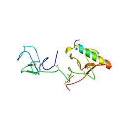
1P1J
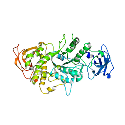
 | | Crystal structure of the 1L-myo-inositol 1-phosphate synthase complexed with NADH | | 分子名称: | 1,4-DIHYDRONICOTINAMIDE ADENINE DINUCLEOTIDE, GLYCEROL, Inositol-3-phosphate synthase, ... | | 著者 | Jin, X, Geiger, J.H. | | 登録日 | 2003-04-12 | | 公開日 | 2003-07-08 | | 最終更新日 | 2024-02-14 | | 実験手法 | X-RAY DIFFRACTION (1.7 Å) | | 主引用文献 | Structures of NAD(+)- and NADH-bound 1-l-myo-inositol 1-phosphate synthase.
Acta Crystallogr.,Sect.D, 59, 2003
|
|
1P1I
 | |
1P1F
 | |


