Loading
PDBj
MenuPDBj@FacebookPDBj@TwitterPDBj@YouTubewwPDB FoundationwwPDB
RCSB PDBPDBeBMRBAdv. SearchSearch help
Search by PDB author
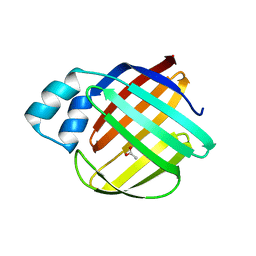
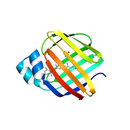
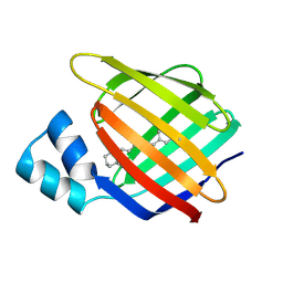
2FRS
DownloadVisualize
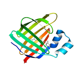
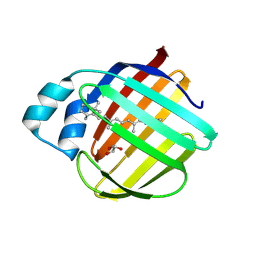
BU of 2frs by Molmil
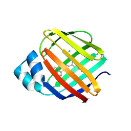
Crystal structure of the f15w mutant of apo-cellular retinoic acid binding protein type ii at 1.51 angstroms resolution
Descriptor: Cellular retinoic acid-binding protein 2, SODIUM ION
Authors:Vaezeslami, S, Geiger, J.H.
Deposit date:2006-01-20
Release date:2006-09-19
Last modified:2024-02-14
Method:X-RAY DIFFRACTION (1.51 Å)
Cite:The structure of Apo-wild-type cellular retinoic acid binding protein II at 1.4 A and its relationship to ligand binding and nuclear translocation.
J.Mol.Biol., 363, 2006
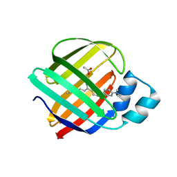
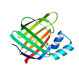
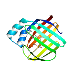
5U6G
DownloadVisualize
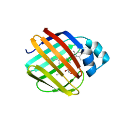
BU of 5u6g by Molmil
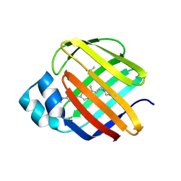
Crystal Structure of the holo Domain-Swapped Dimer mutant Q108K:K40D Human Cellular Retinol Binding Protein II bound with all trans retinal
Descriptor: RETINAL, Retinol-binding protein 2
Authors:Assar, Z, Geiger, J.H.
Deposit date:2016-12-08
Release date:2017-10-18
Last modified:2023-10-04
Method:X-RAY DIFFRACTION (2.6 Å)
Cite:Crystal Structure of the holo Domain-Swapped Dimer mutant Q108K:K40D Human Cellular Retinol Binding Protein II bound with all trans retinal
To Be Published
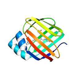
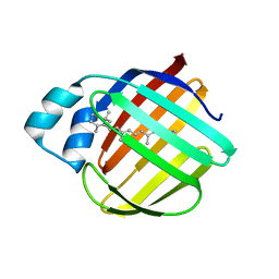
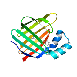
6C7Z
DownloadVisualize
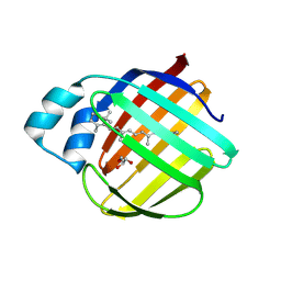
BU of 6c7z by Molmil
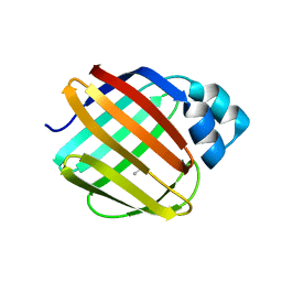
Crystal structure of the Q108K:K40L:T51V:R58F mutant of human Cellular Retinol Binding Protein II in complex with synthetic Ligand Julolidine
Descriptor: (2E,4E)-3-methyl-5-(2,3,6,7-tetrahydro-1H,5H-pyrido[3,2,1-ij]quinolin-9-yl)penta-2,4-dienal, ACETATE ION, Retinol-binding protein 2
Authors:Nosrati, M, Geiger, J.H.
Deposit date:2018-01-23
Release date:2018-04-25
Last modified:2023-10-04
Method:X-RAY DIFFRACTION (1.42 Å)
Cite:A Genetically Encoded Ratiometric pH Probe: Wavelength Regulation-Inspired Design of pH Indicators.
Chembiochem, 19, 2018
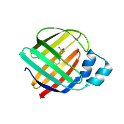
8VZY
DownloadVisualize
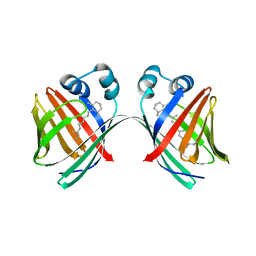
BU of 8vzy by Molmil
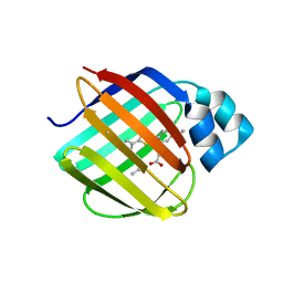
Q108K:K40L:T51V:T53S:R58Y mutant of hCRBPII bound to synthetic fluorophore TD-1V
Descriptor: (2E)-3-{5-[4-(dimethylamino)phenyl]thiophen-2-yl}but-2-enal, ACETATE ION, Retinol-binding protein 2
Authors:Nossoni, Z, Bingham, C.R, Geiger, J.H, Borhan, B.
Deposit date:2024-02-13
Release date:2024-04-03
Method:X-RAY DIFFRACTION (1.34 Å)
Cite:Regulation of Emission via a Protein-Bound Fluorophore
To Be Published
8VZX
DownloadVisualize
BU of 8vzx by Molmil
Q108K:K40L:T53A:R58F mutant of hCRBPII bound to synthetic fluorophore TD-1V
Descriptor: (2E)-3-{5-[4-(dimethylamino)phenyl]thiophen-2-yl}but-2-enal, ACETATE ION, Retinol-binding protein 2
Authors:Nossoni, Z, Bingham, C.R, Geiger, J.H.
Deposit date:2024-02-13
Release date:2024-04-03
Method:X-RAY DIFFRACTION (1.47 Å)
Cite:Regulation of Emission via a Protein-Bound Fluorophore
To Be Published
8VZZ
DownloadVisualize
BU of 8vzz by Molmil
Q108K:K40L:T51V:T53S:Y19W:R58W mutant of hCRBPII bound to synthetic fluorophore TD-1V
Descriptor: (2E)-3-{5-[4-(dimethylamino)phenyl]thiophen-2-yl}but-2-enal, ACETATE ION, Retinol-binding protein 2
Authors:Nosssoni, Z, Bingham, C.R, Geiger, J.H, Borhan, B.
Deposit date:2024-02-13
Release date:2024-04-03
Method:X-RAY DIFFRACTION (1.22 Å)
Cite:Regulation of Emission via a Protein-Bound Fluorophore
To Be Published
8W02
DownloadVisualize
BU of 8w02 by Molmil
Q108K:K40L:T51V:T53S:R58W mutant of hCRBPII bound to synthetic fluorophore TD-1V
Descriptor: (2E)-3-{5-[4-(dimethylamino)phenyl]thiophen-2-yl}but-2-enal, ACETATE ION, Retinol-binding protein 2
Authors:Ghanbarpour, A, Geiger, J.H, Borhan, B.
Deposit date:2024-02-13
Release date:2024-04-03
Method:X-RAY DIFFRACTION (1.499 Å)
Cite:Regulation of Emission via a Protein-Bound Fluorophore
To Be Published
8W00
DownloadVisualize
BU of 8w00 by Molmil
Q108K:K40L:T51V:T53S:Y19W:R58W:L117E mutant of hCRBPII bound to synthetic fluorophore TD-1V
Descriptor: (2E)-3-{5-[4-(dimethylamino)phenyl]thiophen-2-yl}but-2-enal, ACETATE ION, Retinol-binding protein 2
Authors:Nossoni, Z, Bingham, C.R, Geiger, J.H.
Deposit date:2024-02-13
Release date:2024-04-03
Method:X-RAY DIFFRACTION (1.23 Å)
Cite:Regulation of Emission via a Protein-Bound Fluorophore
To Be Published
4EXZ
DownloadVisualize
BU of 4exz by Molmil
Crystal Structure of the Q108K:K40L Mutant of Cellular Retinol Binding Protein Type II in Complex with All-trans-Retinal at 1.7 Angstrom Resolution
Descriptor: ACETATE ION, RETINAL, Retinol-binding protein 2
Authors:Nossoni, Z, Geiger, J.H.
Deposit date:2012-05-01
Release date:2012-12-26
Last modified:2023-09-13
Method:X-RAY DIFFRACTION (1.608 Å)
Cite:Tuning the electronic absorption of protein-embedded all-trans-retinal.
Science, 338, 2012
4EDE
DownloadVisualize
BU of 4ede by Molmil
Crystal Structure of the Q108K:K40L:T51V:T53C:Y19W:R58W:T29L:A33W Mutant of Cellular Retinol Binding Protein Type II in Complex with All-trans-Retinal at 1.4 Angstrom Resolution
Descriptor: ACETATE ION, RETINAL, Retinol-binding protein 2
Authors:Nossoni, Z, Geiger, J.H.
Deposit date:2012-03-27
Release date:2012-12-26
Last modified:2023-09-13
Method:X-RAY DIFFRACTION (1.4 Å)
Cite:Tuning the electronic absorption of protein-embedded all-trans-retinal.
Science, 338, 2012
4EEJ
DownloadVisualize
BU of 4eej by Molmil
Crystal Structure of the Q108K:K40L:T51V:T53C:Y19W:R58W:T29L:Q4R Mutant of Cellular Retinol Binding Protein Type II in Complex with All-trans-Retinal at 1.5 Angstrom Resolution
Descriptor: ACETATE ION, RETINAL, Retinol-binding protein 2
Authors:Nossoni, Z, Geiger, J.H.
Deposit date:2012-03-28
Release date:2012-12-26
Last modified:2023-09-13
Method:X-RAY DIFFRACTION (1.501 Å)
Cite:Tuning the electronic absorption of protein-embedded all-trans-retinal.
Science, 338, 2012
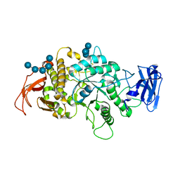
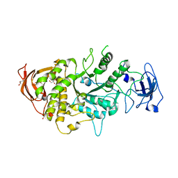
8SDB
DownloadVisualize
BU of 8sdb by Molmil
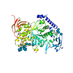
Crystal Structure of E.Coli Branching Enzyme in complex with malto-octose
Descriptor: 1,4-alpha-glucan branching enzyme GlgB, alpha-D-glucopyranose, alpha-D-glucopyranose-(1-4)-alpha-D-glucopyranose, ...
Authors:Bingham, C.R, Nayebi, H, Fawaz, R, Geiger, J.H.
Deposit date:2023-04-06
Release date:2023-07-12
Last modified:2023-08-16
Method:X-RAY DIFFRACTION (3 Å)
Cite:The Structure of Maltooctaose-Bound Escherichia coli Branching Enzyme Suggests a Mechanism for Donor Chain Specificity.
Molecules, 28, 2023
4GKC
DownloadVisualize
BU of 4gkc by Molmil
Crystal structure of Q108K:K40L:T51V:T53C:R58W:T29L:Y19W:Q4A mutant of cellular retinol binding protein II complex with all-trans-retinal at 1.33
Descriptor: RETINAL, Retinol-binding protein 2
Authors:Nossoni, Z, Geiger, J.H.
Deposit date:2012-08-10
Release date:2012-12-26
Last modified:2023-09-13
Method:X-RAY DIFFRACTION (1.3 Å)
Cite:Tuning the electronic absorption of protein-embedded all-trans-retinal.
Science, 338, 2012
4I9S
DownloadVisualize
BU of 4i9s by Molmil
Crystal Structure of the R111K:R132L:Y134F:T54V:R59W Mutant of the Cellular Retinoic Acid Binding Protein Type II in Complex with All-Trans Retinal at 2.58 Angstrom Resolution
Descriptor: Cellular retinoic acid-binding protein 2, RETINAL
Authors:Nosrati, M, Geiger, J.H.
Deposit date:2012-12-05
Release date:2013-10-09
Last modified:2023-09-20
Method:X-RAY DIFFRACTION (2.58 Å)
Cite:Rational Design of a Colorimetric pH Sensor from a Soluble Retinoic Acid Chaperone.
J.Am.Chem.Soc., 135, 2013
4I9R
DownloadVisualize
BU of 4i9r by Molmil
Crystal Structure of the R111K:R132L:Y134F:T54V:R59W:A32W Mutant of the Cellular Retinoic Acid Binding Protein Type II in Complex with All-Trans Retinal at 2.6 Angstrom Resolution
Descriptor: Cellular retinoic acid-binding protein 2, RETINAL
Authors:Nosrati, M, Geiger, J.H.
Deposit date:2012-12-05
Release date:2013-10-09
Last modified:2023-09-20
Method:X-RAY DIFFRACTION (2.6 Å)
Cite:Rational Design of a Colorimetric pH Sensor from a Soluble Retinoic Acid Chaperone.
J.Am.Chem.Soc., 135, 2013
7ML5
DownloadVisualize
BU of 7ml5 by Molmil
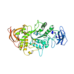
Structure of the Starch Branching Enzyme I (BEI) complexed with maltododecaose from Oryza sativa L
Descriptor: Isoform 2 of 1,4-alpha-glucan-branching enzyme, chloroplastic/amyloplastic, alpha-D-glucopyranose-(1-4)-alpha-D-glucopyranose-(1-4)-alpha-D-glucopyranose-(1-4)-alpha-D-glucopyranose, ...
Authors:Nayebi Gavgani, H, Fawaz, R, Geiger, J.H.
Deposit date:2021-04-27
Release date:2021-11-17
Last modified:2023-10-18
Method:X-RAY DIFFRACTION (2.35 Å)
Cite:A structural explanation for the mechanism and specificity of plant branching enzymes I and IIb.
J.Biol.Chem., 298, 2021
7MS9
DownloadVisualize
BU of 7ms9 by Molmil
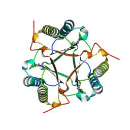
Crystal structure of E114D mutant of Cg10062 with a covalent intermediate of the hydration of acetylenecarboxylic acid
Descriptor: 3-HYDROXY-PROPANOIC ACID, 4-oxalocrotonate tautomerase, SULFATE ION
Authors:Nayebi, G.H, Geiger, J.H, Draths, K.
Deposit date:2021-05-10
Release date:2022-02-02
Last modified:2023-10-18
Method:X-RAY DIFFRACTION (2.2 Å)
Cite:Cg10062 Catalysis Forges a Link between Acetylenecarboxylic Acid and Bacterial Metabolism.
Biochemistry, 60, 2021
4LQ1
DownloadVisualize
BU of 4lq1 by Molmil
Crystal Structure of E.Coli Branching Enzyme in complex with maltohexaose
Descriptor: 1,4-alpha-glucan branching enzyme GlgB, GLYCEROL, alpha-D-glucopyranose-(1-4)-alpha-D-glucopyranose, ...
Authors:Feng, L, Geiger, J.H.
Deposit date:2013-07-17
Release date:2015-04-01
Last modified:2024-02-28
Method:X-RAY DIFFRACTION (2.55 Å)
Cite:Crystal Structures of Escherichia coli Branching Enzyme in Complex with Linear Oligosaccharides.
Biochemistry, 54, 2015
4M6S
DownloadVisualize
BU of 4m6s by Molmil
Crystal structure of the R111K:R132Y:Y134F:T54V:R59W:A32W mutant of the Cellular Retinoic Acid Binding Protein Type II in complex with All-Trans Retinal at 2.38 Angstrom Resolution
Descriptor: Cellular retinoic acid-binding protein 2, RETINAL
Authors:Nosrati, M, Geiger, J.H.
Deposit date:2013-08-10
Release date:2013-10-09
Last modified:2023-09-20
Method:X-RAY DIFFRACTION (2.47 Å)
Cite:Rational Design of a Colorimetric pH Sensor from a Soluble Retinoic Acid Chaperone.
J.Am.Chem.Soc., 135, 2013
4LPC
DownloadVisualize
BU of 4lpc by Molmil
Crystal Structure of E.Coli Branching Enzyme in complex with maltoheptaose
Descriptor: 1,4-alpha-glucan branching enzyme GlgB, GLYCEROL, alpha-D-glucopyranose-(1-4)-alpha-D-glucopyranose, ...
Authors:Feng, L, Geiger, J.H.
Deposit date:2013-07-16
Release date:2015-04-01
Last modified:2024-02-28
Method:X-RAY DIFFRACTION (2.39 Å)
Cite:Crystal Structures of Escherichia coli Branching Enzyme in Complex with Linear Oligosaccharides.
Biochemistry, 54, 2015
4M7M
DownloadVisualize
BU of 4m7m by Molmil
Crystal structure of the R111K:R132Q:Y134F:T54V:R59W:A32W mutant of the Cellular Retinoic Acid Binding Protein Type II in complex with All-Trans Retinal at 2.57 Angstrom Resolution
Descriptor: Cellular retinoic acid-binding protein 2, RETINAL
Authors:Nosrati, M, Geiger, J.H.
Deposit date:2013-08-12
Release date:2013-10-09
Last modified:2014-02-19
Method:X-RAY DIFFRACTION (2.571 Å)
Cite:Rational Design of a Colorimetric pH Sensor from a Soluble Retinoic Acid Chaperone.
J.Am.Chem.Soc., 135, 2013
7LHJ
DownloadVisualize
BU of 7lhj by Molmil
Crystal structure of Q108K:K40L:T51V:T53C:R58W:T29L:Y19W:Q4A mutant of cellular retinol binding protein II complex with 15-cis-retinal
Descriptor: ACETATE ION, GLYCEROL, RETINAL, ...
Authors:Ehyaei, N, Geiger, J.H, Borhan, B.
Deposit date:2021-01-25
Release date:2021-02-10
Last modified:2023-10-18
Method:X-RAY DIFFRACTION (1.26 Å)
Cite:Crystal structure of Q108K:K40L:T51V:T53C:R58W:T29L:Y19W:Q4A mutant of cellular retinol binding protein II complex with 15cis- retinal
To Be Published
7LHN
DownloadVisualize
BU of 7lhn by Molmil
Crystal structure of Q108K:K40L:T51V:T53C:R58W:T29L:Y19W:Q4A mutant of cellular retinol binding protein II complex with all-trans-retinal after exposure to visible light
Descriptor: GLYCEROL, RETINAL, Retinol-binding protein 2
Authors:Ehyaei, N, Geiger, J.H, Borhan, B.
Deposit date:2021-01-25
Release date:2021-04-14
Last modified:2023-10-18
Method:X-RAY DIFFRACTION (2.11 Å)
Cite:Crystal structure of Q108K:K40L:T51V:T53C:R58W:T29L:Y19W:Q4A mutant of cellular retinol binding protein II complex with all-trans-retinal after exposure to visible light
To Be Published
7LHM
DownloadVisualize
BU of 7lhm by Molmil
Crystal structure of Q108K:K40L:T51V:T53C:R58W:T29L:Y19W:Q4A:Q38L mutant of cellular retinol binding protein II complex with all-trans-retinal
Descriptor: ACETATE ION, GLYCEROL, RETINAL, ...
Authors:Ehyaei, N, Geiger, J.H, Borhan, B.
Deposit date:2021-01-25
Release date:2021-04-14
Last modified:2023-10-18
Method:X-RAY DIFFRACTION (1.5 Å)
Cite:Crystal structure of Q108K:K40L:T51V:T53C:R58W:T29L:Y19W:Q4A:Q38L mutant of cellular retinol binding protein II complex with all-trans-retinal
To Be Published
7LHO
DownloadVisualize
BU of 7lho by Molmil
Crystal structure of Q108K:K40L:T51V:T53C:R58W:T29L:Y19W:Q4A mutant of cellular retinol binding protein II complex with all-trans-retinal in the dark
Descriptor: GLYCEROL, RETINAL, Retinol-binding protein 2
Authors:Ehyaei, N, Geiger, J.H, Borhan, B.
Deposit date:2021-01-26
Release date:2021-04-14
Last modified:2023-10-18
Method:X-RAY DIFFRACTION (1.4 Å)
Cite:Crystal structure of Q108K:K40L:T51V:T53C:R58W:T29L:Y19W:Q4A mutant of cellular retinol binding protein II complex with all-trans-retinal in the dark
To Be Published

219140

건을2024-05-01부터공개중

PDB statisticsPDBj update infoContact PDBjnumon