Loading
PDBj
MenuPDBj@FacebookPDBj@TwitterPDBj@YouTubewwPDB FoundationwwPDB
RCSB PDBPDBeBMRBAdv. SearchSearch help
Search by PDB author
8BQE
DownloadVisualize
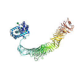
BU of 8bqe by Molmil
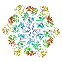
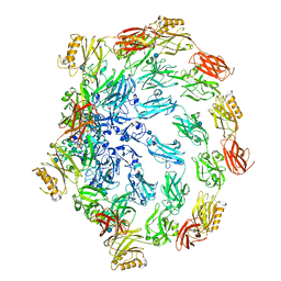
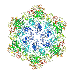
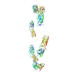
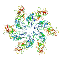
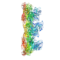
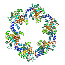
In situ structure of the Caulobacter crescentus S-layer
Descriptor: 4-acetamido-4,6-dideoxy-alpha-D-mannopyranose-(1-3)-beta-D-mannopyranose-(1-3)-4-acetamido-4,6-dideoxy-alpha-D-mannopyranose-(1-3)-4-acetamido-4,6-dideoxy-alpha-D-mannopyranose-(1-3)-beta-D-mannopyranose-(1-3)-4-acetamido-4,6-dideoxy-alpha-D-mannopyranose, CALCIUM ION, S-layer protein rsaA
Authors:von Kuegelgen, A, Bharat, T.
Deposit date:2022-11-21
Release date:2022-12-28
Method:ELECTRON MICROSCOPY (3.5 Å)
Cite:A Bayesian approach to single-particle electron cryo-tomography in RELION-4.0.
Elife, 11, 2022
6Z7P
DownloadVisualize
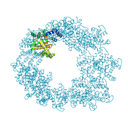
BU of 6z7p by Molmil
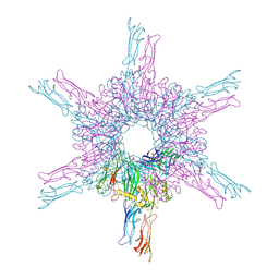
Composite model of the Caulobacter crescentus S-layer bound to the O-antigen of lipopolysaccharide
Descriptor: 4-acetamido-4,6-dideoxy-alpha-D-mannopyranose-(1-3)-4-acetamido-4,6-dideoxy-alpha-D-mannopyranose-(1-3)-beta-D-mannopyranose-(1-3)-4-acetamido-4,6-dideoxy-alpha-D-mannopyranose-(1-3)-4-acetamido-4,6-dideoxy-alpha-D-mannopyranose-(1-3)-beta-D-mannopyranose-(1-3)-4-acetamido-4,6-dideoxy-alpha-D-mannopyranose-(1-3)-4-acetamido-4,6-dideoxy-alpha-D-mannopyranose-(1-3)-beta-D-mannopyranose, CALCIUM ION, S-layer protein
Authors:Bharat, T.A.M, von Kugelgen, A.
Deposit date:2020-06-01
Release date:2020-07-15
Last modified:2024-05-15
Method:ELECTRON MICROSCOPY (4.8 Å)
Cite:In Situ Structure of an Intact Lipopolysaccharide-Bound Bacterial Surface Layer.
Cell, 180, 2020
6T72
DownloadVisualize
BU of 6t72 by Molmil
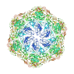
Structure of the RsaA N-terminal domain bound to LPS
Descriptor: 4-acetamido-4,6-dideoxy-alpha-D-mannopyranose-(1-3)-4-acetamido-4,6-dideoxy-alpha-D-mannopyranose-(1-3)-beta-D-mannopyranose-(1-3)-4-acetamido-4,6-dideoxy-alpha-D-mannopyranose-(1-3)-4-acetamido-4,6-dideoxy-alpha-D-mannopyranose-(1-3)-beta-D-mannopyranose-(1-3)-4-acetamido-4,6-dideoxy-alpha-D-mannopyranose-(1-3)-4-acetamido-4,6-dideoxy-alpha-D-mannopyranose-(1-3)-beta-D-mannopyranose-(1-3)-4-acetamido-4,6-dideoxy-alpha-D-mannopyranose-(1-3)-4-acetamido-4,6-dideoxy-alpha-D-mannopyranose-(1-3)-beta-D-mannopyranose, CALCIUM ION, S-layer protein
Authors:von Kuegelgen, A, Bharat, T.A.M.
Deposit date:2019-10-21
Release date:2020-01-15
Last modified:2024-05-22
Method:ELECTRON MICROSCOPY (3.7 Å)
Cite:In Situ Structure of an Intact Lipopolysaccharide-Bound Bacterial Surface Layer.
Cell, 180, 2020
7PTU
DownloadVisualize
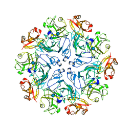
BU of 7ptu by Molmil
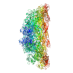
Structure of pentameric S-layer protein from Halofaerax volcanii
Descriptor: Cell surface glycoprotein, beta-D-glucopyranose
Authors:von Kuegelgen, A, Bharat, T.A.M.
Deposit date:2021-09-27
Release date:2021-12-15
Method:ELECTRON MICROSCOPY (3.87 Å)
Cite:Complete atomic structure of a native archaeal cell surface.
Cell Rep, 37, 2021
7PTP
DownloadVisualize
BU of 7ptp by Molmil
In-situ structure of pentameric S-layer protein
Descriptor: Cell surface glycoprotein
Authors:von Kuegelgen, A, Bharat, T.A.M.
Deposit date:2021-09-27
Release date:2021-12-15
Method:ELECTRON MICROSCOPY (11.58 Å)
Cite:Complete atomic structure of a native archaeal cell surface.
Cell Rep, 37, 2021
7PTT
DownloadVisualize
BU of 7ptt by Molmil
In-situ structure of hexameric S-layer protein
Descriptor: Cell surface glycoprotein
Authors:von Kuegelgen, A, Bharat, T.A.M.
Deposit date:2021-09-27
Release date:2021-12-15
Method:ELECTRON MICROSCOPY (7.968 Å)
Cite:Complete atomic structure of a native archaeal cell surface.
Cell Rep, 37, 2021
7PTR
DownloadVisualize
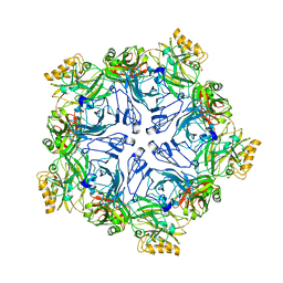
BU of 7ptr by Molmil
Structure of hexameric S-layer protein from Haloferax volcanii archaea
Descriptor: CALCIUM ION, Cell surface glycoprotein, beta-D-glucopyranose
Authors:von Kuegelgen, A, Bharat, T.A.M.
Deposit date:2021-09-27
Release date:2021-12-15
Method:ELECTRON MICROSCOPY (3.46 Å)
Cite:Complete atomic structure of a native archaeal cell surface.
Cell Rep, 37, 2021
8AE1
DownloadVisualize
BU of 8ae1 by Molmil
Structure of trimeric SlpA outer membrane protein
Descriptor: CALCIUM ION, S-layer protein SlpA
Authors:von Kuegelgen, A, Bharat, T.A.M.
Deposit date:2022-07-12
Release date:2022-11-30
Method:ELECTRON MICROSCOPY (3.25 Å)
Cite:A multidomain connector links the outer membrane and cell wall in phylogenetically deep-branching bacteria.
Proc.Natl.Acad.Sci.USA, 119, 2022
8C8L
DownloadVisualize
BU of 8c8l by Molmil
In vitro structure of the Nitrosopumilus maritimus S-layer - Two-fold symmetry (C2)
Descriptor: Cell surface protein
Authors:von Kuegelgen, A, Bharat, T.
Deposit date:2023-01-20
Release date:2024-04-10
Last modified:2024-06-19
Method:ELECTRON MICROSCOPY (2.71 Å)
Cite:Membraneless channels sieve cations in ammonia-oxidizing marine archaea.
Nature, 630, 2024
8C8K
DownloadVisualize
BU of 8c8k by Molmil
In vitro structure of the Nitrosopumilus maritimus S-layer - Six-fold symmetry (C6)
Descriptor: Cell surface protein
Authors:von Kuegelgen, A, Bharat, T.
Deposit date:2023-01-20
Release date:2024-04-10
Last modified:2024-06-19
Method:ELECTRON MICROSCOPY (2.87 Å)
Cite:Membraneless channels sieve cations in ammonia-oxidizing marine archaea.
Nature, 630, 2024
8C8N
DownloadVisualize
BU of 8c8n by Molmil
In situ structure of the Nitrosopumilus maritimus S-layer - Two-fold symmetry (C2)
Descriptor: Cell surface protein
Authors:von Kuegelgen, A, Bharat, T.
Deposit date:2023-01-20
Release date:2024-04-10
Last modified:2024-06-19
Method:ELECTRON MICROSCOPY (3.4 Å)
Cite:Membraneless channels sieve cations in ammonia-oxidizing marine archaea.
Nature, 630, 2024
8CKA
DownloadVisualize
BU of 8cka by Molmil
Deinococcus radidurans HPI S-layer
Descriptor: Hexagonally packed intermediate-layer surface protein
Authors:von Kuegelgen, A, Yamashita, K, Murshudov, G, Bharat, T.
Deposit date:2023-02-14
Release date:2023-04-19
Last modified:2023-04-26
Method:ELECTRON MICROSCOPY (2.54 Å)
Cite:Interdigitated immunoglobulin arrays form the hyperstable surface layer of the extremophilic bacterium Deinococcus radiodurans.
Proc.Natl.Acad.Sci.USA, 120, 2023
8C8M
DownloadVisualize
BU of 8c8m by Molmil
In vitro structure of the Nitrosopumilus maritimus S-layer - Composite map between two and six-fold symmetrised
Descriptor: Cell surface protein
Authors:von Kuegelgen, A, Bharat, T.
Deposit date:2023-01-20
Release date:2024-04-10
Last modified:2024-06-19
Method:ELECTRON MICROSCOPY (2.87 Å)
Cite:Membraneless channels sieve cations in ammonia-oxidizing marine archaea.
Nature, 630, 2024
8C8R
DownloadVisualize
BU of 8c8r by Molmil
In situ structure of the Nitrosopumilus maritimus S-layer - Composite map between C2 and C6
Descriptor: Cell surface protein
Authors:von Kuegelgen, A, Bharat, T.
Deposit date:2023-01-20
Release date:2024-04-17
Last modified:2024-06-19
Method:ELECTRON MICROSCOPY (3.5 Å)
Cite:Membraneless channels sieve cations in ammonia-oxidizing marine archaea.
Nature, 630, 2024
8C8O
DownloadVisualize
BU of 8c8o by Molmil
In situ structure of the Nitrosopumilus maritimus S-layer - Six-fold symmetry (C6)
Descriptor: Cell surface protein
Authors:von Kuegelgen, A, Bharat, T.
Deposit date:2023-01-20
Release date:2024-04-17
Last modified:2024-06-19
Method:ELECTRON MICROSCOPY (3.4 Å)
Cite:Membraneless channels sieve cations in ammonia-oxidizing marine archaea.
Nature, 630, 2024

221716

數據於2024-06-26公開中

PDB statisticsPDBj update infoContact PDBjnumon