Loading
PDBj
MenuPDBj@FacebookPDBj@TwitterPDBj@YouTubewwPDB FoundationwwPDB
RCSB PDBPDBeBMRBAdv. SearchSearch help
Search by PDB author
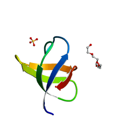
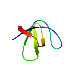
8AH7
DownloadVisualize
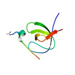
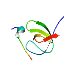
BU of 8ah7 by Molmil
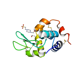
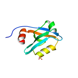
Crystal Structure of the third PDZ domain of PSD-95 protein in the space group P212121 at pH 4.0
Descriptor: SULFATE ION, cDNA FLJ50577, highly similar to Discs large homolog 4
Authors:Camara-Artigas, A, Salinas-Garcia, M.C.
Deposit date:2022-07-20
Release date:2023-03-08
Last modified:2024-02-07
Method:X-RAY DIFFRACTION (1.25 Å)
Cite:pH-Driven Polymorphic Behaviour of the Third PDZ Domain of PSD95: The Role of Electrostatic Interactions
Crystals, 2023
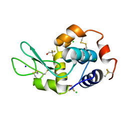
6S7N
DownloadVisualize
BU of 6s7n by Molmil
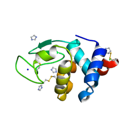
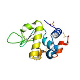
Crystal structure of orthorhombic lysozyme grown at pH 5.5 with a 26% of solvent content
Descriptor: Lysozyme C, SULFATE ION
Authors:Camara-Artigas, A, Plaza-Garrido, M, Salinas-Garcia, M.C.
Deposit date:2019-07-05
Release date:2019-11-20
Last modified:2024-01-24
Method:X-RAY DIFFRACTION (1.2 Å)
Cite:Major conformational changes in the structure of lysozyme obtained from a crystal with a very low solvent content.
Acta Crystallogr.,Sect.F, 75, 2019
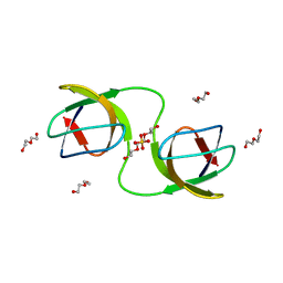
7NER
DownloadVisualize
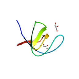
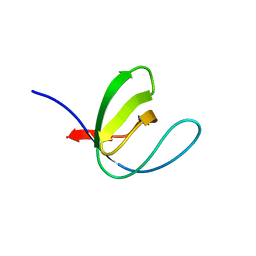
BU of 7ner by Molmil
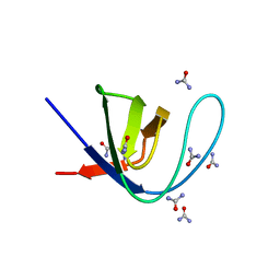
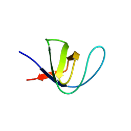
Crystal structure of the v-Src SH3 domain Q128R mutant
Descriptor: SULFATE ION, TETRAETHYLENE GLYCOL, v-Src SH3 domain
Authors:Camara-Artigas, A, Salinas-Garcia, M.C.
Deposit date:2021-02-04
Release date:2021-06-02
Last modified:2024-01-31
Method:X-RAY DIFFRACTION (1.55 Å)
Cite:The impact of oncogenic mutations of the viral Src kinase on the structure and stability of the SH3 domain.
Acta Crystallogr D Struct Biol, 77, 2021
7NET
DownloadVisualize
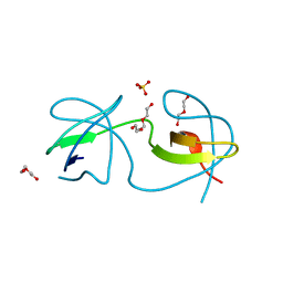
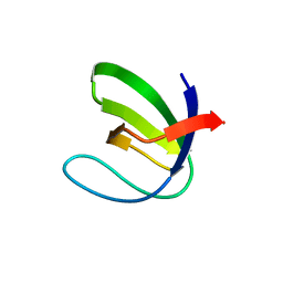
BU of 7net by Molmil
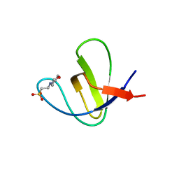
Crystal structure of the v-Src SH3 domain W95R-I96T mutant
Descriptor: DI(HYDROXYETHYL)ETHER, SULFATE ION, TRIETHYLENE GLYCOL, ...
Authors:Camara-Artigas, A, Salinas-Garcia, M.C.
Deposit date:2021-02-04
Release date:2021-06-02
Last modified:2024-01-31
Method:X-RAY DIFFRACTION (1.5 Å)
Cite:The impact of oncogenic mutations of the viral Src kinase on the structure and stability of the SH3 domain.
Acta Crystallogr D Struct Biol, 77, 2021
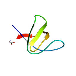
7NES
DownloadVisualize
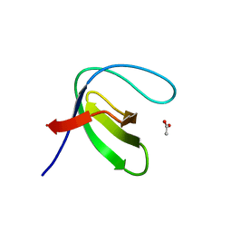
BU of 7nes by Molmil
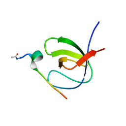
Crystal structure of the v-Src SH3 domain N117D-V124L mutant
Descriptor: GLYCINE, v-Src SH3 domain
Authors:Camara-Artigas, A, Salinas-Garcia, M.C.
Deposit date:2021-02-04
Release date:2021-06-02
Last modified:2024-05-15
Method:X-RAY DIFFRACTION (1.35 Å)
Cite:The impact of oncogenic mutations of the viral Src kinase on the structure and stability of the SH3 domain.
Acta Crystallogr D Struct Biol, 77, 2021
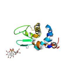
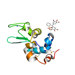
6SYC
DownloadVisualize
BU of 6syc by Molmil
Crystal structure of the lysozyme in presence of bromophenol blue at pH 6.5
Descriptor: CHLORIDE ION, IMIDAZOLE, Lysozyme, ...
Authors:Camara-Artigas, A, Plaza-Garrido, M, Salinas-Garcia, M.C.
Deposit date:2019-09-27
Release date:2020-09-09
Last modified:2024-01-24
Method:X-RAY DIFFRACTION (1.38 Å)
Cite:Lysozyme crystals dyed with bromophenol blue: where has the dye gone?
Acta Crystallogr D Struct Biol, 76, 2020
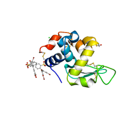
6SYD
DownloadVisualize
BU of 6syd by Molmil
Crystal structure of the lysozyme in presence of bromophenol blue at pH 5.5
Descriptor: Lysozyme, SODIUM ION, bromophenol blue
Authors:Camara-Artigas, A, Plaza-Garrido, M, Salinas-Garcia, M.C.
Deposit date:2019-09-27
Release date:2020-09-09
Last modified:2024-01-24
Method:X-RAY DIFFRACTION (1.1 Å)
Cite:Lysozyme crystals dyed with bromophenol blue: where has the dye gone?
Acta Crystallogr D Struct Biol, 76, 2020
6SYE
DownloadVisualize
BU of 6sye by Molmil
Crystal structure of orthorhombic lysozyme in presence of the dye bromophenol blue at pH 7.0
Descriptor: CHLORIDE ION, Lysozyme C, bromophenol blue
Authors:Camara-Artigas, A, Plaza-Garrido, M, Salinas-Garcia, M.C.
Deposit date:2019-09-27
Release date:2020-09-09
Last modified:2024-01-24
Method:X-RAY DIFFRACTION (0.97 Å)
Cite:Lysozyme crystals dyed with bromophenol blue: where has the dye gone?
Acta Crystallogr D Struct Biol, 76, 2020
7OL7
DownloadVisualize
BU of 7ol7 by Molmil
Crystal structure of Lysozyme in complex with trifluoroethanol: tetragonal form
Descriptor: CHLORIDE ION, Lysozyme, SODIUM ION, ...
Authors:Camara-Artigas, A, Salinas-Garcia, M.C, Plaza-Garrido, M.
Deposit date:2021-05-19
Release date:2022-06-01
Last modified:2024-01-31
Method:X-RAY DIFFRACTION (1.15 Å)
Cite:Crystal structure of Lysozyme in complex with TFE
To be published
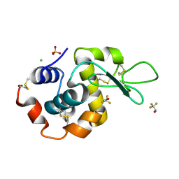
7OL8
DownloadVisualize
BU of 7ol8 by Molmil
Crystal structure of Lysozyme in complex with trifluoroethanol: orthorhombic form
Descriptor: CHLORIDE ION, Lysozyme, SULFATE ION, ...
Authors:Camara-Artigas, A, Salinas-Garcia, M.C, Plaza-Garrido, M.
Deposit date:2021-05-19
Release date:2022-06-01
Last modified:2024-01-31
Method:X-RAY DIFFRACTION (1.15 Å)
Cite:Crystal structure of Lysozyme in complex with TFE
To be published
7OL6
DownloadVisualize
BU of 7ol6 by Molmil
Crystal structure of Lysozyme in complex with Imidazole
Descriptor: CHLORIDE ION, IMIDAZOLE, Lysozyme, ...
Authors:Camara-Artigas, A, Salinas-Garcia, M.C, Plaza-Garrido, M.
Deposit date:2021-05-19
Release date:2022-06-01
Last modified:2024-01-31
Method:X-RAY DIFFRACTION (1.1 Å)
Cite:Crystal structure of Lysozyme in complex with Imidazole
To be published
7OL5
DownloadVisualize
BU of 7ol5 by Molmil
Crystal structure of Lysozyme in complex with Hepes
Descriptor: 4-(2-HYDROXYETHYL)-1-PIPERAZINE ETHANESULFONIC ACID, ISOPROPYL ALCOHOL, Lysozyme
Authors:Camara-Artigas, A, Salinas-Garcia, M.C, Plaza-Garrido, M.
Deposit date:2021-05-19
Release date:2022-06-01
Last modified:2024-01-31
Method:X-RAY DIFFRACTION (0.94 Å)
Cite:Crystal structure of Lysozyme in complex with Hepes
To be published
6TG7
DownloadVisualize
BU of 6tg7 by Molmil
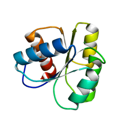
Crystal structure of the CheY in presence of magnesium
Descriptor: Chemotaxis protein CheY, MAGNESIUM ION
Authors:Camara-Artigas, A, Salinas-Garcia, M.C, Alba-Elena, D.
Deposit date:2019-11-15
Release date:2019-12-04
Last modified:2024-01-24
Method:X-RAY DIFFRACTION (1.65 Å)
Cite:Crystal structure of the CheY in presence of magnesium
To be published
7A36
DownloadVisualize
BU of 7a36 by Molmil
Crystal structure of the c-Src SH3 domain mutant V111L-N113S-T114S in 7 M urea
Descriptor: Proto-oncogene tyrosine-protein kinase Src, SODIUM ION, UREA
Authors:Camara-Artigas, A, Plaza-Garrido, M, Salinas-Garcia, M.C.
Deposit date:2020-08-18
Release date:2021-08-25
Last modified:2024-01-31
Method:X-RAY DIFFRACTION (1.5 Å)
Cite:Crystal structure of the c-Src SH3 domain mutant V111L-N113S-T114S in 7 M urea
To be published
7A39
DownloadVisualize
BU of 7a39 by Molmil
Crystal structure of the c-Src SH3 domain mutant V111L-N113S-T114S-Q128E at pH 7.0
Descriptor: Proto-oncogene tyrosine-protein kinase Src, SULFATE ION
Authors:Camara-Artigas, A, Plaza-Garrido, M, Salinas-Garcia, M.C.
Deposit date:2020-08-18
Release date:2021-08-25
Last modified:2024-01-31
Method:X-RAY DIFFRACTION (1.65 Å)
Cite:Crystal structure of the c-Src SH3 domain mutant V111L-N113S-T114S-Q128E at pH 7.0
To be published
7A3E
DownloadVisualize
BU of 7a3e by Molmil
Intertwined dimer of the c-Src SH3 domain mutant T126S
Descriptor: DI(HYDROXYETHYL)ETHER, Proto-oncogene tyrosine-protein kinase Src, SULFATE ION, ...
Authors:Camara-Artigas, A, Plaza-Garrido, M, Salinas-Garcia, M.C.
Deposit date:2020-08-18
Release date:2021-08-25
Last modified:2024-01-31
Method:X-RAY DIFFRACTION (1.52 Å)
Cite:Intertwined dimer of the c-Src SH3 domain mutant T126S
To be published
7A3C
DownloadVisualize
BU of 7a3c by Molmil
Crystal structure of the c-Src SH3 domain mutant L100I at pH 3.0
Descriptor: GLYCEROL, Proto-oncogene tyrosine-protein kinase Src, SULFATE ION
Authors:Camara-Artigas, A, Plaza-Garrido, M, Salinas-Garcia, M.C.
Deposit date:2020-08-18
Release date:2021-08-25
Last modified:2024-01-31
Method:X-RAY DIFFRACTION (1.8 Å)
Cite:Crystal structure of the c-Src SH3 domain mutant L100I at pH 3.0
To be published
7A2T
DownloadVisualize
BU of 7a2t by Molmil
Crystal structure of the Fyn SH3 domain L112V-S114N-S115T-E121L-R123H mutant at pH 4.0
Descriptor: ACETATE ION, Tyrosine-protein kinase Fyn
Authors:Camara-Artigas, A, Plaza-Garrido, M, Salinas-Garcia, M.C.
Deposit date:2020-08-18
Release date:2021-08-25
Last modified:2024-01-31
Method:X-RAY DIFFRACTION (1.22 Å)
Cite:Crystal structure of the Fyn SH3 domain L112V-S114N-S115T-E121L-R123H mutant at pH 4.0
To be published
7A2Y
DownloadVisualize
BU of 7a2y by Molmil
Crystal structure of the Fyn SH3 domain L112V-S114N-S115T-E121L-R123H mutant in complex with VSL12 at pH 4.0
Descriptor: Tyrosine-protein kinase Fyn, VSL12
Authors:Camara-Artigas, A, Plaza-Garrido, M, Salinas-Garcia, M.C.
Deposit date:2020-08-18
Release date:2021-08-25
Last modified:2024-01-31
Method:X-RAY DIFFRACTION (0.97 Å)
Cite:Crystal structure of the Fyn SH3 domain L112V-S114N-S115T-E121L-R123H mutant in complex with VSL12 at pH 4.0
To be published
7A2Z
DownloadVisualize
BU of 7a2z by Molmil
Crystal structure of the Fyn SH3 domain L112V-S114N-S115T-E121L-R123H mutant in complex with VSL12 at pH 6.0
Descriptor: Tyrosine-protein kinase Fyn, VSL12
Authors:Camara-Artigas, A, Plaza-Garrido, M, Salinas-Garcia, M.C.
Deposit date:2020-08-18
Release date:2021-08-25
Last modified:2024-01-31
Method:X-RAY DIFFRACTION (1.14 Å)
Cite:Crystal structure of the Fyn SH3 domain L112V-S114N-S115T-E121L-R123H mutant in complex with VSL12 at pH 6.0
To be published
7A31
DownloadVisualize
BU of 7a31 by Molmil
Crystal structure of the c-Src SH3 domain mutant S94A-T98D-V111L-N113S-T114S at pH 4.5
Descriptor: Proto-oncogene tyrosine-protein kinase Src
Authors:Camara-Artigas, A, Plaza-Garrido, M, Salinas-Garcia, M.C.
Deposit date:2020-08-18
Release date:2021-08-25
Last modified:2024-01-31
Method:X-RAY DIFFRACTION (0.94 Å)
Cite:Crystal structure of the c-Src SH3 domain mutant S94A-T98D-V111L-N113S-T114S at pH 4.5
To be published
7A32
DownloadVisualize
BU of 7a32 by Molmil
Crystal structure of the c-Src SH3 domain mutant S94A-T98D-V111L-N113S-T114S at pH 7.0
Descriptor: GLYCEROL, Proto-oncogene tyrosine-protein kinase Src, SULFATE ION
Authors:Camara-Artigas, A, Plaza-Garrido, M, Salinas-Garcia, M.C.
Deposit date:2020-08-18
Release date:2021-08-25
Last modified:2024-01-31
Method:X-RAY DIFFRACTION (1.15 Å)
Cite:Crystal structure of the c-Src SH3 domain mutant S94A-T98D-V111L-N113S-T114S at pH 7.0
To be published
7A35
DownloadVisualize
BU of 7a35 by Molmil
Crystal structure of the c-Src SH3 domain mutant V111L-N113S-T114S at pH 7.0
Descriptor: Proto-oncogene tyrosine-protein kinase Src, SULFATE ION
Authors:Camara-Artigas, A, Plaza-Garrido, M, Salinas-Garcia, M.C.
Deposit date:2020-08-18
Release date:2021-08-25
Last modified:2024-01-31
Method:X-RAY DIFFRACTION (1.314 Å)
Cite:Crystal structure of the c-Src SH3 domain mutant V111L-N113S-T114S at pH 7.0
To be published
7A38
DownloadVisualize
BU of 7a38 by Molmil
Crystal structure of the c-Src SH3 domain mutant V111L-N113S-T114S-Q128E at pH 6.0
Descriptor: 2-(N-MORPHOLINO)-ETHANESULFONIC ACID, Proto-oncogene tyrosine-protein kinase Src, SULFATE ION
Authors:Camara-Artigas, A, Plaza-Garrido, M, Salinas-Garcia, M.C.
Deposit date:2020-08-18
Release date:2021-08-25
Last modified:2024-01-31
Method:X-RAY DIFFRACTION (1.62 Å)
Cite:Crystal structure of the c-Src SH3 domain mutant V111L-N113S-T114S-Q128E at pH 6.0
To be published
7A2X
DownloadVisualize
BU of 7a2x by Molmil
Crystal structure of the Fyn SH3 domain L112V-S114N-S115T-E121L-R123H mutant in complex with VSL12 at pH 5.0
Descriptor: Tyrosine-protein kinase Fyn, VSL12 high affinity synthetic peptide acetylated in the amino-terminus
Authors:Camara-Artigas, A, Plaza-Garrido, M, Salinas-Garcia, M.C.
Deposit date:2020-08-18
Release date:2021-08-25
Last modified:2024-01-31
Method:X-RAY DIFFRACTION (0.92 Å)
Cite:Crystal structure of the Fyn SH3 domain L112V-S114N-S115T-E121L-R123H mutant in complex with VSL12 at pH 5.0
To be published


123>

222415

數據於2024-07-10公開中

PDB statisticsPDBj update infoContact PDBjnumon