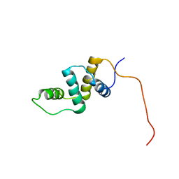
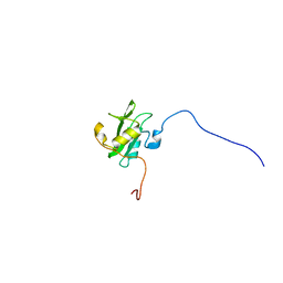
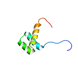
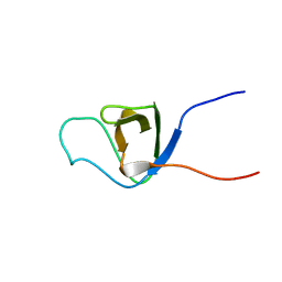
2CPN
 
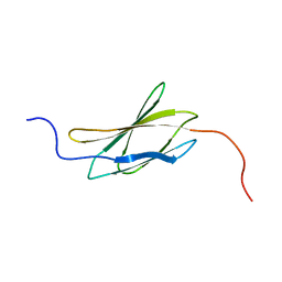
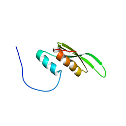 | | Solution structure of the second dsRBD of TAR RNA-binding protein 2 | | Descriptor: | TAR RNA-binding protein 2 | | Authors: | Nagata, T, Muto, Y, Inoue, M, Kigawa, T, Terada, T, Shirouzu, M, Yokoyama, S, RIKEN Structural Genomics/Proteomics Initiative (RSGI) | | Deposit date: | 2005-05-19 | | Release date: | 2005-11-19 | | Last modified: | 2024-05-29 | | Method: | SOLUTION NMR | | Cite: | Solution structure of the second dsRBD of TAR RNA-binding protein 2
To be Published
|
|
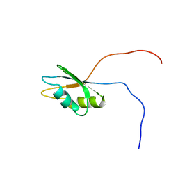
2CR8
 | |
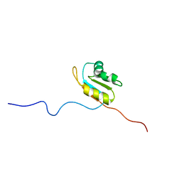
2COT
 | |
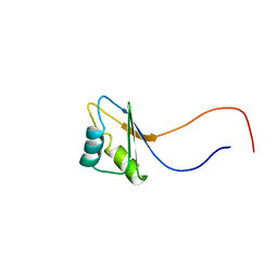
2CPT
 | |
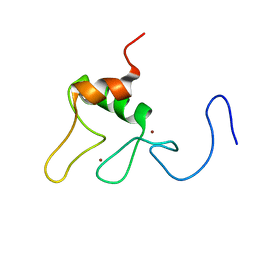
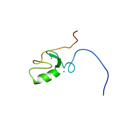
2CO8
 | | Solution structures of the LIM domain of human NEDD9 interacting protein with calponin homology and LIM domains | | Descriptor: | NEDD9 interacting protein with calponin homology and LIM domains, ZINC ION | | Authors: | Sato, M, Tomizawa, T, Saito, K, Koshiba, S, Inoue, M, Kigawa, T, Yokoyama, S, RIKEN Structural Genomics/Proteomics Initiative (RSGI) | | Deposit date: | 2005-05-17 | | Release date: | 2005-11-17 | | Last modified: | 2024-05-29 | | Method: | SOLUTION NMR | | Cite: | Solution structures of the LIM domain of human NEDD9 interacting protein with calponin homology and LIM domains
To be Published
|
|
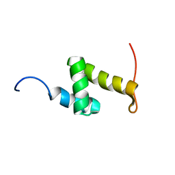
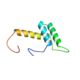
2COP
 | | Solution structure of RSGI RUH-040, an ACBP domain from human cDNA | | Descriptor: | Acyl-Coenzyme A binding domain containing 6 | | Authors: | Ruhul Momen, A.Z.M, Hirota, H, Hayashi, F, Yokoyama, S, RIKEN Structural Genomics/Proteomics Initiative (RSGI) | | Deposit date: | 2005-05-18 | | Release date: | 2005-11-18 | | Last modified: | 2024-05-29 | | Method: | SOLUTION NMR | | Cite: | Solution structure of RSGI RUH-040, an ACBP domain from human cDNA
To be Published
|
|
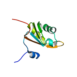
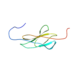
2CPD
 | | Solution structure of the RNA recognition motif of human APOBEC-1 complementation factor, ACF | | Descriptor: | APOBEC-1 stimulating protein | | Authors: | Nagata, T, Muto, Y, Inoue, M, Kigawa, T, Terada, T, Shirouzu, M, Yokoyama, S, RIKEN Structural Genomics/Proteomics Initiative (RSGI) | | Deposit date: | 2005-05-19 | | Release date: | 2005-11-19 | | Last modified: | 2024-05-29 | | Method: | SOLUTION NMR | | Cite: | Solution structure of the RNA recognition motif of human APOBEC-1 complementation factor, ACF
To be Published
|
|
2CPX
 | | solution structure of RNA binding domain in Hypothetical protein FLJ11016 | | Descriptor: | Hypothetical protein FLJ11016 | | Authors: | Tsuda, K, Muto, Y, Inoue, M, Kigawa, T, Terada, T, Shirouzu, M, Yokoyama, S, RIKEN Structural Genomics/Proteomics Initiative (RSGI) | | Deposit date: | 2005-05-19 | | Release date: | 2005-11-19 | | Last modified: | 2024-05-29 | | Method: | SOLUTION NMR | | Cite: | solution structure of RNA binding domain in Hypothetical protein FLJ11016
To be Published
|
|
2CQH
 | | Solution structure of the RNA binding domain of IGF-II mRNA-binding protein 2 | | Descriptor: | IGF-II mRNA-binding protein 2 isoform a | | Authors: | Suzuki, S, Muto, Y, Inoue, M, Kigawa, T, Terada, T, Shirouzu, M, Yokoyama, S, RIKEN Structural Genomics/Proteomics Initiative (RSGI) | | Deposit date: | 2005-05-20 | | Release date: | 2005-11-20 | | Last modified: | 2024-05-29 | | Method: | SOLUTION NMR | | Cite: | Solution structure of the RNA binding domain of IGF-II mRNA-binding protein 2
To be Published
|
|
2CRH
 | |
2CS3
 | | Solution structure of the zf-C3HC4 domain of human KIAA1865 | | Descriptor: | Protein C14orf4, ZINC ION | | Authors: | Inoue, K, Saitoh, K, Hayashi, F, Kigawa, T, Yokoyama, S, RIKEN Structural Genomics/Proteomics Initiative (RSGI) | | Deposit date: | 2005-05-20 | | Release date: | 2005-11-20 | | Last modified: | 2024-05-29 | | Method: | SOLUTION NMR | | Cite: | Solution structure of the zf-C3HC4 domain of human KIAA1865
To be Published
|
|
2CP9
 | | Solution structure of RSGI RUH-042, a UBA domain from human mitochondrial elongation factor Ts | | Descriptor: | Elongation factor Ts, mitochondrial | | Authors: | Ohashi, W, Hirota, H, Izumi, K, Yoshida, M, Yokoyama, S, RIKEN Structural Genomics/Proteomics Initiative (RSGI) | | Deposit date: | 2005-05-19 | | Release date: | 2005-11-19 | | Last modified: | 2024-05-29 | | Method: | SOLUTION NMR | | Cite: | Solution structure of RSGI RUH-042, a UBA domain from human mitochondrial elongation factor Ts
To be Published
|
|
2CQ2
 | | solution structure of RNA binding domain in Hypothetical protein LOC91801 | | Descriptor: | hypothetical protein LOC91801 | | Authors: | Tsuda, K, Muto, Y, Inoue, M, Kigawa, T, Terada, T, Shirouzu, M, Yokoyama, S, RIKEN Structural Genomics/Proteomics Initiative (RSGI) | | Deposit date: | 2005-05-19 | | Release date: | 2005-11-19 | | Last modified: | 2024-05-29 | | Method: | SOLUTION NMR | | Cite: | solution structure of RNA binding domain in Hypothetical protein LOC91801
To be Published
|
|
2CRA
 | | Solution structure of the homeobox domain of human homeo box B13 | | Descriptor: | Homeobox protein Hox-B13 | | Authors: | Seimiya, K, Hayashi, F, Nagashima, T, Yoshida, M, Yokoyama, S, RIKEN Structural Genomics/Proteomics Initiative (RSGI) | | Deposit date: | 2005-05-20 | | Release date: | 2005-11-20 | | Last modified: | 2024-05-29 | | Method: | SOLUTION NMR | | Cite: | Solution structure of the homeobox domain of human homeo box B13
To be published
|
|
2CT2
 | | Solution Structure of the RING domain of the Tripartite motif protein 32 | | Descriptor: | Tripartite motif protein 32, ZINC ION | | Authors: | Miyamoto, K, Tochio, N, Sato, M, Koshiba, S, Inoue, M, Kigawa, T, Yokoyama, S, RIKEN Structural Genomics/Proteomics Initiative (RSGI) | | Deposit date: | 2005-05-23 | | Release date: | 2005-11-23 | | Last modified: | 2024-05-29 | | Method: | SOLUTION NMR | | Cite: | Solution Structure of the RING domain of the Tripartite motif protein 32
To be Published
|
|
2CUF
 | | Solution structure of the homeobox domain of the human hypothetical protein FLJ21616 | | Descriptor: | FLJ21616 protein | | Authors: | Ohnishi, S, Kigawa, T, Tomizawa, T, Koshiba, S, Inoue, M, Yokoyama, S, RIKEN Structural Genomics/Proteomics Initiative (RSGI) | | Deposit date: | 2005-05-26 | | Release date: | 2005-11-26 | | Last modified: | 2024-05-29 | | Method: | SOLUTION NMR | | Cite: | Solution structure of the homeobox domain of the human hypothetical protein FLJ21616
To be Published
|
|
2CRM
 | |
2CSI
 | |
2CSZ
 | | Solution Structure of the RING domain of the Synaptotagmin-like protein 4 | | Descriptor: | Synaptotagmin-like protein 4, ZINC ION | | Authors: | Miyamoto, K, Sato, M, Koshiba, S, Inoue, M, Kigawa, T, Yokoyama, S, RIKEN Structural Genomics/Proteomics Initiative (RSGI) | | Deposit date: | 2005-05-23 | | Release date: | 2005-11-23 | | Last modified: | 2024-05-29 | | Method: | SOLUTION NMR | | Cite: | Solution Structure of the RING domain of the Synaptotagmin-like protein 4
To be Published
|
|
2CWN
 | | Crystal structure of mouse transaldolase | | Descriptor: | Transaldolase | | Authors: | Handa, N, Arai, R, Nishino, A, Uchikubo, T, Takemoto, C, Morita, S, Kinoshita, Y, Nagano, Y, Uda, H, Terada, T, Shirouzu, M, Yokoyama, S, RIKEN Structural Genomics/Proteomics Initiative (RSGI) | | Deposit date: | 2005-06-22 | | Release date: | 2005-12-22 | | Last modified: | 2023-10-25 | | Method: | X-RAY DIFFRACTION (2.1 Å) | | Cite: | Crystal structure of mouse transaldolase
To be Published
|
|
2CX6
 | |
2CXL
 | | RUN domain of Rap2 interacting protein x, crystallized in I422 space group | | Descriptor: | rap2 interacting protein x | | Authors: | Kukimoto-Niino, M, Umehara, T, Murayama, K, Shirouzu, M, Yokoyama, S, RIKEN Structural Genomics/Proteomics Initiative (RSGI) | | Deposit date: | 2005-06-30 | | Release date: | 2005-12-30 | | Last modified: | 2024-10-09 | | Method: | X-RAY DIFFRACTION (3.2 Å) | | Cite: | Crystal Structure of the RUN Domain of the RAP2-interacting Protein x
J.Biol.Chem., 281, 2006
|
|
2CT7
 | | Solution Structure of the IBR domain of the RING finger protein 31 protein | | Descriptor: | RING finger protein 31, ZINC ION | | Authors: | Miyamoto, K, Koshiba, S, Tomizawa, T, Inoue, M, Kigawa, T, Yokoyama, S, RIKEN Structural Genomics/Proteomics Initiative (RSGI) | | Deposit date: | 2005-05-23 | | Release date: | 2005-11-23 | | Last modified: | 2024-05-29 | | Method: | SOLUTION NMR | | Cite: | Solution Structure of the IBR domain of the RING finger protein 31 protein
To be Published
|
|
2CUM
 | | The solution structure of the 33rd fibronectin type III domain of human Tenascin-X | | Descriptor: | Tenascin-X | | Authors: | Tochio, N, Koshiba, S, Inoue, M, Kigawa, T, Yokoyama, S, RIKEN Structural Genomics/Proteomics Initiative (RSGI) | | Deposit date: | 2005-05-26 | | Release date: | 2005-11-26 | | Last modified: | 2024-05-29 | | Method: | SOLUTION NMR | | Cite: | The solution structure of the 33rd fibronectin type III domain of human Tenascin-X
To be Published
|
|
2CYD
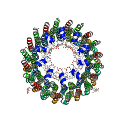 | | Crystal structure of Lithium bound rotor ring of the V-ATPase from Enterococcus hirae | | Descriptor: | 1,2-DIPALMITOYL-PHOSPHATIDYL-GLYCEROLE, LITHIUM ION, UNDECYL-MALTOSIDE, ... | | Authors: | Murata, T, Yamato, I, Kakinuma, Y, Shirouzu, M, Walker, J.E, Yokoyama, S, RIKEN Structural Genomics/Proteomics Initiative (RSGI) | | Deposit date: | 2005-07-06 | | Release date: | 2006-06-27 | | Last modified: | 2024-03-13 | | Method: | X-RAY DIFFRACTION (2.8 Å) | | Cite: | Crystal structure of Lithium bound rotor ring of the V-ATPase from Enterococcus hirae
To be Published
|
|


