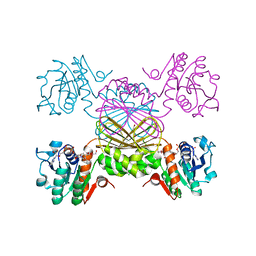
6PQH
 
 | |
6P61
 | |
6P81
 | |
6PBL
 | |
3MD7
 | |
6PTG
 | |
6PZJ
 | |
3PY6
 | |
4WSH
 | |
4XEL
 | |
4XGN
 | |
4XFJ
 | |
4XGI
 | |
4XFK
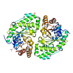
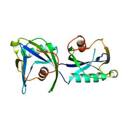
 | | Crystal structure of Leucine-, Isoleucine-, Valine-, Threonine-, and Alanine-binding protein from Brucella ovis | | Descriptor: | ACETATE ION, Putative branched chain amino acid ABC transporter, periplasmic amino acid-binding protein, ... | | Authors: | Seattle Structural Genomics Center for Infectious Disease (SSGCID) | | Deposit date: | 2014-12-27 | | Release date: | 2015-03-11 | | Last modified: | 2024-02-28 | | Method: | X-RAY DIFFRACTION (1.3 Å) | | Cite: | Crystal structure of Leucine-, Isoleucine-, Valine-, Threonine-, and Alanine-binding protein from Brucella ovis
to be published
|
|
4YMI
 | |
4YWN
 | |
4YWJ
 | |
3QH8
 | |
3PY5
 | |
4KYX
 | |
4LW8
 | |
3LUZ
 | |
4Q6U
 | |
7S5N
 | |
7S5M
 | |


