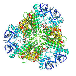
7N1L
 
 | |
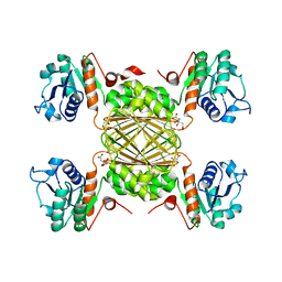
7N6S
 | |
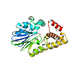
7N7S
 | |
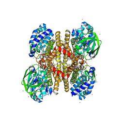
7N56
 | |
8D1X
 | |
8D57
 | |
8D2Z
 | |
3PFD
 | |
3NJB
 | |
3OIB
 | |
3P96
 | |
4Z04
 | |
4ZR8
 | |
4ZJU
 | |
5BNZ
 | |
5WNN
 | |
5BQ2
 | |
5BR9
 | |
6WHJ
 | |
6WQM
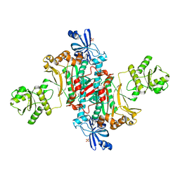
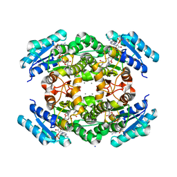
 | | Crystal structure of 2,3,4,5-tetrahydropyridine-2-carboxylate N-succinyltransferase from Bartonella henselae | | Descriptor: | 2,3,4,5-tetrahydropyridine-2,6-dicarboxylate N-succinyltransferase, CHLORIDE ION | | Authors: | Seattle Structural Genomics Center for Infectious Disease (SSGCID) | | Deposit date: | 2020-04-29 | | Release date: | 2020-05-13 | | Last modified: | 2023-10-18 | | Method: | X-RAY DIFFRACTION (2.15 Å) | | Cite: | Crystal structure of 2,3,4,5-tetrahydropyridine-2-carboxylate N-succinyltransferase from Bartonella henselae
to be published
|
|
6WOM
 | |
6XR5
 | |
7ULH
 | |
7ULZ
 | |
7V0H
 | |


