Loading
PDBj
MenuPDBj@FacebookPDBj@TwitterPDBj@YouTubewwPDB FoundationwwPDB
RCSB PDBPDBeBMRBAdv. SearchSearch help
Search by PDB author
7TJ3
DownloadVisualize
BU of 7tj3 by Molmil
Crystal structure of a dihydrofolate reductase folA from Stenotrophomonas maltophilia bound to NADP and p218
Descriptor: 1,2-ETHANEDIOL, 3-(2-{3-[(2,4-diamino-6-ethylpyrimidin-5-yl)oxy]propoxy}phenyl)propanoic acid, Dihydrofolate reductase, ...
Authors:Seattle Structural Genomics Center for Infectious Disease (SSGCID)
Deposit date:2022-01-14
Release date:2022-11-23
Last modified:2023-10-25
Method:X-RAY DIFFRACTION (1.55 Å)
Cite:Crystal structure of a dihydrofolate reductase folA from Stenotrophomonas maltophilia bound to NADP and p218
To Be Published
6UDF
DownloadVisualize
BU of 6udf by Molmil
Crystal structure of Enoyl-[acyl-carrier-protein] reductase [NADH] (InhA) from Mycobacterium kansasii
Descriptor: Enoyl-[acyl-carrier-protein] reductase [NADH], FORMIC ACID
Authors:Seattle Structural Genomics Center for Infectious Disease (SSGCID)
Deposit date:2019-09-19
Release date:2019-10-16
Last modified:2023-10-11
Method:X-RAY DIFFRACTION (1.95 Å)
Cite:Crystal stucture of Enoyl-[acyl-carrier-protein] reductase [NADH] (InhA) from Mycobacterium kansasii
to be published
6UJ5
DownloadVisualize
BU of 6uj5 by Molmil
Crystal structure of CAB1 Pantothenate Kinase from Saccharomyces cerevisiae
Descriptor: MAGNESIUM ION, Pantothenate kinase CAB1
Authors:Seattle Structural Genomics Center for Infectious Disease (SSGCID)
Deposit date:2019-10-02
Release date:2020-10-07
Last modified:2024-03-13
Method:X-RAY DIFFRACTION (1.8 Å)
Cite:Crystal structure of CAB1 Pantothenate Kinase from Saccharomyces cerevisiae
to be published
6UJK
DownloadVisualize
BU of 6ujk by Molmil
Crystal Structure of a Probable short-chain type dehydrogenase/reductase (Rv1144) from Mycobacterium tuberculosis with bound NAD
Descriptor: 1,2-ETHANEDIOL, 2-AMINO-2-HYDROXYMETHYL-PROPANE-1,3-DIOL, NICOTINAMIDE-ADENINE-DINUCLEOTIDE, ...
Authors:Seattle Structural Genomics Center for Infectious Disease (SSGCID)
Deposit date:2019-10-03
Release date:2019-10-16
Last modified:2023-10-11
Method:X-RAY DIFFRACTION (1.2 Å)
Cite:Crystal Structure of a Probable short-chain type dehydrogenase/reductase (Rv1144) from Mycobacterium tuberculosis with bound NAD
to be published
6ULO
DownloadVisualize
BU of 6ulo by Molmil
Structure of an N-terminally truncated uncharacterized protein from Leptospira interrogans serogroup Icterohaemorrhagiae serovar Copenhageni (strain Fiocruz L1-130)
Descriptor: CHLORIDE ION, Uncharacterized protein
Authors:Seattle Structural Genomics Center for Infectious Disease (SSGCID)
Deposit date:2019-10-08
Release date:2019-10-16
Last modified:2024-03-13
Method:X-RAY DIFFRACTION (1.3 Å)
Cite:Structure of an N-terminally truncated uncharacterized protein from Leptospira interrogans serogroup Icterohaemorrhagiae serovar Copenhageni (strain Fiocruz L1-130)
to be published
6UK3
DownloadVisualize
BU of 6uk3 by Molmil
Crystal Structure of S-adenosyl-L-homocysteine hydrolase from Acanthamoeba castellanii with bound NAD and Adenosine
Descriptor: ACETATE ION, ADENOSINE, Adenosylhomocysteinase, ...
Authors:Seattle Structural Genomics Center for Infectious Disease (SSGCID)
Deposit date:2019-10-04
Release date:2019-10-16
Last modified:2023-10-11
Method:X-RAY DIFFRACTION (1.4 Å)
Cite:Crystal Structure of S-adenosyl-L-homocysteine hydrolase from Acanthamoeba castellanii with bound NAD and Adenosine
to be published
6TYJ
DownloadVisualize
BU of 6tyj by Molmil
Crystal structure of zinc-bound Hemerythrin HHE cation binding domain-containing protein (soak): Rv2633c homolog from Mycobacterium kansasii
Descriptor: Hemerythrin HHE cation binding domain protein, ZINC ION
Authors:Seattle Structural Genomics Center for Infectious Disease (SSGCID)
Deposit date:2019-08-09
Release date:2020-01-22
Last modified:2023-10-11
Method:X-RAY DIFFRACTION (1.6 Å)
Cite:Crystal structure of zinc-bound Hemerythrin HHE cation binding domain-containing protein (soak): Rv2633c homolog from Mycobacterium kansasii
to be published
6UCZ
DownloadVisualize
BU of 6ucz by Molmil
Crystal structure of dihydropteroate synthase from Anaplasma phagocytophilum with bound 6-hydroxymethylpterin-monophosphate
Descriptor: 1,2-ETHANEDIOL, ACETATE ION, CITRIC ACID, ...
Authors:Seattle Structural Genomics Center for Infectious Disease (SSGCID)
Deposit date:2019-09-18
Release date:2019-10-16
Last modified:2023-10-11
Method:X-RAY DIFFRACTION (2 Å)
Cite:Crystal structure of dihydropteroate synthase from Anaplasma phagocytophilum with bound 6-hydroxymethylpterin-monophosphate
To be Published
6UDE
DownloadVisualize
BU of 6ude by Molmil
Crystal structure of Glycerol kinase from Elizabethkingia anophelis NUHP1 in complex with ADP and glycerol
Descriptor: ADENOSINE-5'-DIPHOSPHATE, GLYCEROL, Glycerol kinase, ...
Authors:Seattle Structural Genomics Center for Infectious Disease (SSGCID)
Deposit date:2019-09-19
Release date:2019-10-16
Last modified:2023-10-11
Method:X-RAY DIFFRACTION (1.95 Å)
Cite:Crystal structure of Glycerol kinase from Elizabethkingia anophelis NUHP1 in complex with ADP and glycerol
TO BE PUBLISHED
6UH2
DownloadVisualize
BU of 6uh2 by Molmil
Crystal Structure of Short Chain Dehydrogenase from Leptospira borgpetersenii serovar Hardjo-bovis (Strain JB197) with bound NAD+
Descriptor: NICOTINAMIDE-ADENINE-DINUCLEOTIDE, Short chain dehydrogenase
Authors:Seattle Structural Genomics Center for Infectious Disease (SSGCID)
Deposit date:2019-09-26
Release date:2019-10-09
Last modified:2023-10-11
Method:X-RAY DIFFRACTION (1.9 Å)
Cite:TBD
TO BE PUBLISHED
6ULD
DownloadVisualize
BU of 6uld by Molmil
Crystal structure of serine hydroxymethyltransferase from Mycobacterium tuberculosis with bound PLP forming a Schiff base with substrate Serine in one monomer and PLP forming a Schiff base with product Glycine in the other monomer
Descriptor: (4S)-2-METHYL-2,4-PENTANEDIOL, GLYCINE, PYRIDOXAL-5'-PHOSPHATE, ...
Authors:Seattle Structural Genomics Center for Infectious Disease (SSGCID)
Deposit date:2019-10-07
Release date:2019-10-16
Last modified:2023-10-11
Method:X-RAY DIFFRACTION (1.5 Å)
Cite:Crystal structure of serine hydroxymethyltransferase from Mycobacterium tuberculosis with bound PLP forming a Schiff base with substrate Serine in one monomer and PLP forming a Schiff base with product Glycine in the other monomer
to be published
6UDG
DownloadVisualize
BU of 6udg by Molmil
Crystal structure of a Probable thiol peroxidase from Elizabethkingia anophelis NUHP1
Descriptor: Thiol peroxidase
Authors:Seattle Structural Genomics Center for Infectious Disease (SSGCID)
Deposit date:2019-09-19
Release date:2019-10-16
Last modified:2024-04-03
Method:X-RAY DIFFRACTION (2.65 Å)
Cite:Crystal structure of a Probable thiol peroxidase from Elizabethkingia anophelis NUHP1
TO BE PUBLISHED
7ULH
DownloadVisualize
BU of 7ulh by Molmil
Crystal Structure of a Short chain dehydrogenase from Mycobacterium avium 104
Descriptor: Short chain dehydrogenase
Authors:Seattle Structural Genomics Center for Infectious Disease, Seattle Structural Genomics Center for Infectious Disease (SSGCID)
Deposit date:2022-04-04
Release date:2022-04-13
Last modified:2023-10-18
Method:X-RAY DIFFRACTION (2.6 Å)
Cite:Crystal Structure of Crystal Structure of a Short chain dehydrogenase from Mycobacterium avium 104
to be published
7ULZ
DownloadVisualize
BU of 7ulz by Molmil
Crystal Structure of Methionine-tRNA ligase / Methionyl-tRNA synthetase (MetRS) from Pseudomonas aeruginosa PAO1
Descriptor: METHIONINE, Methionine--tRNA ligase, SULFATE ION
Authors:Seattle Structural Genomics Center for Infectious Disease (SSGCID)
Deposit date:2022-04-05
Release date:2022-04-27
Last modified:2023-10-18
Method:X-RAY DIFFRACTION (2.8 Å)
Cite:Crystal Structure of Methionine-tRNA ligase / Methionyl-tRNA synthetase (MetRS) from Pseudomonas aeruginosa PAO1
to be published
7V0H
DownloadVisualize
BU of 7v0h by Molmil
Crystal Structure of Putative glucose 1-dehydrogenase from Burkholderia cenocepacia in complex with NADP and a potential reaction product
Descriptor: (2R)-2-(hydroxymethyl)pentanedioic acid, CALCIUM ION, GLYCEROL, ...
Authors:Seattle Structural Genomics Center for Infectious Disease, Seattle Structural Genomics Center for Infectious Disease (SSGCID)
Deposit date:2022-05-10
Release date:2022-06-08
Last modified:2023-10-18
Method:X-RAY DIFFRACTION (1.55 Å)
Cite:Crystal Structure of Putative glucose 1-dehydrogenase from Burkholderia cenocepacia in complex with NADP and a potential reaction product
to be published
4DZ3
DownloadVisualize
BU of 4dz3 by Molmil
Crystal structure of a Peptidyl-prolyl cis-trans isomerase with surface mutation M61H from Burkholderia pseudomallei complexed with FK506
Descriptor: 1,2-ETHANEDIOL, 8-DEETHYL-8-[BUT-3-ENYL]-ASCOMYCIN, ACETATE ION, ...
Authors:Seattle Structural Genomics Center for Infectious Disease (SSGCID)
Deposit date:2012-02-29
Release date:2012-03-28
Last modified:2023-09-13
Method:X-RAY DIFFRACTION (2 Å)
Cite:A structural biology approach enables the development of antimicrobials targeting bacterial immunophilins.
Antimicrob.Agents Chemother., 58, 2014
3T7C
DownloadVisualize
BU of 3t7c by Molmil
Crystal structure of carveol dehydrogenase from Mycobacterium avium bound to NAD
Descriptor: Carveol dehydrogenase, NICOTINAMIDE-ADENINE-DINUCLEOTIDE
Authors:Seattle Structural Genomics Center for Infectious Disease (SSGCID)
Deposit date:2011-07-29
Release date:2011-08-17
Last modified:2023-09-13
Method:X-RAY DIFFRACTION (1.95 Å)
Cite:Mycofactocin-associated mycobacterial dehydrogenases with non-exchangeable NAD cofactors.
Sci Rep, 7, 2017
3SX2
DownloadVisualize
BU of 3sx2 by Molmil
Crystal structure of a putative 3-ketoacyl-(acyl-carrier-protein) reductase from Mycobacterium paratuberculosis in complex with NAD
Descriptor: (4S)-2-METHYL-2,4-PENTANEDIOL, NICOTINAMIDE-ADENINE-DINUCLEOTIDE, Putative 3-ketoacyl-(acyl-carrier-protein) reductase
Authors:Seattle Structural Genomics Center for Infectious Disease (SSGCID)
Deposit date:2011-07-14
Release date:2011-08-03
Last modified:2023-09-13
Method:X-RAY DIFFRACTION (1.5 Å)
Cite:Mycofactocin-associated mycobacterial dehydrogenases with non-exchangeable NAD cofactors.
Sci Rep, 7, 2017
3TK1
DownloadVisualize
BU of 3tk1 by Molmil
Crystal structure of a MeaB and Rv1496 ortholog from Mycobacterium thermoresistible bound to GDP
Descriptor: CHLORIDE ION, GUANOSINE-5'-DIPHOSPHATE, Membrane ATPase/protein kinase
Authors:Seattle Structural Genomics Center for Infectious Disease (SSGCID)
Deposit date:2011-08-25
Release date:2011-10-05
Last modified:2023-09-13
Method:X-RAY DIFFRACTION (2.4 Å)
Cite:Crystal structures of Mycobacterial MeaB and MMAA-like GTPases.
J.Struct.Funct.Genom., 16, 2015
3U0B
DownloadVisualize
BU of 3u0b by Molmil
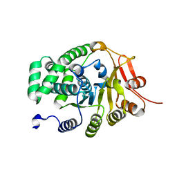
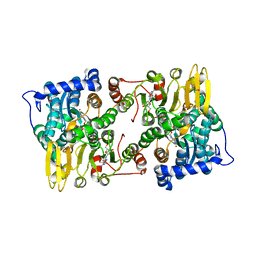
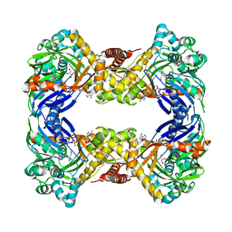
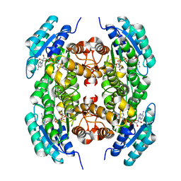
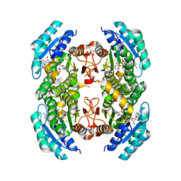
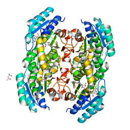
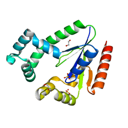
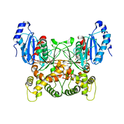
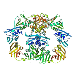
Crystal structure of an oxidoreductase from Mycobacterium smegmatis
Descriptor: Oxidoreductase, short chain dehydrogenase/reductase family protein, SODIUM ION
Authors:Arakaki, T.L, Staker, B.L, Clifton, M.C, Abendroth, J, Seattle Structural Genomics Center for Infectious Disease (SSGCID)
Deposit date:2011-09-28
Release date:2011-10-05
Last modified:2024-07-17
Method:X-RAY DIFFRACTION (1.7 Å)
Cite:Structural and functional characterization of FabG4 from Mycolicibacterium smegmatis.
Acta Crystallogr.,Sect.F, 80, 2024
3TSC
DownloadVisualize
BU of 3tsc by Molmil
Crystal structure of short chain dehydrogenase MAP_2410 from Mycobacterium paratuberculosis bound to NAD
Descriptor: (4S)-2-METHYL-2,4-PENTANEDIOL, NICOTINAMIDE-ADENINE-DINUCLEOTIDE, Putative oxidoreductase
Authors:Seattle Structural Genomics Center for Infectious Disease (SSGCID)
Deposit date:2011-09-12
Release date:2011-10-05
Last modified:2023-09-13
Method:X-RAY DIFFRACTION (2.05 Å)
Cite:Mycofactocin-associated mycobacterial dehydrogenases with non-exchangeable NAD cofactors.
Sci Rep, 7, 2017
8DV0
DownloadVisualize
BU of 8dv0 by Molmil
Crystal Structure of a Dephospho-CoA kinase from Rickettsia felis
Descriptor: 1,2-ETHANEDIOL, Dephospho-CoA kinase, SULFATE ION
Authors:Seattle Structural Genomics Center for Infectious Disease (SSGCID)
Deposit date:2022-07-27
Release date:2022-09-28
Last modified:2023-10-18
Method:X-RAY DIFFRACTION (1.4 Å)
Cite:Crystal Structure of a Dephospho-CoA kinase from Rickettsia felis
to be published
8DTC
DownloadVisualize
BU of 8dtc by Molmil
Crystal Structure of Glucokinase with bound glucose from Acanthamoeba castellanii
Descriptor: 1,2-ETHANEDIOL, Glucokinase, SULFATE ION, ...
Authors:Seattle Structural Genomics Center for Infectious Disease, Seattle Structural Genomics Center for Infectious Disease (SSGCID)
Deposit date:2022-07-25
Release date:2022-08-10
Last modified:2024-04-03
Method:X-RAY DIFFRACTION (2.25 Å)
Cite:Crystal Structure of Glucokinase with bound glucose from Acanthamoeba castellanii
to be published
8DU0
DownloadVisualize
BU of 8du0 by Molmil
Crystal Structure of NADP bound GDP-L-fucose synthase from Brucella ovis
Descriptor: GDP-L-fucose synthase, GLYCEROL, NADP NICOTINAMIDE-ADENINE-DINUCLEOTIDE PHOSPHATE
Authors:Seattle Structural Genomics Center for Infectious Disease, Seattle Structural Genomics Center for Infectious Disease (SSGCID)
Deposit date:2022-07-26
Release date:2022-08-10
Last modified:2024-04-03
Method:X-RAY DIFFRACTION (2.1 Å)
Cite:Crystal Structure of NADP bound GDP-L-fucose synthase from Brucella ovis
to be published
8DT6
DownloadVisualize
BU of 8dt6 by Molmil
Crystal Structure of DNA Polymerase III beta subunit from Elizabethkingia anophelis
Descriptor: Beta sliding clamp, CALCIUM ION, CHLORIDE ION, ...
Authors:Seattle Structural Genomics Center for Infectious Disease, Seattle Structural Genomics Center for Infectious Disease (SSGCID)
Deposit date:2022-07-25
Release date:2022-08-10
Last modified:2024-04-03
Method:X-RAY DIFFRACTION (2.35 Å)
Cite:Crystal Structure of DNA Polymerase III beta subunit from Elizabethkingia anophelis
to be published

225681

數據於2024-10-02公開中

PDB statisticsPDBj update infoContact PDBjnumon