Loading
PDBj
MenuPDBj@FacebookPDBj@TwitterPDBj@YouTubewwPDB FoundationwwPDB
RCSB PDBPDBeBMRBAdv. SearchSearch help
Search by PDB author
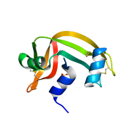
1CJQ
DownloadVisualize
BU of 1cjq by Molmil
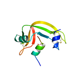
X-RAY CRYSTALLOGRAPHIC STUDIES OF THE DENATURATION OF THE DENATURATION OF RIBONUCLEASE S.
Descriptor: PROTEIN (RIBONUCLEASE S), SULFATE ION
Authors:Ratnaparkhi, G.S, Varadarajan, R.
Deposit date:1999-04-19
Release date:1999-05-07
Last modified:2023-08-09
Method:X-RAY DIFFRACTION (3 Å)
Cite:X-ray crystallographic studies of the denaturation of ribonuclease S.
Proteins, 36, 1999
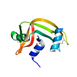
1CJR
DownloadVisualize
BU of 1cjr by Molmil
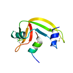
X-RAY CRYSTALLOGRAPHIC STUDIES OF DENATURATION IN RIBONUCLEASE S
Descriptor: PROTEIN (RIBONUCLEASE S), SULFATE ION
Authors:Ratnaparkhi, G.S, Varadarajan, R.
Deposit date:1999-04-19
Release date:1999-05-07
Last modified:2023-12-27
Method:X-RAY DIFFRACTION (2.3 Å)
Cite:X-ray crystallographic studies of the denaturation of ribonuclease S.
Proteins, 36, 1999
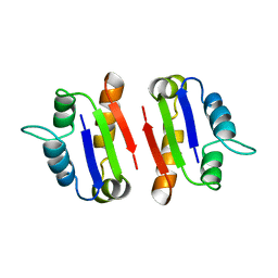
1A19
DownloadVisualize
BU of 1a19 by Molmil
BARSTAR (FREE), C82A MUTANT
Descriptor: BARSTAR
Authors:Ratnaparkhi, G.S, Varadarajan, R.
Deposit date:1997-12-25
Release date:1998-04-08
Last modified:2024-05-22
Method:X-RAY DIFFRACTION (2.76 Å)
Cite:Discrepancies between the NMR and X-ray structures of uncomplexed barstar: analysis suggests that packing densities of protein structures determined by NMR are unreliable.
Biochemistry, 37, 1998
1RNU
DownloadVisualize
BU of 1rnu by Molmil
REFINEMENT OF THE CRYSTAL STRUCTURE OF RIBONUCLEASE S. COMPARISON WITH AND BETWEEN THE VARIOUS RIBONUCLEASE A STRUCTURES
Descriptor: RIBONUCLEASE S, SULFATE ION
Authors:Kim, E.E, Varadarajan, R, Wyckoff, H.W, Richards, F.M.
Deposit date:1992-02-19
Release date:1994-01-31
Last modified:2019-08-14
Method:X-RAY DIFFRACTION (1.6 Å)
Cite:Refinement of the crystal structure of ribonuclease S. Comparison with and between the various ribonuclease A structures.
Biochemistry, 31, 1992
1RNV
DownloadVisualize
BU of 1rnv by Molmil
REFINEMENT OF THE CRYSTAL STRUCTURE OF RIBONUCLEASE S. COMPARISON WITH AND BETWEEN THE VARIOUS RIBONUCLEASE A STRUCTURES
Descriptor: RIBONUCLEASE S, SULFATE ION
Authors:Kim, E.E, Varadarajan, R, Wyckoff, H.W, Richards, F.M.
Deposit date:1992-02-19
Release date:1994-01-31
Last modified:2019-08-14
Method:X-RAY DIFFRACTION (1.6 Å)
Cite:Refinement of the crystal structure of ribonuclease S. Comparison with and between the various ribonuclease A structures.
Biochemistry, 31, 1992
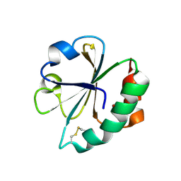
2EIR
DownloadVisualize
BU of 2eir by Molmil
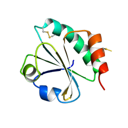
Design of Disulfide-linked Thioredoxin Dimers and Multimers Through Analysis of Crystal Contacts
Descriptor: COPPER (II) ION, Thioredoxin 1
Authors:Kobayashi, M.
Deposit date:2007-03-13
Release date:2007-09-11
Last modified:2023-10-25
Method:X-RAY DIFFRACTION (2.5 Å)
Cite:Design of Disulfide-linked Thioredoxin Dimers and Multimers Through Analysis of Crystal Contacts
J.Mol.Biol., 372, 2007
2EIO
DownloadVisualize
BU of 2eio by Molmil
Design of Disulfide-linked Thioredoxin Dimers and Multimers Through Analysis of Crystal Contacts
Descriptor: Thioredoxin 1
Authors:Kobayashi, M.
Deposit date:2007-03-13
Release date:2007-09-11
Last modified:2023-10-25
Method:X-RAY DIFFRACTION (2.6 Å)
Cite:Design of Disulfide-linked Thioredoxin Dimers and Multimers Through Analysis of Crystal Contacts
J.Mol.Biol., 372, 2007
2EIQ
DownloadVisualize
BU of 2eiq by Molmil
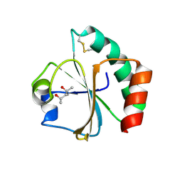
Design of Disulfide-linked Thioredoxin Dimers and Multimers Through Analysis of Crystal Contacts
Descriptor: (4S)-2-METHYL-2,4-PENTANEDIOL, COPPER (II) ION, Thioredoxin 1
Authors:Kobayashi, M.
Deposit date:2007-03-13
Release date:2007-09-11
Last modified:2023-10-25
Method:X-RAY DIFFRACTION (1.9 Å)
Cite:Design of Disulfide-linked Thioredoxin Dimers and Multimers Through Analysis of Crystal Contacts
J.Mol.Biol., 372, 2007
<12


222926

數據於2024-07-24公開中

PDB statisticsPDBj update infoContact PDBjnumon