Loading
PDBj
MenuPDBj@FacebookPDBj@TwitterPDBj@YouTubewwPDB FoundationwwPDB
RCSB PDBPDBeBMRBAdv. SearchSearch help
Search by PDB author
1LMT
DownloadVisualize
BU of 1lmt by Molmil
STRUCTURE OF A CONFORMATIONALLY CONSTRAINED ARG-GLY-ASP SEQUENCE INSERTED INTO HUMAN LYSOZYME
Descriptor: 2-acetamido-2-deoxy-beta-D-glucopyranose-(1-4)-2-acetamido-2-deoxy-beta-D-glucopyranose-(1-4)-2-acetamido-2-deoxy-beta-D-glucopyranose, CHLORIDE ION, HUMAN LYSOZYME
Authors:Matsushima, M, Song, H.
Deposit date:1995-01-13
Release date:1995-03-31
Last modified:2020-07-29
Method:X-RAY DIFFRACTION (1.6 Å)
Cite:Structure of a conformationally constrained Arg-Gly-Asp sequence inserted into human lysozyme.
J.Biol.Chem., 270, 1995
3B1J
DownloadVisualize
BU of 3b1j by Molmil
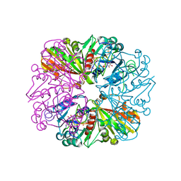
Crystal structure of Glyceraldehyde-3-Phosphate Dehydrogenase complexed with CP12 in the presence of copper from Synechococcus elongatus
Descriptor: COPPER (II) ION, CP12, Glyceraldehyde 3-phosphate dehydrogenase (NADP+), ...
Authors:Matsumura, H, Kai, A, Inoue, T.
Deposit date:2011-07-04
Release date:2012-01-11
Last modified:2023-11-01
Method:X-RAY DIFFRACTION (2.2 Å)
Cite:Structure Basis for the Regulation of Glyceraldehyde-3-Phosphate Dehydrogenase Activity via the Intrinsically Disordered Protein CP12.
Structure, 19, 2011
3B1K
DownloadVisualize
BU of 3b1k by Molmil
Crystal structure of Glyceraldehyde-3-Phosphate Dehydrogenase complexed with CP12 in the absence of copper from Synechococcus elongatus
Descriptor: CP12, Glyceraldehyde 3-phosphate dehydrogenase (NADP+), NICOTINAMIDE-ADENINE-DINUCLEOTIDE
Authors:Matsumura, H, Kai, A, Inoue, T.
Deposit date:2011-07-04
Release date:2012-01-11
Last modified:2023-11-01
Method:X-RAY DIFFRACTION (3.302 Å)
Cite:Structure Basis for the Regulation of Glyceraldehyde-3-Phosphate Dehydrogenase Activity via the Intrinsically Disordered Protein CP12.
Structure, 19, 2011
4OXF
DownloadVisualize
BU of 4oxf by Molmil
Structure of ECP in complex with citrate ions at 1.50 Angstroms
Descriptor: CITRIC ACID, Eosinophil cationic protein, FE (III) ION
Authors:Blanco, J.A, Boix, E, Moussaoui, M, Salazar, V.A.
Deposit date:2014-02-05
Release date:2015-03-04
Last modified:2023-12-27
Method:X-RAY DIFFRACTION (1.5 Å)
Cite:Structure of ECP in complex with citrate ions at 1.50 Angstroms
To be published
3C4W
DownloadVisualize
BU of 3c4w by Molmil
Crystal Structure of G protein coupled receptor kinase 1 bound to ATP and magnesium chloride at 2.7A
Descriptor: ADENOSINE-5'-TRIPHOSPHATE, CHLORIDE ION, MAGNESIUM ION, ...
Authors:Singh, P, Tesmer, J.J.G.
Deposit date:2008-01-30
Release date:2008-03-11
Last modified:2023-08-30
Method:X-RAY DIFFRACTION (2.7 Å)
Cite:Structures of rhodopsin kinase in different ligand states reveal key elements involved in G protein-coupled receptor kinase activation.
J.Biol.Chem., 283, 2008
3C4Z
DownloadVisualize
BU of 3c4z by Molmil
Crystal structure of G protein coupled receptor kinase 1 bound to ADP and magnesium chloride at 1.84A
Descriptor: ADENOSINE-5'-DIPHOSPHATE, CHLORIDE ION, MAGNESIUM ION, ...
Authors:Singh, P, Tesmer, J.J.G.
Deposit date:2008-01-30
Release date:2008-03-11
Last modified:2023-08-30
Method:X-RAY DIFFRACTION (1.84 Å)
Cite:Structures of rhodopsin kinase in different ligand states reveal key elements involved in G protein-coupled receptor kinase activation.
J.Biol.Chem., 283, 2008
<12


221051

數據於2024-06-12公開中

PDB statisticsPDBj update infoContact PDBjnumon